Showing posts with label Dermatitis. Show all posts

Periorbital dermatitis (periocular eczema)

A 45-year-old woman is seen in the allergy clinic with a complaint of red, itchy, scaly rash around the eyes for 5 months. No eye itching but she has occasional sneezing and a history of allergic rhinitis.

Examination is positive for dry skin and macular rash around both eyes, below the orbits.

What is the most likely diagnosis?

Periorbital dermatitis. Differential diagnosis includes atopic dermatitis, contact dermatitis, nonspecific dermatitis.

What would you suggest?

TRUE patch test for contact dermatitis.

If negative, then a skin test with airborne allergens could be considered.

For mild exacerbations, use topical hydrocortisone 1% for up to 1-3 weeks or Elidel. Use topical moisturizer such as Eucerin for dry areas.

Summary

Periorbital dermatitis is common and frequently difficult to treat. Patients with periorbital dermatitis often suffer severely because their disease is in such a visible location.

Predominant causes of periorbital dermatitis are:

- allergic contact dermatitis, 32-44%
- atopic eczema, 14-25%
- airborne contact dermatitis, 2-10%
- irritant contact dermatitis, 8-9%
- less frequent causes for secondary eczematous periocular skin lesions were periorbital rosacea, allergic conjunctivitis or psoriasis vulgaris

Risk factors include female gender, atopic skin diathesis and age of 40 years and older.

Common causes of periorbital allergic contact dermatitis are leave-on cosmetic products (face cream, eye shadow) and eye drops with the typical allergens being fragrances, preservatives and drugs.

Exact identification of relevant contact allergens and allergen elimination are essential for successful treatment.

Calcineurin inhibitors are the first-line therapy for facial atopic eczema.


Contact Dermatitis - Approach to Treatment (click to enlarge the image).

References

Periorbital dermatitis: causes, differential diagnoses and therapy. J Dtsch Dermatol Ges. 2010 Mar;8(3):159-66. doi: 10.1111/j.1610-0387.2009.07216.x. Epub 2009 Sep 14.
http://www.ncbi.nlm.nih.gov/pubmed/19751221
http://www.feingold.org/Research/PDFstudies/Feser2010.pdf

Textile Dermatitis and Contact Dermatitis

Author: V. Dimov, M.D., Allergist/Immunologist, Assistant Professor at University of Chicago
Reviewer: S. Randhawa, M.D., Allergist/Immunologist, Assistant Professor at NSU

A 53-year-old female is in the clinic for evaluation of her complaints of feeling of skin "burning", discomfort and mild erythema with different clothes. These symptoms mostly affect the lower part of the body but also occasionally the upper extremities. They started approximately 6 years ago after she used a hair dye with quaternary ammonium. An allergist at the time performed contact allergy testing with T.R.U.E. Test and, as per patient, this test was positive for cobalt, nickel and quaternary ammonium. It has been a struggle for her to avoid the use of products with quaternary ammonium, also known as benzalkonium chloride, which is widely used as a disinfectant. She is able to tolerate pure cotton clothes, but she has difficulty finding those in the stores.

Past medical history

Her past medical history is remarkable for the above-referenced symptoms of contact dermatitis/textile dermatitis, as well as anxiety and panic attacks.

Drug allergies include Bactrim with rash and also possiblle allergic reactions with Augmentin, Levaquin and amoxicillin, and pruritus but no rash with Motrin and Cipro.

Her current medications include Ativan b.i.d.

Her family history and social history are unremarkable.

The physical examination is normal.

Procedures: CBC, differential and CMP were ordered.

What is the most likely diagnosis?

This is a patient with a history of contact dermatitis with positive T.R.U.E. Test to cobalt, nickel and quaternary ammonium, which is also know as benzalkonium chloride, with a suspected diagnosis of textile dermatitis. Interestingly, she does not actually have a skin rash. It is mostly manifested by burning sensation and pruritus with some mild erythema. However, the presence of rash is important for the conclusive diagnosis of dermatitis. In any case, textile dermatitis can be caused by irritant reactions to textile fibers or contact allergy to textile dyes and finishing chemicals. It is important to be aware that dispersed dyes can also cause a reaction in sensitive patients.

What would you suggest in terms of diagnostic tests?

We recommended that she has CBC with differential count and CMP for basic screening for other causes of the sensation of skin "burning" and pruritus. Also, we advised her to take loratadine 10 mg p.o. daily, and to use "All Clear" detergent without any perfumes to wash her clothing. She will request the T.R.U.E. Test results from her previous allergist. There is currently no evidence of environmental sensitization to trees, gasses, mold and no evidence of symptoms for allergic rhinitis, conjunctivitis, asthma or food allergy and we decided to postpone skin prick testing to environmental allergens and food allergens at this time because it is not likely that IgE mediated allergy plays a role in this clinical setting. After we review the T.R.U.E. Test results, it may be reasonable to refer her to a dermatologist who can perform an expanded T.R.U.E. test for more contact allergens (50 or 74 rather than the initial 25 allergens included in T.R.U.E.) to see if any other allergens play a role. It is recommended to avoid the triggering substances or textiles that cause irritation in this patient.

The patch test for contact dermatitis is expensive. The cost is $30 per potential allergen, and a 20-allergen TRUE test would cost around $160-600. The CPT code is 95044, it is paid only contact dermatitis is listed as ICD-9 code for the visit (source: http://www.aaaai.org/ask-the-expert/coding-for-the-Atopy-Patch-Test.aspx). A typical charge is in the range of $160 (professional fee of $40, facility fee $120).

Related reading

Thin-Layer Rapid-Use Epicutaneous Test (patch test) misses allergens in 12.5% of patients with contact dermatitis http://goo.gl/nqUsn

Published: 07/12/2010
Updated: 02/12/2012

Mind Maps: Atopic Dermatitis

Author: V. Dimov, M.D., Allergist/Immunologist and Assistant Professor at University of Chicago
Reviewer: S. Randhawa, M.D., Allergist/Immunologist


Atopic Dermatitis Treatment - Illustrated (click here for full size image).


Allergic (atopic) march (click here to enlarge the image).

Related reading

A simplified, stepwise algorithm for the treatment of AD. Allergy, Asthma & Clinical Immunology 2011 7(Suppl 1):S4.
Adult atopic eczema - from patient's and doctor's practical point of view - BMJ, 2011.

Published: 01/24/2010
Updated: 02/03/2012

Urticaria after sexual activity

Author: V. Dimov, M.D., Allergist/Immunologist and Assistant Professor at University of Chicago
Reviewer: S. Randhawa, M.D., Allergist/Immunologist and Assistant Professor at NSU

A 25-year-old Caucasian male is at the allergy clinic for evaluation of chronic urticaria for the last one and a half years, precipitated by sexual activity, hot showers, exercise, pressure and touch. The urticarial lesions appear on his trunk, arms, and face. He reports no angioedema, shortness of breath (SOB), wheezing, abdominal pain or any other systemic symptoms. There is no history of loss of consciousness or reaction to cold weather.

His primary physician mentioned that the patient has had wheals/rash on his abdomen after having sex with his wife. The rash occurs almost every time after sex and lasts several minutes. It begins on his back and wraps around to his abdomen but has occasionally been present on his neck. It shows up during of slightly after sex and resolves within one hour afterward. He states that his wife has attempted to stop use of various lotions, body creams etc. and rarely wears perfume. His physician suspected that the rash might be due to contact dermatitis to some cosmetic product used by the patient's wife. CBCD, ESR and ANA were all within normal limits. He was referred for skin testing.

The patient shows a picture of the rash from his cell phone:


Urticarial lesions on the trunk. Image source: Wikipedia, public domain.

Past Medical History (PMH)

Negative, apart from urticaria for one and a half years.

Medications

None.

Physical examination

Normal.

What is the most likely diagnosis?

Chronic urticaria? What type?
Dermatographism?
Contact dermatitis?

What diagnostic test would you suggest?

Pressure test for dermatographic urticaria with a tongue depressor.

What happened?

The test for dermatographism was strongly positive on both forearms:


Dermatographic urticaria is sometimes called "skin writing". Image source: Wikipedia, public domain.

What treatment would you suggest?

The patient has chronic urticaria and dermatographism. The most likely explanation of his symptoms is dermatographism, although physical urticaria, and specifically cholinergic urticaria cannot be completely ruled out. Vibratory urticaria is typically observed at the friction site rather than in widespread distribution and is therefore unlikely in his case.

Contact dermatitis or a reaction to any cosmetic products is unlikely with his clinical history. Skin prick testing or patch testing is not indicated.

The patient was prescribed a H1- and H2- blocker, loratidine 10 mg po daily and ranitidine 150 mg po daily. He is to follow up with the allergy clinic in 3 months.

Summary

There is a mnemonic for fifferential diagnosis of chronic urticaria: VIP

Vasculitis, confirmed by biopsy
Idiopathic, 75% of patients
Physical, benign

Physical urticaria

Physical urticaria is defined as hives provoked by physical stimulus such as:

CDC S (mnemonic)

Cold urticaria due to cooling the skin
Dermographism due to stroking the skin
Cholinergic urticaria due to exercise, emotion, or heat
Solar urticaria due to sun exposure

Physical urticaria can be confirmed by challenge testing, and is best treated symptomatically by avoidance of provocative stimuli and antihistamines.

Testing procedures for diagnosis of physical urticarias depend on the cause (stimulus):

- Dermographism: Stroking with narrow object, e.g. a tongue depressor
- Cold urticaria: ice cube test
- Heat urticaria: test tube water at 44°C (111°F)
- Pressure urticaria: Sandbag test or a bag with heavy books (Middleton's Allergy textbook, 2 volumes)
- Vibratory urticaria: vibration with laboratory vortex for four minutes
- Cholinergic urticaria: exercise for 15-20 minutes or leg immersion in 44°C (111°F) bath
- Aquagenic urticaria: challenge with tap water at various temperatures

References

Urticaria: A Short Review. V. Dimov. Clinical Notes in Allergy and Immunology.
Chronic Idiopathic Urticaria. Current Opinion in Allergy and Clinical Immunology, Medscape, 2003.

Related reading

Flickr user: "I have a weird allergy called "Dermatography" where the skin welts up from contact"
"Young mother must wrap up all year round because she is allergic to the cold"http://goo.gl/w25WB - Cold urticaria in Daily Mail.
Self-referential illustration of dermatographic urticaria. Wikipedia, Creative Commons Attribution-Share Alike 3.0 Unported license.

Published: 03/10/2010
Updated: 03/10/2012

Interleukin 33 (IL-33)

Author: V. Dimov, M.D., Allergist/Immunologist, Assistant Professor, University of Chicago
Reviewer: S. Randhawa, M.D.

Interleukin 33 (IL-33) is a cytokine belonging to the IL-1 superfamily, it induces cells to produce type 2 cytokines.

Interleukin-1 superfamily

Interleukin-1 superfamily includes:

- IL-1α
- IL-1β and IL-1RA
- IL1F5, IL1F6, IL1F7, IL1F8, IL1F9, and IL1F10
- IL-33
- IL-18

IL-1α, IL-1β, and IL-1RA have been renamed IL-1F1, IL-1F2, and IL-1F3. IL-33 is also called IL-1F11.

Whereas IL-1 and IL-18 promote proinflammatory and TH1-associated responses, IL-33 induces the production of TH2-associated cytokines.

IL-33 was previously named NF-HEV nuclear factor (NF) in high endothelial venules (HEVs) since it was originally identified in this location. IL-33 is also called IL-1F11 to note its place as a member of IL-1 superfamily (IL-1F11).

IL-33 binds to its receptors triggering NF-κB and MAP kinase signaling pathways that drive production of type 2 cytokines (IL-5 and IL-13) from Th2 cells.

Receptors for IL-33

IL-33 binds to 2 receptors:

- ST2 (IL1RL1)
- IL-1 Receptor Accessory Protein (IL1RAP)

ST2 (IL1RL1)

IL1RL1 stands for IL-1 receptor-like 1 protein. Mutations in the gene for IL1RL1 (ST2) have been linked to atopic dermatitis and asthma.

ST2 is also called IL1RL1, T1, DER4 and Fit-2. ST2 is highly expressed on mast cells and on TH2 cells.

The ST2 gene encodes two isoforms of ST2 proteins:

- ST2L, a transmembrane form
- soluble ST2 (sST2), a secreted form that can serve as a decoy receptor of IL-33

High levels of sST2 has been found in the sera of patients with acute asthma.

IL-33 is markedly elevated in the serum of patients during an anaphylactic shock and in atopic human tissue.

IL-33 activates:

- Phospholipase D1 (PLD1)
- Sphingosine kinase 1 (SPHK1)

Phospholipase D1 (PLD1)

Phospholipase D1 (PLD1) catalyzes the hydrolysis of phosphatidylcholine (PC) to produce phosphatidic acid and choline. PC-specific PLD1 affects numerous cellular pathways, including signal transduction, membrane trafficking, and the regulation of mitosis.

Sphingosine-1-phosphate (S1P) and Sphingosine kinase 1 (SPHK1)

Sphingosine-1-phosphate (S1P) is a signaling sphingolipid. Various stimuli increase cellular levels of S1P by activation of sphingosine kinase (SPHK), the enzyme that catalyzes the phosphorylation of sphingosine.

S1P regulates angiogenesis, vascular stability and permeability.

S1P is a major regulator of trafficking of T- and B-cells. S1P interaction with its receptor S1PR1 is needed for the egress of immune cells from the lymphoiod organs (such as thymus and lymph nodes). Some immunosuppessants such as FTY720 do not let lymphocytes leave the lymph nodes through blockage of S1PR. Fingolimod works in multiple sclerosis by binding to S1P receptors and acting as highly potent antagonist (NEJM, 2012).

In the presence of IgE, IL-33 activates mast cell degranulation through phospholipase
D1 and sphingosine kinase-1.

IL-33 expression is increased in patients with anaphylaxis and atopic dermatitis.

IL-33 is a potential therapeutic target against allergy. IL-33/ST2 pathway may provide new therapeutic targets for allergic rhinitis and asthma (http://goo.gl/3utyB).

NFkB dependent and independent cytokines and chemokines

It has been suggested that whilst the production of pro-inflammatory cytokines in mast
cells is NFkB dependent, the induction of Th2 cytokines is NFkB independent.

For example, IL-1, IL-3, IL-6, TNF-alpha, MIP-2, MCP-1, and MIP-1 syntheses are dependent on NF-kB activity. IL-5, IL-13, Eotaxin-2, RANTES or TARC syntheses are are independent of the NF-kB pathway.

Abbreviations

Chemokine (C-X-C motif) ligand 2 (CXCL2) is a small cytokine belonging to the CXC chemokine family that is also called macrophage inflammatory protein 2-alpha (MIP2-alpha), Growth-regulated protein beta (Gro-beta) and Gro oncogene-2 (Gro-2).

Chemokine (C-C motif) ligand 2 (CCL2) is a small cytokine belonging to the CC chemokine family that is also known as monocyte chemotactic protein-1 (MCP-1).

Chemokine (C-C motif) ligand 4 is also known as CCL4 and MIP-1.

Chemokine (C-C motif) ligand 24 (CCL24) is a small cytokine belonging to the CC chemokine family that is also known as Myeloid progenitor inhibitory factor 2 (MPIF-2) and Eosinophil chemotactic protein 2 (Eotaxin-2). CCL24 interacts with chemokine receptor CCR3 to induce chemotaxis in eosinophils.

Chemokine (C-C motif) ligand 5 also known as CCL5 or RANTES. CCL5 was earlier called Regulated upon Activation, Normal T-cell Expressed, and Secreted, abbreviated RANTES.

Chemokine (C-C motif) ligand 17 (CCL17) is a small cytokine belonging to the CC chemokine family that is also known as thymus and activation regulated chemokine (TARC).

IL-33 role in anaphylaxis

In the presence of IgE, IL-33 induces anaphylactic shock by rapidly activating mast cell degranulation. Although mast cell activation and degranulation by IL-33 is independent of the presence of T or B cells, the presence of preformed IgE is critical. In vitro the IgE sensitization need to be for at least 16 hours, and shorter sensitization durations failed to primed the mast cells for IL-33–mediated degranulation.

What is the most potent chemokine (chemoattractant) for eosinophils?

(A) IL-5
(B) IL-8
(C) LTB4
(D) eotaxin
(E) IL-4
(F) IL-13

Answer: D.

Eosinophil chemotactic protein 2 (Eotaxin-2) is the most potent chemoattractant for eosinophils but IL-5 is the most specific stimulant of their production.

References

The cytokine interleukin-33 mediates anaphylactic shock. Pushparaj PN, Tay HK, H'ng SC, Pitman N, Xu D, McKenzie A, Liew FY, Melendez AJ. Proc Natl Acad Sci U S A. 2009 Jun 16;106(24):9773-8. Epub 2009 Jun 8.

IL-33 and its receptor ST2 play important roles in allergic rhinitis http://goo.gl/xYCga

Published: 08/25/2009
Updated: 01/07/2012

Irritant contact dermatitis to acne medication that contains benzoyl peroxide

Author: V. Dimov, M.D., Fellow, Creighton University Division of Allergy & Immunology
Reviewer: S. Randhawa, M.D., Fellow, LSU (Shreveport) Department of Allergy & Immunology

A 25-year-old Caucasian female developed worsening symptoms of facial dermatitis during the last 3 months. She started using over-the-counter hydrocortisone cream yesterday and her symptoms are better now. This is the third episode of similar complaints in the last 3 months. She reports long term use of the acne cream Proactiv, which contains benzyl peroxide, and she also uses Eucerin lotion on her face.

Medications

Her medications include Allegra (fexofenadine), Flonase (fluticasone nasal) and levothyroxine. Proactiv and Eucerin.

Past medical history

She has a long history of allergic rhinitis and conjunctivitis.

Physical examination

The physical examination is positive for facial erythema with some eyelid eczema.

What is the most likely diagnosis?

Facial dermatitis related to Proactiv, containing benzoyl peroxide. The etiology of the dermatitis is either irritant or allergic contact dermatitis to benzoyl

peroxide. Considering that she had two prior episodes that resolved and the rash is not worsening now, the most likely explanation for her condition is irritant contact dermatitis to benzoyl peroxide.

What treatment would you suggest?

The patient was advised to use hydrocortisone at the lowest of concentration of 1%, daily, on the face, for a week. She had to stop using Proactiv immediately. She could still use Eucerin daily.

Final diagnosis

Irritant contact dermatitis to acne medication that contains benzoyl peroxide.

Benzoyl peroxide (BP) is shown to be a weak allergen. In a study of 25 guinea pigs, only 5 were sensitized in the TINA test. BP is a skin irritant. However, only 11 of 155 acne patients had clinical signs of intolerance, which settled despite continued use in 10 cases.

References

Purpuric contact dermatitis to benzoyl peroxide. van Joost T, van Ulsen J, Vuzevski VD, Naafs B, Tank B. J Am Acad Dermatol. 1990 Feb;22(2 Pt 2):359-61.

Allergic contact angioedema to benzoyl peroxide. Minciullo PL, Patafi M, Giannetto L, Ferlazzo B, Trombetta D, Saija A, Gangemi S. J Clin Pharm Ther. 2006 Aug;31(4):385-7.

Allergic and irritant potential of benzoyl peroxide. Haustein UF, Tegetmeyer L, Ziegler V. Contact Dermatitis. 1985 Oct;13(4):252-7.

Contact dermatitis due to benzoyl peroxide. Morelli R, Lanzarini M, Vincenzi C, Reggiani M. Contact Dermatitis. 1989 Mar;20(3):238-9.

Related YouTube videos


Published: 01/30/2010
Updated: 01/30/2010

It's not a drug reaction but a fungal infection in an immunosuppressed patient (tinea corporis)

Author: V. Dimov, M.D., Allergist/Immunologist and Assistant Professor at University of Chicago
Reviewer: S. Randhawa, M.D., Allergist/Immunologist and Assistant Professor at LSU (Shreveport) Department of Allergy and Immunology

A 37-year-old Caucasian male with severe asthma and allergic rhinitis. He was initially placed on omalizumab but developed an anaphylactic reaction and his treatment regimen was changed to cyclosporin. He was not able to tolerate cyclosporin and has been on methotrexate for six weeks. He felt much better after switching to methotrexate. His shortness of breath improved and he was able to exercise again.

However, within a week of starting methotrexate, he noted tiny skin lesions of on his both feet. He did not pay any attention until about a week earlier when he started to notice that the lesions on his feet had expanded. They itch and he also noticed new lesions located on the left thigh and knee area, and on his left forearm.

Past medical history (PMH)

Asthma, allergic rhinitis.

Medications

Pulmicort three puffs b.i.d., Combivent prn, Zyflo 1200 mg po b.i.d., Prevacid 20 mg po b.i.d., Flonase two sprays in each nostril b.i.d., prednisone 7.5 mg po every other day. Methotrexate 15 mg every week on Thursdays.

Physical examination

Temperature 97.9, heart rate 86, respiratory rate 14, blood pressure 122/78.
Skin: He has multiple erythematous plagues which affect both feet up to the size of 2 x 3 cm with central clearing and advancing edge. He also has fungal lesions between his toes. No clear fungal infection of his nails. He also has 7 cm ringworm lesion on the inner aspect of his left knee with central clearing and a blanching edge. He has 3 cm ringworm lesion on his left forearm with central clearing and advancing edge.
Eyes: Normal. Ears: Cerumen, otherwise normal. Nose: Pale boggy turbinates on both sides. Throat: Normal.
Respiratory system clear to auscultation bilaterally.
Cardiovascular system: Clear S1, S2.
Abdomen: Soft, non-tender, non-distended. Extremities: No edema.


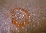
Ringworm on the arm, tinea corporis due to Trichophyton mentagrophytes. Image source: Wikipedia, CDC/Dr. Lucille K. Georg, public domain.

What is the most likely diagnosis?

Tinea corporis (ringworm) in an immunosupressed patient with severe asthma who is treated with prednisone and methotrexate.

What treatment would you recommend for this patient?

His liver function tests were normal a few weeks ago. He was prescribed Terbinafine (Lamisil) 250 mg po daily for two weeks. He is to return to our clinic in 3 weeks.

If a patient has a fungal infection of the toenails, the duration of Terbinafine (Lamisil) is 12 weeks as opposed to 2 weeks in our case.

Final diagnosis

Tinea corporis (ringworm) in an immunosupressed patient.

References

Topical Treatment of Common Superficial Tinea Infections. AFP, 2002.
Diagnosis and Management of Common Tinea Infections. AFP, 1998.
Tinea Corporis. eMedicine Specialties, 2008.

Published: 02/26/2009
Updated: 02/26/2009

Cutaneous mastocytosis in a 3-month-old girl

Author: B. Chahine, M.D., Allergist/Immunologist
Reviewer: V. Dimov, M.D., Allergist/Immunologist

A 3-month AAF presented to the hospital with a persistent rash that started at 2 weeks of age. The rash started on the neck and was described as ‘fluid filled blisters that burst and spread to other parts of the body’. Her pediatrician treated her empirically for scabies without improvement.

Associated with her progressive skin lesions, was a low-grade fever of 100-100.4 F. She returned to her PCP who treated her for impetigo but the antibiotic therapy did not resolve her progressive rash, and therefore was she referred to the hospital.

Past medical history (PMH)

GERD. Born at term, 6 lbs 2 oz, vaginal delivery without complications. Tolerating cow milk formula since birth. Immunizations: Received her Hep. B at birth. Her 2 mo. vaccines were deferred due to the rash.

Medications

Zantac po, Permethrin topical, Clindamycin po, Altebax topical ointment.

Social history (SH)

Lives with her mother, mat. GM and brother. No sick contacts, no pets. No smoking exposure.

No tobacco or alcohol use.

Family history (FH)

The mother is 20 yo, infected with HPV and has an abnormal pap smear. No other illnesses or STD. The father and paternal GM have SLE. Her brother is a healthy 4-year-old.

Physical examination

VSS
Gen: Alert well fed baby, healthy appearing, in no discomfort.
ENT: no scleral icterus, no conjunctival injection. No nasal mucosal edema. Tonsils present, no hypertrophy, no exudates.
Chest: CTA bl, RRR.
Abdomen: Soft, no distension, + BS, no organomegaly. Small reducible periumbilical hernia.
Skin: Bullous 0.5-1 cm lesions in multiple stages of healing involving face trunk and extremities. Ruptured hyperpigmented lesions healing without evidence of superinfection.

What laboratory tests would you order?

CBCD, CMP, Blood cx, HSV titers and VZV titers were all normal.

What happened?

The patient was placed in isolation and started on empiric treatment with Acyclovir for HSV. Her rubtured bullae were treated with topical antibiotics. She was started on antihistamines for her pruritis. Skin biopsy and Tzanck test performed.

What happened next?

Skin biopsy showed cutaneous (bullous) mastocytosis.

Final diagnosis

Cutaneous mastocytosis

Summary

Mastocytosis is characterized by an excessive number of apparently normal mast cells in the skin and, occasionally, in other organs. Characteristic skin lesions, called urticaria pigmentosa, are present in most patients, but clinical presentation can vary from a pruritic rash to unexplained collapse and sudden death. These lesions are typically tan to red-brown macules that appear on the trunk and spread symmetrically. Patients with mastocytosis often have a long history of chronic and acute symptoms that were unrecognized as mastocytosis. Skin lesions may or may not accompany systemic mastocytosis. Systemic disease may involve the gastrointestinal tract, the bone marrow or other organs.

Drug therapy is initiated to stabilize mast cell membranes, to reduce the severity of the attacks and to block the action of inflammatory mediators.

Mastocytosis should be suspected and the diagnostic criteria applied in patients without skin lesions if one or more of the following features is present: unexplained ulcer disease ormalabsorption, radiographic or technetium 99 bone scan abnormalities, hepatomegaly, splenomegaly, lymphadenopathy, peripheral blood abnormalities, and unexplained flushing or anaphylaxis. Elevations in the levels of plasma or urinary histamine, or histamine metabolites, urine prostaglandin D2 metabolites, plasma thromboxane B2, plasma (total) mast cell tryptase, or IL-6 are not definitive diagnostic findings, but are consistent with the diagnosis of mastocytosis

A principal objective of treatment in all categories of mastocytosis is the control of mast cell mediator-induced signs and symptoms. The mainstay of therapy is histamine H1 and H2 blockers and the avoidance of triggering factors such as minor skin trauma, hot or cold water that can lead to massive systemic histamine release and systemic symptoms.

Most prominent among these are systemic hypotension, gastric hypersecretion, GI cramping, and pruritus.

H1-receptor antagonists such as the classic antihistamine hydroxyzine, or non-sedating antihistamines such as loratadine or fexofenadine, are instituted to reduce pruritus and flushing. If this is insufficient, the addition of an H2 antagonist, such as ranitidine, cimetidine or famotidine, may be beneficial. One approach is to administer a non-sedating antihistamine during the day, and a potent sedating antihistamine at bedtime. Even with these medications, many patients continue to complain of musculoskeletal pain, headaches, and flushing, in part because of the inability of histamine antagonists to block the effects of high levels of histamine and because of the presence of other mast cell mediators. There may thus be some value in adding aleukotriene-modifying agent.

Disodium cromoglycate (cromolyn sodium) inhibits degranulation of mast cells and has some efficacy in mastocytosis, particularly in the relief of GI complaints.

Epinephrine is used to treat episodes of systemic hypotension. Patients should be taught to administer this medication themselves. If subcutaneous epinephrine is inadequate, intensive therapy for systemic hypotension, as for anaphylaxis, should be instituted. Patients with recurrent episodes of hypotension may be given H1 or H2 antihistamines to reduce the severity of attacks. Episodes of profound hypotension may be spontaneous but have also been observed after insect stings and administration of contrast media.

References

Mastocytosis. eMedicine Specialties > Dermatology > Benign Neoplasms, 2008.
Cutaneous and Systemic Manifestations of Mastocytosis. AFP, 1999.
Mastocytosis. Merck Manual.
Middleton's Allergy: Principles and Practice, 7th ed (chapter 60).
Mastocytosis in Children Is Associated with Mutations in c-KIT. Nature, 2010.
Neuropeptide blood levels correlate with mast cell load in patients with mastocytosis http://goo.gl/vlQhm
Mastocytosis. NEJM blog, 2011.
Mastocytosis - Where are we now? World Allergy Organization summary, 2012.
Proposed diagnostic algorithm for patients with suspected mastocytosis. Allergy, 2014 http://buff.ly/1hZ4ayz

Mast Cell Disorders (presentation on Google drive):


Published: 03/24/2009
Updated: 03/12/2012

An "Allergic Rash" Which Turned Out to be Seborrheic Dermatitis

Author: V. Dimov, M.D., Allergist/Immunologist and Assistant Professor at University of Chicago
Reviewer: S. Randhawa, M.D., Allergist/Immunologist and Assistant Professor at LSU (Shreveport) Department of Allergy and Immunology

A 21-year-old Caucasian male called the allergy clinic to make an appointment for an "allergic skin rash" affecting his face, upper torso and right arm. The rash started above his left eye 2 years ago and spread gradually. It is marked by pruritus even during the night. He has used an aloe vera-based cream and mineral oil daily for the last year without significant improvement. His symptoms are slightly worse in the fall and when he has a hot shower.

The patients wants to have allergy testing to determine what is the cause if his rash.

PMH

Unremarkable.

Social history

Non-smoker; denies IV drug use and high-risk sexual behavior.

Medications

An aloe vera-based cream and mineral oil daily.

Physical examination

VSS, temperature 97.2, heart rate 79, respiratory rate 14, blood pressure 102/60
Thin male in no apparent distress.
Skin: Maculo-papular rash affecting his face and upper back and shoulder blades. Dry scales, oily skin, yellow crusts and scratch marks.
Eyes: Normal.
External auditory canal: Skin scales. Tympanic membranes: Normal.
Nose: Normal.
Lymph nodes: not palpable.
Respiratory system: Clear to auscultation bilaterally.
Cardiovascular system: Clear S1S2.
Gastrointestinal system: +BS, NT, ND.
Extremities: No cyanosis, clubbing, or edema.


Seborrheic dermatitis. Image source: Wikipedia, GNU Free Documentation License.

What is the most likely diagnosis?

This is a male with seborrheic dermatitis with evidence of secondary infections, both fungal and likely impetigo as evidenced by the yellow crusts around the scratch marks.

What laboratory workup would you order?

An HIV test.

He denies having sex with men, IV drug abuse, blood transfusions or any history of positive HIV testing in the past and he declines an HIV test now. We feel that the most likely diagnosis is seborrheic dermatitis with secondary infection.

What treatment would you recommend for this patient?

The patient was reassured that his rash is not of allergic origin and therefore further testing with skin prick tests or specific IgE is not recommended.

He was prescribed:

Augmentin (amoxicillin clavulanate) 875 mg po b.i.d. for 10 days.
Prednisone 40 mg po daily for four days, then stop.
Hydrocortisone cream 1% cream t.i.d., apply topically for no longer than a month.
Nizoral shampoo 2% applied daily to the scalp, apply for 5 minutes, then rinse.
Nizoral 2% cream applied b.i.d. for four weeks.

If there is no response to this treatment at the one-month follow-up appointment, the patient will be referred to dermatology.

Final diagnosis

Seborrheic dermatitis

What did we learn from this case?

Seborrheic dermatitis is a chronic inflammatory disorder which affects areas of the head and trunk where sebaceous glands are most prominent. Scalp seborrhea varies from mild dandruff to dense, diffuse, adherent scale.

Lipophilic yeasts of the Malassezia genus contribute to development of this condition.

Treatment of associated fungal infection is an integral part of the management of moderate to severe seborrheic dermatitis.

References

Treatment of Seborrheic Dermatitis. Johnson et al. Am Fam Physician 2000;61:2703-10,2713-4.
Seborrheic Dermatitis. American Academy of Dermatology.
Seborrheic Dermatitis. Samuel Selden, eMedicine Specialties, Dermatology, 2007.
Dermatitis. Merck Manual.
Seborrheic Dermatitis. Luigi Naldi, M.D., and Alfredo Rebora, M.D. NEJM, Volume 360:387-396 January 22, 2009 Number 4.

Published: 12/23/2008
Updated: 01/21//2009

Semen Allergy: Diagnosis and Treatment

Author: V. Dimov, M.D., Assistant Professor, Allergist/Immunologist, University of Chicago; A. Bewtra, M.D., M.B.B.S., Professor, Creighton University Division of Allergy & Immunology
Reviewer: S. Randhawa, M.D., Allergist/Immunologist, Fort Lauderdale, Florida

A 26-year-old Caucasian female is referred to the allergy clinic for skin prick testing for suspected semen allergy. For the last 5 years, she reports symptoms when she has unprotected intercourse with her husband. She has no symptoms when she has a protected intercourse. The typical symptoms are burning, swelling, and redness outside the vaginal area that start 10 to 15 minutes after intercourse and last from hours to days. The patient reports that on the first day of intercourse, she will have symptoms for a few hours, but if she has intercourse the very next day, then the symptoms are worse and last for days rather than hours. She and her husband expressed an interest in conceiving and had already seen an OB/GYN physician who prescribed Clomid (Clomiphene). Since then the patient had a menstrual period and she is here for skin prick testing to semen allergy. Her husband brought a container with previously collected semen fluid to the clinic.

Past medical history (PMH)

Mild asthma, allergic rhinitis.

Medications

Veramyst (fluticasone) nasal spray, albuterol prn (uses very rarely), Clomid, loratidine (stopped six days ago for the skin prick testing).

Physical examination

Stable vital signs (VSS), normal physical examination.

What would you do next?

Skin prick testing with positive control with histamine, negative control with normal saline and semen fluid.

Test results: The patient’s skin prick testing showed a positive skin control with histamine, a wheal of 5 x 5 mm, a flare of 15 x 15 mm, a negative control with saline 0 x 0 mm for both wheal and flare, and and a positive reaction to the semen fluid with 4 x 3 mm for the wheal and 10 x 7 mm for the flare. We read the test after 15 minutes.

What would you recommend for this patient?

The patient has a diagnosis of semen allergy confirmed by medical history, symptoms timeline and positive skin prick testing.

In order to avoid symptoms, we advised the patient and her husband to have protected sexual intercourse with condoms in the future.

In terms of conceiving a baby, we recommend that she should work closely with her OB/GYN and once she begins to have ovulatory cycles, as determined by a pelvic ultrasound, the plan is for her to be placed on prednisone for 7 to 10 days around the expected time of ovulation. A possible consideration, if this does not work, would be intrauterine insemination with washed semen.

After she finalizes her plan about conception, we will change her intranasal steroid to budesonide, which is a category B medication in pregnancy. She is to follow up prn with us regarding further follow-up with her OB/GYN and the allergy clinic here.

Final diagnosis

Semen allergy.

What did we learn from this case?

Women with seminal plasma protein allergy (SPPA) have an immunologic response to human semen. The immunological mechanism of semen allergy is a type I hypersensitivity reactions.

Symptoms vary from local inflammation and pruritus to systemic anaphylaxis after exposure. The first case was documented in Germany in 1958.

Patients with SPPA often have recurrent vaginitis associated with intercourse and are unresponsive to traditional therapies. Semen allergy should be on the list of differential diagnoses for recurrent vaginitis in sexually active women. SPPA may also present as ‘vulvar vestibulitis syndrome’ or ‘burning semen syndrome’.

The gold standard of diagnosis is absence of symptoms with condom use.

Treatment may involve artificial insemination for those seeking pregnancy, oral corticosteroids, immunotherapy, or antihistamines, rather than use of a condom or abstinence.

Systemic and localized reactions can be prevented by use of condoms. However, partners should always be instructed on the use of an automatic epinephrine injector (EpiPen) and it should be available.

Conception

In patients who wish to conceive, and/or condoms are unacceptable, immunotherapy with seminal fluid can be performed. Alternatively, patients can conceive by artificial insemination with washed spermatozoa.

Here are the suggested diagnostic steps from AAAAI Ask the Expert and Dr. Bernstein (http://buff.ly/16yXcPD):

1) Test by PST to whole seminal fluid. This requires letting the ejaculate liquefy for 30 min and centrifuge for 10 minutes to separate seminal fluid from spermatozoa. Also test sexual partner as a negative control. You do not have to sterilize the seminal fluid for prick testing. Intracutaneous testing to whole seminal fluid should not be performed as it will cause an irritant response.

2) Both the patient and her sexual partner should be screened for STD's.

3) Some women have trouble with all men whereas other have symptoms with only one.

4) For localized seminal plasma hypersensitivity, skin testing is not always concordant with serologic testing. Patients can send their serum and their sexual partner's serum and a 5 day pooled ejaculate to Dr. Bernstein's laboratory to test for sIgE to whole seminal plasma.

5) There is cross reactivity between dog allergens and seminal plasma.

6) Patients with localized seminal plasma hypersensitivity should first undergo intravaginal graded challenge; dilute whole seminal fluid with sterile water to 1:100,000 dilution and instill 10 cc of volume intravaginally every 10-15 minutes up to a `1:1 dilution to see if this alleviates symptoms. There are reported cases that this may be effective. If not then the patient may be a candidate for subcutaneous desensitization to relevant fractionated seminal plasma proteins.


References

Allergy to coitus. Jones WR. Aust N Z J Obstet Gynaecol. 1991 May;31(2):137-41.
Human Seminal Plasma Protein Allergy: A Diagnosis Rarely Considered. Barbara GlaserLudman. Journal of Obstetric, Gynecologic, & Neonatal Nursing, Volume 28 Issue 4, Pages 359 - 363, 2006.
Seminal Fluid. AAAAI Ask the Expert, 2003.
Immunologic disorders of the female and male reproductive tract. Annals of Allergy, Asthma & Immunology, Volume 108, Issue 6 , Pages 390-395, June 2012.
IgE-Mediated Allergy against Human Seminal Plasma. Weidinger S, Ring J, Köhn J. Immunology of Gametes and Embryo Implantation. Chem Immunol Allergy. Basel, Karger, 2005, vol 88, pp 128-138.
Allergy to human seminal plasma: a presentation of six cases. Kroon S. Acta Derm Venereol. 1980;60(5):436-9.
Diagnosis and treatment of human seminal plasma hypersensitivity. Lee-Wong M, Collins JS, Nozad C, Resnick DJ. Obstet Gynecol. 2008 Feb;111(2 Pt 2):538-9.
Anaphylaxis to husband's seminal plasma and treatment by local desensitization. Lee J, Kim S, Kim M, Chung YB, Huh JS, Park CM, Lee KH, Kim JH. Clin Mol Allergy. 2008 Dec 5;6:13.
Management of seminal fluid hypersensitivity reactions. AAAAI Ask the Expert, 2011.
The Diagnosis and Management of Anaphylaxis Practice Parameter: 2010 Update. J Allergy Clin Immunol 2010; 126(3):477-522. The segment dealing with seminal fluid allergy begins on page 480.e17, under the heading "Seminal Fluid Anaphylaxis."

Related reading

Frequent Sex Cures Women's Semen Allergy. Fox News, 11/2008.
Woman discovers she's allergic to her husband. Daily Mail, 2009.
A 5-Year Followup of Human Seminal Plasma Allergy http://bit.ly/M3mDMX
Involvement of the dog allergen Can f 5 in a Case of Human Seminal Plasma Allergy http://goo.gl/cQSvj
Successful intravaginal graded challenge after a systemic reaction with skin prick testing to seminal fluid http://buff.ly/XKq4Ak
Vaginal pain with intercourse: is it seminal fluid allergy? AAAAI Ask the Expert answers: http://buff.ly/16yXcPD
Skin test may not be reliable for diagnosis of sensitization to seminal fluid proteins. Caused by PGs rather than IgE http://buff.ly/1wES08b

Published: 12/23/2008
Updated: 05/07/2013

How to Diagnose Latex Allergy

Author: V. Dimov, M.D., Allergist/Immunologist and Assistant Professor at University of Chicago
Reviewer: S. Randhawa, M.D., Allergist/Immunologist and Assistant Professor at NSU

A 22-year-old African American female is referred to the clinic for suspected latex allergy. She is to start work as a nurse at the local medical center and she is referred by occupational medicine. The patient has a history of intolerance to latex. Three years ago, she was taking care of her ailing grandmother and was using latex gloves when she reacted with “bumps” on her hands. She also reports that she used an eyelash extender with latex-based glue 5 months ago and she had eyelid swelling after she removed the eyelashes. She also reports history of irritation and itching in the vaginal area when using condoms. A month ago, she was at the hairdresser who was applying hair extenders to her scalp using a latex-based glue. Within six minutes of applying the extenders, she started to complain of feeling a lump in her throat; she started to have shortness of breath, and hives which affected her face, arms, legs, and back. She was sent to the ER where she was given an injection of Benadryl and injection of corticosteroid. Her symptoms resolved within an hour. She has not used the same hair extenders with latex glue or the eyelash application with latex base since then. She denies any reactions to food that may closely react with latex allergens, including kiwi, papaya, passion fruit, bananas, or almonds.

Past medical history (PMH)

Negative.

Medications

None.

Social history

Nonsmoker. No pets. Nursing student.

Family history

No history of allergic disease or reaction to latex.

Physical examination

Vital signs stable. Normal nose and throat exam. Respiratory system: Clear to auscultation bilaterally. Cardiovascular system: Clear S1, S2. Abdomen: Soft, non-tender, non-distended. Extremities: no edema. Skin: no rashes.

What tests would you suggest?

Skin prick testing with latex. If negative, ImmunoCAP test for specific IgE for latex allergens.


Sample form for latex skin prick testing (click to enlarge).

What happened?


Test results (click to enlarge).

Skin prick testing with latex was done using the standard protocol at the clinic. She reacted to histamine with 5 x 5 mm wheal, erythema 20 x 15 mm. She reacted to "wet glove one" with a wheal 5 x 6 mm, erythema 25 x 25 mm. She reacted to "wet glove one solution" with a 3 x 7 mm, erythema 15 x 20 mm. She reacted to "wet glove 2" with a wheal 3 x 10 mm, erythema 15 x 20 mm. She reacted to "wet glove 2 solution" with a wheal 1 x 1 mm, erythema 2 x 2 mm. She reacted to "dry glove" with wheal 2 x 2 mm, erythema 3 x 3 mm. The reaction to saline was negative.

What treatment would you suggest?

This is a patient with latex allergy and she was advised to avoid all latex products including gloves, latex-based glue, hair extenders, condoms, etc. She was advised to discuss workplace avoidance measures and also to avoid foods, which may cross react with latex. She was prescribed an EpiPen and education was provided how to use the EpiPen. Considering the relatively strong reaction to latex during the skin prick test, we gave her Alavert (loratadine) oral dissolving tablet 10 mg x 1 and she was discharged from the clinic without symptoms. A comprehensive list of latex-containing products and cross-reactive foods was provided to the patient.

Final diagnosis

Latex allergy.

Summary

Latex is most often referred to the cytoplasmic exudate of the Hevea brasiliensis tree, hence the name Hev b allergens. There are more than 250 latex proteins but only 13 proteins have been characterized and designated as Hev b allergens. Skin prick reactivity to Hev b 5, 6, 7 identifies 93% of workers allergic to latex.


Latex being collected from a "wounded" rubber tree. Image source: Wikipedia, public domain.


Hevea brasiliensis (Rubber Tree). Image source: Wikipedia, public domain.


Latex allergy, a mind map diagram (click to enlarge).

Tests to rule out latex allergy

- Skin prick test with the liquid from the soaked latex glove, and then if that is negative, apply a wet glove to the forearm and prick through the glove. If all of the above are negative using the same gloves the patient has used in the past it would make the diagnosis of latex allergy unlikely. This should be followed by a challenge when appropriate.

- Specific IgE blood test may be helpful but the skin testing is the preferred diagnostic method. sIgE for latex allergy has much lower sensitivity than previously reported - sensitivity was 35%; specificity 98% (Ann of Allergy, Asthma, Imm, 2012).  Sensitivity, specificity, NPV, and PPV of latex-specific IgE assay are 91, 72, 96 and 50%, respectively (http://goo.gl/9gX0F).

- Pre- and post- pulmonary function tests as objective confirmation of obstruction if a patients complains of shortness of breath but did not exhibit visible cutaneous manifestations

An approach to performing a test for immediate hypersensitivity to latex:

1. Prick test with the solution obtained from a soaked latex glove left in saline for one hour. Await 15 minutes.
2. If test 1. is negative, place a wet latex glove on the arm for 15 minutes.
3. If there is no response to test 2., prick through the wet latex glove. Await 15 minutes.
4. Simultaneously draw an ELISA for IgE-anti-latex (ImmunoCAP). Await the results of the ELISA before clearing for the surgical procedure.

Evaluation of patient with possible latex allergy. AAAAI Ask the Expert, 2012.

35% of sensitized patients develop allergic reactions to fruits and vegetables that contain proteins that cross-react with latex allergens (passion fruit, kiwi, avocado, banana, chestnuts, papaya, mango, tomato, and wheat).

Patients with history of anaphylaxis should be prescribed emergency epinephrine kits.

References

Latex Allergy: A Short Review. V. Dimov. Allergy Cases, 2008.
Latex Allergy. AFP, 1998.
Latex Allergy. eMedicine Specialties > Emergency Medicine > Allergy & Immunology, 2008.
Latex allergy. Constance H Katelaris. MJA 2006; 185 (6): 339.
Hidden Hazard: Hospitals Target Lurking Latex. WSJ, 02/2008 (subscription may be required).
IgE-mediated latex allergy - An exciting and instructive piece of allergy history. http://goo.gl/i5Tn
Profilin may be a pan-allergen among plants that crossreacts between pollen, fruits, vegetables and latex http://goo.gl/ZUPRQ

Multiple choice questions

Chapter 58: Latex Allergy. Allergy and Immunology Review Corner: Chapter 58 of Pediatric Allergy: Principles & Practices, edited by Donald Y.M. Leung, et al.

Patient information

Latex Allergy. National Institutes of Health.
Latex allergy. Mayo Clinic staff.
Tips to Remember: Latex allergy. AAAAI.

Related reading

Knowing your allergies can save your life - there are different types of latex allergy. Arizona Daily Sun, 2011.
Anaphylaxis caused by latex surgical gloves immediately after starting surgery. Korean J Anesthesiol. 2010 Dec;59(Suppl):S99-S102.
Latex Medical Gloves: Time for a Reappraisal. Int Arch Allergy Immunol 2011;156:234-246 (DOI: 10.1159/000323892).
Diagnosis of latex allergy. AAAAI Ask the Expert, 2011.
Contact dermatitis to latex surgical gloves? There are limited choices for non-latex gloves: vinyl or nitrile. AAAAI, 2011. Hypersensitivity reactions due to nitrile gloves - presence of latex was the cause of the symptoms (JACI, 2012).
Latex immunotherapy: state of the art. Guidelines do not consider allergy to latex as indication to desensitization http://goo.gl/j5UaA

Published: 04/15/2009
Updated: 07/15/2012

Correct diagnosis is keratosis pilaris rather than an "allergic rash"

Author: V. Dimov, M.D., Allergist/Immunologist and Assistant Professor at University of Chicago
Reviewer: S. Randhawa, M.D., Allergist/Immunologist and Assistant Professor at LSU (Shreveport) Department of Allergy and Immunology

A 15-year-old Caucasian female is self-referred to the allergy clinic because of an "allergic rash" for six years affecting the dorsal aspect of both arms, occasionally the back and the legs. She also has history ofdermatographism but does not report any symptoms of asthma, allergic rhinitis, or food allergies. She also reports itching with the rash including during the night and for that, she takeshydroxyzine at night. The rash has been ongoing for six years with waxing and waning episodes throughout the years. The rash present throughout all seasons. She occasionally uses baking soda in the bathtub and she feels that it helps the rash. Skin prick testing was done by another allergist 6 months ago and was positive for cat. The patient saw a dermatologist 2 years ago who did a biopsy, but she does not remember the diagnosis, the biopsy report was "normal."

Past medical history (PMH)

Chronic rash for 6 years.

Medications

Hydroxyzine one tablet po q.h.s. triamcinalone 0.1% cream on the affected areas prn.

Social history

Passive tobacco smoke exposure. Pets: She has two cats and three dogs.

Family history

Father with chronic skin rashes without a clear diagnosis.

Physical examination

Vital signs stable.
Skin: There are erythematous papules the size of 1 to 2 mm, that affect the dorsal aspects of both forearms with the visual effect of sandpaper. There are occasional areas of dry skin and similarerythematous papules on both lower extremities.
Normal nose and throat exam. Respiratory system: Clear to auscultation bilaterally. Cardiovascular system: Clear S1, S2. Abdomen: Soft, non-tender, non-distended. Extremities: no edema.


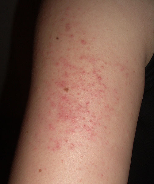
Keratosis pilaris rubra on the right upper arm. Image source: Wikipedia, Irja, Creative Commons Attribution ShareAlike 2.0 License.

Does she have an allergic condition? What is the most likely diagnosis?

Keratosis pilaris.

What tests would you suggest?

No diagnostic tests are currently indicated. Keratosis pilaris is a clinical diagnosis.

What treatment would you suggest?

Keratosis pilaris is a common genetic condition for which local treatments can be used. A brochure was provided to the patient and she was instructed to use a Lac-Hydrin and other topical treatments available in the brochure. She was advised to use triamcinalone only sparingly for her keratosis pilaris lesions and to follow with a dermatologist for dermal microabrasion in the spring so she can have a cosmetically acceptable appearance to her during the summer months when she wears short sleeves.

What happened?

There is no evidence of any other allergic disease at the moment and therefore we are going to follow up with the patient prn when she needs to see us. She was recommended to follow up with a dermatologist for her keratitis pilaris.

Final diagnosis

Keratosis pilaris

References

Keratosis Pilaris. eMedicine Specialties > Dermatology > Diseases of the Adnexa.
Keratosis pilaris. DermNet NZ.

Patient information

Keratosis pilaris. MayoClinic.com reprints.
Keratosis pilaris. U.S. National Library of Medicine.

Glytone KP Kit is an OTC therapy:


Published: 03/03/2009
Updated: 02/03/2015

Latex Allergy: Brief Review

Author: V. Dimov, M.D., Allergist/Immunologist and Assistant Professor at University of Chicago
Reviewer: S. Randhawa, M.D., Allergist/Immunologist and Assistant Professor at NSU

Latex is an emulsion of polymer microparticles and it may be natural or synthetic. Natural rubber latex (NRL) is the milky sap of many plants that coagulates on exposure to air.


Natural rubber latex (NRL) being collected from a "wounded" rubber tree. Image source: Wikipedia, public domain.

Latex is most often referred to the cytoplasmic exudate of the Hevea brasiliensis tree, hence the name Hev b allergens. There are more than 250 latex proteins but only 13 proteins have been characterized and designated as Hev b allergens. Skin prick reactivity to Hev b 5, 6, 7 identifies 93% of workers allergic to latex.


Hevea brasiliensis (Rubber Tree). Image source: Wikipedia, public domain.

Use of universal precautions for prevention of blood-borne infections results in exposure of health care workers to latex proteins via latex gloves. Latex has been around for more than a century but its use exploded with the AIDS epidemic in the 1980s. The material was found to strong enough to hold up after hours of surgery and offered the best protection against blood-borne diseases. The use of gloves increased almost 20 times between 1987 and 1997 (from 12 billion pairs to 200 billion).


Latex allergy, a mind map diagram (click to enlarge).

Risk factors

Risk of latex allergy is related to duration of exposure and cumulative use of latex-containing gloves. Corn starch powder in the gloves acts as an airborne vehicle for adherent latex proteins. There is a well-defined dose-response relationship between duration and numbers of gloves used and prevalence of latex sensitization. For example, in a study of dental hygienist students, none of first-year and 10% of 4th-year students were skin test−positive to latex extract.

Approximately 1% of newly hired health care workers are skin prick test−positive to latex. Hand washing disrupts the skin barrier and facilitate sensitization to latex.

Patients affected by latex allergy are especially those who have been exposed through multiple surgeries.

Symptoms

Less than 1% of the general population is sensitized to latex (the scientifically correct name is natural rubber latex). In contrast, 3-17% of health-care workers are sensitized. Latex may trigger allergic reactions in as many as one in 10 people who are exposed to it. By some estimates, 15% of medical workers are allergic to latex.

“Latex allergy” is defined as presence latex−specific IgE with symptoms of IgE-mediated reaction to latex:
- contact urticaria begins minutes after putting on latex gloves in 11% of sensitized workers
- nasal symptoms and wheezing in 15%. Respiratory symptoms are caused by latex proteins that adhere to cornstarch powder and disperse during glove changes.

Cross-reactions with fruits and vegetables

35% of sensitized patients develop allergic reactions to fruits and vegetables that contain proteins that cross-react with latex allergens:

- bell pepper and olive (Hev b 2)
- kiwi, potato (Hev b 5)
- avocado, banana, chestnut (Hev b 6)
- tomato and potato (Hev b 7)
- wheat
- papaya
- mango

Diagnosis of Latex Allergy

Skin prick test (SPT) with a latex extract is more sensitive and specific than commercial specific IgE immunoassays. However, there are no standardized skin test extracts and the allergists have to prepare the extracts for SPT themselves.

sIgE for latex allergy has much lower sensitivity than previously reported - sensitivity was 35%; specificity 98% (Ann of Allergy, Asthma, Imm, 2012). Sensitivity, specificity, NPV, and PPV of latex-specific IgE assay are 91, 72, 96 and 50%, respectively (http://goo.gl/9gX0F).

An approach to performing a test for immediate hypersensitivity to latex:

1. Prick test with the solution obtained from a soaked latex glove left in saline for one hour. Await 15 minutes.
2. If test 1. is negative, place a wet latex glove on the arm for 15 minutes.
3. If there is no response to test 2., prick through the wet latex glove. Await 15 minutes.
4. Simultaneously draw an ELISA for IgE-anti-latex (ImmunoCAP). Await the results of the ELISA before clearing for the surgical procedure.

Evaluation of patient with possible latex allergy. AAAAI Ask the Expert, 2012.

Treatment

Avoidance of exposure by a transfer to a latex-safe area in the hospital.

Control of ambient exposure by use of non−natural latex gloves and powder-free gloves by co-workers.

Future dental and surgical procedures should be performed in latex-safe facilities. Patients with history of anaphylaxis should be prescribed emergency epinephrine kits.

Johns Hopkins Hospital announced in January 2008 that it plans to end the use of all latex gloves and almost all latex medical products. It has switched to sterile neoprene and polyisoprene gloves for all surgery despite the fact that the new gloves cost 30-50% more.

Latex allergy is rarely seen in the U.S currently due to the diminished use of latex gloves in health care.

References


How to Diagnose Latex AllergyLatex Allergy. AFP, 1998.
Latex Allergy. eMedicine Specialties > Emergency Medicine > Allergy & Immunology, 2008.
Allergy and Immunology MKSAP, 3rd edition.
Latex allergy. Constance H Katelaris. MJA 2006; 185 (6): 339.
Hidden Hazard: Hospitals Target Lurking Latex. WSJ, 02/2008 (subscription may be required).
Latex avoidance in children with spina bifida prevents latex sensitization and latex allergy. http://www3.interscience.wiley.com/journal/123592239/abstract
IgE-mediated latex allergy - An exciting and instructive piece of allergy history.http://goo.gl/i5Tn
Profilin may be a pan-allergen among plants that crossreacts between pollen, fruits, vegetables and latex http://goo.gl/ZUPRQ

Multiple choice questions

Chapter 58: Latex Allergy. Allergy and Immunology Review Corner: Chapter 58 of Pediatric Allergy: Principles & Practices, edited by Donald Y.M. Leung, et al.

Patient information

Latex Allergy. National Institutes of Health.
Latex allergy. Mayo Clinic staff.
Tips to Remember: Latex allergy. AAAAI.

Related reading

Knowing your allergies can save your life - there are different types of latex allergy. Arizona Daily Sun, 2011.
Anaphylaxis caused by latex surgical gloves immediately after starting surgery. Korean J Anesthesiol. 2010 Dec;59(Suppl):S99-S102.
Latex Allergy in OR Workers Decline - from 14.1% in 1998 to 3.9% in 2009. AAAAI 2011 Meeting.
Latex Medical Gloves: Time for a Reappraisal. Int Arch Allergy Immunol 2011;156:234-246 (DOI: 10.1159/000323892)
Contact dermatitis to latex surgical gloves? There are limited choices for non-latex gloves: vinyl or nitrile. AAAAI, 2011. Hypersensitivity reactions due to nitrile gloves - presence of latex was the cause of the symptoms (JACI, 2012).
Latex immunotherapy: state of the art. Guidelines do not consider allergy to latex as indication to desensitization http://goo.gl/j5UaA

Published: 02/04/2008
Updated: 07/15/2012