Author: V. Dimov, M.D., Allergist/Immunologist and Assistant Professor at University of Chicago
Reviewer: S. Randhawa, M.D., Allergist/Immunologist and Assistant Professor at NSU, Fort Lauderdale
A 25-year-old female was referred for evaluation of urticaria and lip angioedema after being treated with terbinafine (Lamisil) for 14 days for ringworm. One day after the last dose of terbinafine, she developed hives that progressed to affect her whole body. No systemic symptoms. She developed lip swelling 2 days later and the hives lasted a total of 3 days. They resolved with cetiririzine and oral steroid.
Past Medical History, Past Surgical History: Negative.
Current Medications: None.
Family Medical History and Social History: Not contributory.
Review Of Systems: 12/14 systems were reviewed, negative apart from history of present illness above.
Physical Examination: Unremarkable, no hives.
What is the most likely diagnosis?
Acute urticaria (hives) and lip angioedema (swelling) likely due to adverse reaction to terbinafine (Lamisil). The reaction can happen soon after stopping the medication, or after taking it for weeks. There is no evidence of chronic urticaria, blistering or systemic symptoms. No hives or angioedema now. Food allergy is unlikely with her history, but we can do a skin test for food and aiborne allergens for completeness.
What management would you suggest?
Take Claritin or Zyrtec 10 mg po daily for 2 weeks. Do not take terbinafine (Lamisil) or related medications in the future. An allergy to terbinafine (Lamisil) was added to her allergies list in EMR.
A graded dose drug challenge can be considered in the future if she must take terbinafine and there are no alternative options.
Summary
Terbinafine is an allylamine antifungal agent widely used to treat dermatophyte onychomycosis and dermatomycoses. Br J Dermatol. included 10 case reports of severe cutaneous adverse reactions associated with terbinafine therapy which required discontinuation of the antifungal agent: erythema multiforme, erythroderma, severe urticaria, pityriasis rosea and worsening of pre-existing psoriasis.
References:
Cutaneous adverse effects associated with terbinafine therapy: 10 case reports and a review of the literature. Gupta AK, Lynde CW, Lauzon GJ, Mehlmauer MA, Braddock SW, Miller CA, Del Rosso JQ, Shear NH. Br J Dermatol. 1998 Mar;138(3):529-32.
Association between terbinafine and arthralgia, fever and urticaria: symptoms or syndrome?
van Puijenbroek EP, Egberts AC, Meyboom RH, Leufkens HG. Pharmacoepidemiol Drug Saf. 2001 Mar-Apr;10(2):135-42.
Published: 08-20-2013
Updated: 09-17-2013
Showing posts with label Urticaria. Show all posts
Showing posts with label Urticaria. Show all posts

Hives (urticaria) and angioedema - patient information
Author: V. Dimov, M.D., Allergist/Immunologist and Assistant Professor at University of Chicago
Reviewer: S. Randhawa, M.D., Allergist/Immunologist and Assistant Professor at NSU
Hives (also called urticaria) are red, itchy, raised areas of the skin that can range in size and appear anywhere on your body. They often move from place to place.
Acute urticaria
Hives (urticaria) affects up to 20 percent of people at some point in their lives. More than two-thirds of cases of urticaria are self-limited (acute).
Most common hives are acute, where food or drug allergies are triggers. These hives usually go away within a few days. Hives that are due to foods, for example, do not last two weeks. They usually last a matter of hours.
Food or drug reactions are a common cause of acute hives and/or angioedema. Viral or bacterial infection can also trigger hives in both adults and children. Many patients have idiopathic urticaria which means that no cause can be found.
If the cause of your hives can be identified, you should avoid that trigger. With acute hives, some drugs or foods may take days to leave the body, so your allergist may prescribe antihistamines to relieve your symptoms until that happens.
Chronic urticaria
In cases of chronic urticaria (hives), people may suffer for months to years. Fifty percent of patients with chronic urticaria do not have hives after one year. However, 40 percent of patients with chronic urticaria may have autoimmune urticaria and in this case the hives typically last longe than one year. A board-certified allergist can help deteremine if you have chronic urticaria and if it is autoimmune or not.
Physical urticaria are hives resulting from a non-allergic source: rubbing of the skin, cold, heat, physical exertion or exercise, pressure and direct exposure to sunlight. Some patients have idiopathic urticaria which means that no cause can be found.
While hives are unpleasant and troublesome, there are steps you can take to treat them.
An allergist/immunologist, often referred to as an allergist, is the most qualified physician to treat allergic diseases. An allergist can determine which condition you have and develop a treatment plan to help you feel better.
Angioedema
Angioedema is a swelling of the deeper layers of the skin that sometimes occurs with hives. Angioedema is usually not red or itchy. It may be painful. The areas often involved are the eyelids, lips, tongue, hands and feet.
Angioedema (AE) can be allergic or non-allergic. There are 5 types of non-allergic angioedema (AE):
- acquired AE
- hereditary AE (HAE), genetic, affects several family members
- ACE-inhibitor induced AE, due to a blood pressure medication
- idiopathic AE, it can occur with chronic urticaria, cause is unknown
- pseudoallergic AE, e.g. reaction to pain medications such as NSAIDs
References
Allergic Skin Conditions. AAAAI.
Published: 11/28/2011
Updated: 04/15/2012
Anti-FceR1 Autoantibodies in Chronic Urticaria
Author: V. Dimov, M.D., Assistant Professor, Allergist/Immunologist, University of Chicago
Reviewer: S. Randhawa, M.D., Allergist/Immunologist, Fort Lauderdale, FL
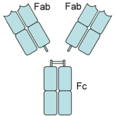
An antibody has Fab (fragment, antigen-binding) and Fc (fragment, crystalizable) regions. Fc receptors bind to the Fc region. Image source: Wikipedia, public domain.

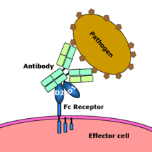
Fc receptor interaction with an antibody-coated microbial pathogen. Image source: Wikipedia, public domain.

Anti-FceR1 autoantibodies in chronic autoimmune urticaria: IgG against FceRI (receptor for IgE) (click to enlarge the image).
CD203c is a basophil activation marker known to be upregulated by cross-linking of the Fc (epsilon) RI alpha receptor and may serve as a useful marker to identify these patients. CD203 is expressed specifically on basophils, mast cells and their CD34+ progenitor cells and is upregulated by cross-linking of the FcεR1.
Sera from patients with chronic urticaria significantly upregulate basophil CD203c expression as measured by flow cytometry. Activated basophils from whole blood are identified by flow cytometry on the basis of the presence of CD203c on high-expressing IgE positive cells. CD203c expression correlates with basophil histamine release and the size of the autologous serum skin test.
Test code: IGERAB (it can be ordered through the Mayo Clinic lab service). Reference Lab: Sent to National Jewish via Mayo Clinic.
Test description: Anti-Fce Receptor Assay. More info: Anti-IgE Receptor I antibody, anti-CD203c, Chronic urticaria assay.
Method: Flow Cytometry
Reference range: greater than 2%, or by report.
Specimen Required: Draw blood in a plain, red-top tube (serum gel tube is not acceptable). Allow blood to clot and separate within one hour. Send 1 mL of serum frozen.
Specimen requirements: 1 ml of serum
Transport requirements: Sera should be shipped frozen and received in the laboratory within 24 hours after drawing. Days test is performed: Mondays. Turn around time: 1 week.
CPT code: 88184, 88185
References
Chronic urticaria sera increase basophile CD203c expression. Yasnowsky KM, Dreskin SC, Efaw B, Schoen D, Vedanthan PK, Alam R, Harbeck RJ. J Allergy Clin Immunol 2006;117:1430-4.
Clinical Reference Laboratories at National Jewish info sheet (PDF).
Clinical Reference Laboratories at National Jewish - 2007 newsletter (PDF).
Clinical Reference Laboratories at National Jewish - 2007 newsletter (PDF).
Anti-IgE Receptor Antibody - Mayo Clinic test catalog.
Tests for "autoimmune" urticaria - AAAAI Ask the Expert, 2011.
Tests for "autoimmune" urticaria - AAAAI Ask the Expert, 2011.
Published: 01/07/2011
Updated: 11/16/2011
Pruritus secondary to opioids without evidence of allergic reaction after a graded drug challenge with oxycodone
Author: V. Dimov, M.D., Allergist/Immunologist and Assistant Professor at University of Chicago
Reviewer: S. Randhawa, M.D., Allergist/Immunologist and Assistant Professor at NSU
A 24-year-old Caucasian female with a history of recurrent back pain and several back surgeries is referred for symptoms of urticaria and pruritus secondary to multiple pain medications during the last 3 to 4 years. She reports reactions to morphine, codeine, oxycodone, Topamax, Ultram, Celebrex and sulfa. In all cases hives occur soon after taking the medication and they never last more than 24 hours. The hives disappear after taking an antihistamine. She also has a history of panic attacks. During those episodes on a few occasions she had hives and had trouble breathing, but no real episodes of throat closing, low blood pressure, abdominal or respiratory symptoms during the episodes of urticaria with taking opioids.
Past medical history (PMH)
Recurrent back pain and several back surgeries, urticaria and pruritus secondary to multiple medications.
Medications
Elavil, Lexapro, ibuprofen p.r.n.
Social history and family history
Unremarkable.
Physical Examination
Normal.
What is the most likely diagnosis?
This is a patient with evidence of intolerance to opioids manifested by histamine release and acute urticarial episodes which typically resolve with or without antihistamines within 24 hours. There is no evidence of allergic or pseudoallergic reaction. There is no evidence of anaphylaxis or life-threatening reactions to opioids.
What tests would you suggest?
Graded drug challenge with oxycodone (short-acting opioid).
What happened next?
There was a suspicion that she may be allergic to opioids and several other medications as listed above. However, this is unlikely with her history - pruritus induced by opioid medications is quite common, and it occurs in about 12% to 15% of patients taking opioids. It is mediated both by peripheral release of histamine by the mast cells in the skin and also by central effect via the mu receptors. Antihistamines are not particularly effective, but they are worth a therapeutic trial, and that was done in this patient.
The patient returned for graded drug challenge 10 days after the initial visit. The current medications included Zyrtec 10 mg p.o. daily and she came with a prescription of oxycodone 5 mg capsules to be taken 4 to 6 times a day p.r.n. pain.
She had been on Zyrtec 10 mg p.o. daily for the last 4 days. We performed a graded drug challenge, starting with a dose of 0.078 mg of oxycodone dissolved in water. The patient was able to tolerate that. After the first and second dose of the medication, she developed pruritus affecting her extremities, face, the back of the head and the abdomen, but no hives, and no other changes in her physical condition.
She was able to tolerate up to 5 mg of oxycodone (total dose) in increments according to the drug challenge protocol. She had pruritus which worsened and improved spontaneously. With the dose of 1.25 mg of oxycodone, we gave her cetirizine 10 mg p.o. x1 in addition to the dose she had taken in the morning, and she reported no improvement in the pruritus with that.
Approximately 2 hours after the first dose of oxycodone, we provided a second dose of 5 mg of oxycodone, and the patient continued to have pruritus, but her blood pressure was stable, and there were no other symptoms. However, she reported feeling somnolent and dizzy, and she was advised to have lunch, which she did, with some improvement. There was no significant change in the blood pressure or other vital signs.
In summary, this is a patient with intolerance to oxycodone manifested by pruritus and feelings of dizziness and somnolence which again are quite common, with somnolence occurring between 23% to 24% of patients in general, dizziness 13% to 16%, and pruritus in 12% to 13%. She did not have hypotension, nausea, or vomiting. She stayed at the clinic for 2 hours after the challenge to recover from the somnolence, but there were no allergic symptoms.
Summary
The pruritus secondary to opioids experienced by this patient is most likely centrally mediated and antihistamines may be of limited effect. From an allergy and immunology perspective, there is no evidence of allergy to opioid medications, and there is no contraindication to starting methadone at this point as recommended by the pain management specialist. However, considering that she has reactions in terms of somnolence, dizziness and headache, it is probably best to give her the first dose at the physician's office and also start with a low dose of the opioid pain medication.
Final diagnosis
Pruritus secondary to opioids without evidence of allergic reaction after a graded drug challenge with oxycodone.
Classification of adverse reactions to drugs: "SOAP III" mnemonic (click to enlarge the image):

Adverse drug reactions (ADRs) affect 10–20% of hospitalized patients and 25% of outpatients.
Rule of 10s in ADR
10% of patients develop ADR
10% of these are due to allergy
10% of these lead to anaphylaxis
10% of these lead to death
There is a difference between graded dose challenge and rapid desensitization. Minimum requirements for rapid desensitization: 1-on-1 RN, CPR/ACLS, crash cart, Epi at bedside, anesthesia/code team, allergist 3 minutes from bedside.
References
To be updated.
Published: 07/03/2010
Updated: 04/03/2012
Reviewer: S. Randhawa, M.D., Allergist/Immunologist and Assistant Professor at NSU
A 24-year-old Caucasian female with a history of recurrent back pain and several back surgeries is referred for symptoms of urticaria and pruritus secondary to multiple pain medications during the last 3 to 4 years. She reports reactions to morphine, codeine, oxycodone, Topamax, Ultram, Celebrex and sulfa. In all cases hives occur soon after taking the medication and they never last more than 24 hours. The hives disappear after taking an antihistamine. She also has a history of panic attacks. During those episodes on a few occasions she had hives and had trouble breathing, but no real episodes of throat closing, low blood pressure, abdominal or respiratory symptoms during the episodes of urticaria with taking opioids.
Past medical history (PMH)
Recurrent back pain and several back surgeries, urticaria and pruritus secondary to multiple medications.
Medications
Elavil, Lexapro, ibuprofen p.r.n.
Social history and family history
Unremarkable.
Physical Examination
Normal.
What is the most likely diagnosis?
This is a patient with evidence of intolerance to opioids manifested by histamine release and acute urticarial episodes which typically resolve with or without antihistamines within 24 hours. There is no evidence of allergic or pseudoallergic reaction. There is no evidence of anaphylaxis or life-threatening reactions to opioids.
What tests would you suggest?
Graded drug challenge with oxycodone (short-acting opioid).
Table 1. Graded drug challenge with oxycodone
Q 15 minutes
0.078125 mg
0.15625 mg
0.3125 mg
0.625 mg
1.25 mg
2.5 mg
5 mg = full capsule
What happened next?
There was a suspicion that she may be allergic to opioids and several other medications as listed above. However, this is unlikely with her history - pruritus induced by opioid medications is quite common, and it occurs in about 12% to 15% of patients taking opioids. It is mediated both by peripheral release of histamine by the mast cells in the skin and also by central effect via the mu receptors. Antihistamines are not particularly effective, but they are worth a therapeutic trial, and that was done in this patient.
The patient returned for graded drug challenge 10 days after the initial visit. The current medications included Zyrtec 10 mg p.o. daily and she came with a prescription of oxycodone 5 mg capsules to be taken 4 to 6 times a day p.r.n. pain.
She had been on Zyrtec 10 mg p.o. daily for the last 4 days. We performed a graded drug challenge, starting with a dose of 0.078 mg of oxycodone dissolved in water. The patient was able to tolerate that. After the first and second dose of the medication, she developed pruritus affecting her extremities, face, the back of the head and the abdomen, but no hives, and no other changes in her physical condition.
She was able to tolerate up to 5 mg of oxycodone (total dose) in increments according to the drug challenge protocol. She had pruritus which worsened and improved spontaneously. With the dose of 1.25 mg of oxycodone, we gave her cetirizine 10 mg p.o. x1 in addition to the dose she had taken in the morning, and she reported no improvement in the pruritus with that.
Approximately 2 hours after the first dose of oxycodone, we provided a second dose of 5 mg of oxycodone, and the patient continued to have pruritus, but her blood pressure was stable, and there were no other symptoms. However, she reported feeling somnolent and dizzy, and she was advised to have lunch, which she did, with some improvement. There was no significant change in the blood pressure or other vital signs.
In summary, this is a patient with intolerance to oxycodone manifested by pruritus and feelings of dizziness and somnolence which again are quite common, with somnolence occurring between 23% to 24% of patients in general, dizziness 13% to 16%, and pruritus in 12% to 13%. She did not have hypotension, nausea, or vomiting. She stayed at the clinic for 2 hours after the challenge to recover from the somnolence, but there were no allergic symptoms.
Summary
The pruritus secondary to opioids experienced by this patient is most likely centrally mediated and antihistamines may be of limited effect. From an allergy and immunology perspective, there is no evidence of allergy to opioid medications, and there is no contraindication to starting methadone at this point as recommended by the pain management specialist. However, considering that she has reactions in terms of somnolence, dizziness and headache, it is probably best to give her the first dose at the physician's office and also start with a low dose of the opioid pain medication.
Final diagnosis
Pruritus secondary to opioids without evidence of allergic reaction after a graded drug challenge with oxycodone.
Classification of adverse reactions to drugs: "SOAP III" mnemonic (click to enlarge the image):
Adverse drug reactions (ADRs) affect 10–20% of hospitalized patients and 25% of outpatients.
Rule of 10s in ADR
10% of patients develop ADR
10% of these are due to allergy
10% of these lead to anaphylaxis
10% of these lead to death
There is a difference between graded dose challenge and rapid desensitization. Minimum requirements for rapid desensitization: 1-on-1 RN, CPR/ACLS, crash cart, Epi at bedside, anesthesia/code team, allergist 3 minutes from bedside.
References
To be updated.
Published: 07/03/2010
Updated: 04/03/2012
Typical laboratory workup for chronic idiopathic urticaria and angioedema
Author: V. Dimov, M.D., Allergist/Immunologist and Assistant Professor at University of Chicago
Reviewer: S. Randhawa, M.D., Allergist/Immunologist and Assistant Professor at NSU
A 36-year-old male is at the clinic for evaluation of chronic urticaria and angioedema for the last 8 years. The initial workup 6-7 years ago was negative. He has hives every 2 days, and angioedema symptoms every month. He reports severe angioedema symptoms - mostly facial swelling - with "throat closing" 2-3 times per year, and then he needs to use his EpiPen. He reports no respiratory or abdominal symptoms and has no family history of angioedema. He takes NSAIDs for joint pain.
Past medical history
Chronic urticaria and angioedema, depression.
Medications
EpiPen PRN, loratidine 10 mg po daily (ran out of loratidine 10 days ago).
Physicial examination
Urticarial lesions on both arms and trunk.
Skin prick testing: negative.
Physical urticaria testing: no dermatographism
What is the most likely diagnosis?
Chronic urticaria and angioedema.
What would you recommend?
Cetirizine 10 mg po bid.
Ranitidine 150 mg po bid.
Instructed to use EpiPen in case of severe angioedema or respiratory symptoms. He does not want to take daily steroids. We asked him to stop NSAIDs and use acetaminophen (Tylenol) instead.
Laboratory tests
- CBCD
- ESR
- CMP
- Thyroid function tests (TFTs), for example, TSH, T4, and thyroid autoantibodies (antimicrosomal and antithyroglobulin antibodies)
Chronic spontaneous urticaria (CSU) is defined as the presence of urticaria with daily or almost daily symptoms for 6 weeks or more. CSU affects 0.1%-0.8% of the population. http://buff.ly/1rDwQ4P
Twitter comments
Reviewer: S. Randhawa, M.D., Allergist/Immunologist and Assistant Professor at NSU
A 36-year-old male is at the clinic for evaluation of chronic urticaria and angioedema for the last 8 years. The initial workup 6-7 years ago was negative. He has hives every 2 days, and angioedema symptoms every month. He reports severe angioedema symptoms - mostly facial swelling - with "throat closing" 2-3 times per year, and then he needs to use his EpiPen. He reports no respiratory or abdominal symptoms and has no family history of angioedema. He takes NSAIDs for joint pain.
Past medical history
Chronic urticaria and angioedema, depression.
Medications
EpiPen PRN, loratidine 10 mg po daily (ran out of loratidine 10 days ago).
Physicial examination
Urticarial lesions on both arms and trunk.
Skin prick testing: negative.
Physical urticaria testing: no dermatographism
What is the most likely diagnosis?
Chronic urticaria and angioedema.
What would you recommend?
Cetirizine 10 mg po bid.
Ranitidine 150 mg po bid.
Instructed to use EpiPen in case of severe angioedema or respiratory symptoms. He does not want to take daily steroids. We asked him to stop NSAIDs and use acetaminophen (Tylenol) instead.
Laboratory tests
- CBCD
- ESR
- CMP
- Thyroid function tests (TFTs), for example, TSH, T4, and thyroid autoantibodies (antimicrosomal and antithyroglobulin antibodies)
- Total IgE
- Chronic urticaria index (positive if greater than 10) is a proprietary index, a positive result that makes autoimmune urticaria more likely
- C1q, C4, C2 levels
- C1-esterase inhibitor - qualitative and quantitative
- CH50, total hemolytic complement
- H. pylori workup, for example, H. pylori IgG (blood test). Diagnosis of Helicobacter pylori infection: 13C urea breath test or the stool antigen test as “test and treat strategy”. BMJ, 2012.
- ANA
- RF

Diagnosis of Chronic Urticaria (click to enlarge the image).

Anti-FceR1 autoantibodies in chronic autoimmune urticaria: IgG against FceRI (receptor for IgE) (click to enlarge the image).
- Chronic urticaria index (positive if greater than 10) is a proprietary index, a positive result that makes autoimmune urticaria more likely
- C1q, C4, C2 levels
- C1-esterase inhibitor - qualitative and quantitative
- CH50, total hemolytic complement
- H. pylori workup, for example, H. pylori IgG (blood test). Diagnosis of Helicobacter pylori infection: 13C urea breath test or the stool antigen test as “test and treat strategy”. BMJ, 2012.
- ANA
- RF
Diagnosis of Chronic Urticaria (click to enlarge the image).
Anti-FceR1 autoantibodies in chronic autoimmune urticaria: IgG against FceRI (receptor for IgE) (click to enlarge the image).
Why test for FceR1?
Approximately 45% of patients with chronic urticaria have an IgG autoantibody directed to the alpha-subunit of the high-affinity IgE receptor (chronic autoimmune urticaria, CAU) leading to cutaneous mast cell and basophil activation. Treatment of allergic asthma with omalizumab produces rapid reduction in free IgE levels and subsequent decrease in Fc epsilon RI expression on mast cells and basophils. In CAU, cross-linking of IgE receptors by autoantibody would be less likely, reducing cell activation and urticaria/angioedema.
In a study of 12 patients, Mean Urticaria Activity Score (UAS) declined significantly from baseline to the final 4 weeks of omalizumab treatment. Seven patients achieved complete symptom resolution. In 4 patients, mean UAS decreased, but urticaria persisted. One patient did not respond. Rescue medication use was reduced significantly, and quality of life improved (JACI, 2008).
Chronic spontaneous urticaria (CSU) is defined as the presence of urticaria with daily or almost daily symptoms for 6 weeks or more. CSU affects 0.1%-0.8% of the population. http://buff.ly/1rDwQ4P
References
Treatment of chronic autoimmune urticaria with omalizumab. Kaplan AP, Joseph K, Maykut RJ, Geba GP, Zeldin RK. J Allergy Clin Immunol. 2008 Sep;122(3):569-73.
Chronic urticaria: diagnosis and management. Khan DA. Allergy Asthma Proc. 2008 Sep-Oct;29(5):439-46.
Twitter comments
@Stolib: You check IgE levels or H. pylori studies in your chronic idiopathic urticaria (CIU) patients?
@Allergy: Depends on the clinical scenario: Typical laboratory workup for chronic idiopathic urticaria and angioedema http://goo.gl/rgDs
@Stolib: That is not how I was trained nor how I practice. I usually only lab pts only when they have failed antihistamines.
@Allergy: What if they have Hp infection that drives the process? By the time I see the CIU pts many of them have failed oral H1/H2 blockers.
@Stolib: Depends on what you considering failing antihistamines. I don't bother with HP unless symptoms suggest otherwise
@Allergy: Typical antihitsamine therapy in CIU: cetirizine 10 mg po bid and ranitidine 150 mg po bid. Would you like to describe your approach briefly, and I will publish it on AllergyCases.org, you retain authorship, of course.
@Stolib: My approach to CIU is outlined pretty well in this article written by my training program director: http://bit.ly/idYVip
@Allergy: Thank you for the reference - it's a free access article by David Khan. I will include it on AllergyCases.org http://goo.gl/JTlef
Published: 05/12/2010
Updated: 02/07/2012
Updated: 02/07/2012
Pressure-related urticaria and angioedema
Author: V. Dimov, M.D., Allergist/Immunologist and Assistant Professor at University of Chicago
Reviewer: S. Randhawa, M.D., Allergist/Immunologist and Assistant Professor at LSU (Shreveport) Department of Allergy and Immunology
A 64-year-old male is here for evaluation of chronic urticaria and angioedema. His first symptoms appeared when he was 17-year-old and worsened in the last 1.5 years.
He has hives daily and angioedema symptoms affecting his lips 1-2/week. He also has complaints of delayed pressure urticaria affecting his hands, feet and gluteus (after prolonged sitting).
First episode of of urticaria when he was 16 years old that lasted for 2 months. Then urticaria began again 18 months ago and "cleared" in 6 months, after stopping lisinopril, only to reappear 5 months later. He has had chronic pressure urticaria on his hands and feet, and the gluteal area through the years. He does not want any steriods because they cause him to "blow up like a balloon."
Past medical history
GERD, Sleep Apnea, Diabetes Mellitus, Hypertension
Medications
Carvediol, cetirizine 10 mg po bid, Epi-Pen 0.3 mg/0.3 ml prn, losartan, omeprazole, ranitidine 150 mg po bid, insulin.
Physical examination
Skin: single hive - R forearm, 4-5 hives - back.
Laboratory results
CBC+DIFF: no eosinophilia.
Colonoscopy - approx. 3 years ago - normal as per patient.
CBCD and CMP - WNL.
ANA - neg.
ESR - 29.
What further testing would you recommend?
Testing for dermatographic reaction and pressure urticaria.
Physical urticaria testing:
- No dermatographic reaction to scratch with the tongue spatula on the volar surface of the forearm.
- Pressure urticaria test (he had approximately 15-pound bag over his right shoulder for 18 minutes) - he developed erythema and 3 hives in the area but no pruritus. We asked him to report delayed pressure urticaria if he develops any.
What is the most likely diagnosis?
Chronic urticaria and angioedema.
What would you recommend at this point?
Cetirizine 10 mg po bid.
Ranitidine 150 mg po bid.
Instructed to use EpiPen in case of severe angioedema or respiratory symptoms.
Laboratory workup:
- Total IgE
- Thyroid function tests (TFTs), for example, TSH, T4, and thyroid autoantibodies (antimicrosomal and antithyroglobulin antibody)
- Anti-FceR1 Autoantibodies
- Chronic urticaria index (positive if greater than 10) is a proprietary index that makes the diagnosis of autoimmune urticaria more likely
- C1q, C4, C2 levels
- C1-esterase inhibitor - qualitative and quantitative
- CH50, total hemolytic complement
- H. pylori workup, for example, H. pylori IgG (blood test)
- RF
Return to the clinic in 1 month.
Summary
Chronic spontaneous urticaria (CSU) is defined as the presence of urticaria with daily or almost daily symptoms for 6 weeks or more. CSU affects 0.1%-0.8% of the population. http://buff.ly/1rDwQ4P
Published: 05/12/2010
Updated: 05/12/2011
- Chronic urticaria index (positive if greater than 10) is a proprietary index that makes the diagnosis of autoimmune urticaria more likely
- C1q, C4, C2 levels
- C1-esterase inhibitor - qualitative and quantitative
- CH50, total hemolytic complement
- H. pylori workup, for example, H. pylori IgG (blood test)
- RF
Return to the clinic in 1 month.
Summary
Chronic spontaneous urticaria (CSU) is defined as the presence of urticaria with daily or almost daily symptoms for 6 weeks or more. CSU affects 0.1%-0.8% of the population. http://buff.ly/1rDwQ4P
Testing procedures for diagnosis of physical urticarias depend on the cause (stimulus):
- Dermographism: Stroking with narrow object, e.g. a tongue depressor
- Cold urticaria: ice cube test
- Heat urticaria: test tube water at 44°C (111°F)
- Pressure urticaria: Sandbag test or a bag with heavy books (Middleton's Allergy textbook, 2 volumes)
- Vibratory urticaria: vibration with laboratory vortex for four minutes
- Cholinergic urticaria: exercise for 15-20 minutes or leg immersion in 44°C (111°F) bath
- Aquagenic urticaria: challenge with tap water at various temperatures
References
Related reading
"Palmaris slapus abdominus" - more commonly known as dermatographic urticariahttp://goo.gl/JMM6e
Updated: 05/12/2011
Neutrophilic Urticaria
Author: V. Dimov, M.D., Allergist/Immunologist and Assistant Professor at University of Chicago
Reviewer: S. Randhawa, M.D., Allergist/Immunologist and Assistant Professor at LSU (Shreveport) Department of Allergy and Immunology
A 48-year-old man is in the clinic today for a follow-up of chronic urticaria and delayed pressure urticaria and angioedema. A biopsy done 3 years showed neutrophils infiltrating the skin lesions without evidence of vasculitis.
He has been on Dapson 150 mg po daily and Doxepin 25 mg po qhs for 2.5 years and this regimen has decreased the intensity if his symptoms by about 25%. He had no effect with high dose oral antihistamines and Singulair in the past. He currently has hives daily, 5-15 lesions that resolve spontaneously, and no night symptoms. He has pressure-related angiodema on his back after porolonged recumbent position (sleep) approximately once a week. He reports no systemic symptoms, no shortness of breath, chest pain or fever.
Medications
Dapson 150 mg po daily and Doxepin 25 mg po qhs.
Physical examination
He has urticarial lesions on his arms and trunk (15-17 lesions). The rest of the examination is normal.
What is the most likely diagnosis?
Chronic urticaria and delayed pressure urticaria and angioedema. We discussed different therapeutic options and their side effects and he would like to stay on the current treatment regimen.
Chronic spontaneous urticaria (CSU) is defined as the presence of urticaria with daily or almost daily symptoms for 6 weeks or more. CSU affects 0.1%-0.8% of the population. http://buff.ly/1rDwQ4P
Published: 05/12/2010
Updated: 05/12/2010
Reviewer: S. Randhawa, M.D., Allergist/Immunologist and Assistant Professor at LSU (Shreveport) Department of Allergy and Immunology
A 48-year-old man is in the clinic today for a follow-up of chronic urticaria and delayed pressure urticaria and angioedema. A biopsy done 3 years showed neutrophils infiltrating the skin lesions without evidence of vasculitis.
He has been on Dapson 150 mg po daily and Doxepin 25 mg po qhs for 2.5 years and this regimen has decreased the intensity if his symptoms by about 25%. He had no effect with high dose oral antihistamines and Singulair in the past. He currently has hives daily, 5-15 lesions that resolve spontaneously, and no night symptoms. He has pressure-related angiodema on his back after porolonged recumbent position (sleep) approximately once a week. He reports no systemic symptoms, no shortness of breath, chest pain or fever.
Medications
Dapson 150 mg po daily and Doxepin 25 mg po qhs.
Physical examination
He has urticarial lesions on his arms and trunk (15-17 lesions). The rest of the examination is normal.
What is the most likely diagnosis?
Chronic urticaria and delayed pressure urticaria and angioedema. We discussed different therapeutic options and their side effects and he would like to stay on the current treatment regimen.
Chronic spontaneous urticaria (CSU) is defined as the presence of urticaria with daily or almost daily symptoms for 6 weeks or more. CSU affects 0.1%-0.8% of the population. http://buff.ly/1rDwQ4P
References
Published: 05/12/2010
Updated: 05/12/2010
Urticaria after sexual activity
Author: V. Dimov, M.D., Allergist/Immunologist and Assistant Professor at University of Chicago
Reviewer: S. Randhawa, M.D., Allergist/Immunologist and Assistant Professor at NSU
A 25-year-old Caucasian male is at the allergy clinic for evaluation of chronic urticaria for the last one and a half years, precipitated by sexual activity, hot showers, exercise, pressure and touch. The urticarial lesions appear on his trunk, arms, and face. He reports no angioedema, shortness of breath (SOB), wheezing, abdominal pain or any other systemic symptoms. There is no history of loss of consciousness or reaction to cold weather.
His primary physician mentioned that the patient has had wheals/rash on his abdomen after having sex with his wife. The rash occurs almost every time after sex and lasts several minutes. It begins on his back and wraps around to his abdomen but has occasionally been present on his neck. It shows up during of slightly after sex and resolves within one hour afterward. He states that his wife has attempted to stop use of various lotions, body creams etc. and rarely wears perfume. His physician suspected that the rash might be due to contact dermatitis to some cosmetic product used by the patient's wife. CBCD, ESR and ANA were all within normal limits. He was referred for skin testing.
The patient shows a picture of the rash from his cell phone:

Urticarial lesions on the trunk. Image source: Wikipedia, public domain.
Past Medical History (PMH)
Negative, apart from urticaria for one and a half years.
Medications
None.
Physical examination
Normal.
What is the most likely diagnosis?
Chronic urticaria? What type?
Dermatographism?
Contact dermatitis?
What diagnostic test would you suggest?
Pressure test for dermatographic urticaria with a tongue depressor.
What happened?
The test for dermatographism was strongly positive on both forearms:

Dermatographic urticaria is sometimes called "skin writing". Image source: Wikipedia, public domain.
What treatment would you suggest?
The patient has chronic urticaria and dermatographism. The most likely explanation of his symptoms is dermatographism, although physical urticaria, and specifically cholinergic urticaria cannot be completely ruled out. Vibratory urticaria is typically observed at the friction site rather than in widespread distribution and is therefore unlikely in his case.
Contact dermatitis or a reaction to any cosmetic products is unlikely with his clinical history. Skin prick testing or patch testing is not indicated.
The patient was prescribed a H1- and H2- blocker, loratidine 10 mg po daily and ranitidine 150 mg po daily. He is to follow up with the allergy clinic in 3 months.
Summary
There is a mnemonic for fifferential diagnosis of chronic urticaria: VIP
Vasculitis, confirmed by biopsy
Idiopathic, 75% of patients
Physical, benign
Physical urticaria
Physical urticaria is defined as hives provoked by physical stimulus such as:
CDC S (mnemonic)
Cold urticaria due to cooling the skin
Dermographism due to stroking the skin
Cholinergic urticaria due to exercise, emotion, or heat
Solar urticaria due to sun exposure
Physical urticaria can be confirmed by challenge testing, and is best treated symptomatically by avoidance of provocative stimuli and antihistamines.
References
Reviewer: S. Randhawa, M.D., Allergist/Immunologist and Assistant Professor at NSU
A 25-year-old Caucasian male is at the allergy clinic for evaluation of chronic urticaria for the last one and a half years, precipitated by sexual activity, hot showers, exercise, pressure and touch. The urticarial lesions appear on his trunk, arms, and face. He reports no angioedema, shortness of breath (SOB), wheezing, abdominal pain or any other systemic symptoms. There is no history of loss of consciousness or reaction to cold weather.
His primary physician mentioned that the patient has had wheals/rash on his abdomen after having sex with his wife. The rash occurs almost every time after sex and lasts several minutes. It begins on his back and wraps around to his abdomen but has occasionally been present on his neck. It shows up during of slightly after sex and resolves within one hour afterward. He states that his wife has attempted to stop use of various lotions, body creams etc. and rarely wears perfume. His physician suspected that the rash might be due to contact dermatitis to some cosmetic product used by the patient's wife. CBCD, ESR and ANA were all within normal limits. He was referred for skin testing.
The patient shows a picture of the rash from his cell phone:

Urticarial lesions on the trunk. Image source: Wikipedia, public domain.

Past Medical History (PMH)
Negative, apart from urticaria for one and a half years.
Medications
None.
Physical examination
Normal.
What is the most likely diagnosis?
Chronic urticaria? What type?
Dermatographism?
Contact dermatitis?
What diagnostic test would you suggest?
Pressure test for dermatographic urticaria with a tongue depressor.
What happened?
The test for dermatographism was strongly positive on both forearms:

Dermatographic urticaria is sometimes called "skin writing". Image source: Wikipedia, public domain.

What treatment would you suggest?
The patient has chronic urticaria and dermatographism. The most likely explanation of his symptoms is dermatographism, although physical urticaria, and specifically cholinergic urticaria cannot be completely ruled out. Vibratory urticaria is typically observed at the friction site rather than in widespread distribution and is therefore unlikely in his case.
Contact dermatitis or a reaction to any cosmetic products is unlikely with his clinical history. Skin prick testing or patch testing is not indicated.
The patient was prescribed a H1- and H2- blocker, loratidine 10 mg po daily and ranitidine 150 mg po daily. He is to follow up with the allergy clinic in 3 months.
Summary
There is a mnemonic for fifferential diagnosis of chronic urticaria: VIP
Vasculitis, confirmed by biopsy
Idiopathic, 75% of patients
Physical, benign
Physical urticaria
Physical urticaria is defined as hives provoked by physical stimulus such as:
CDC S (mnemonic)
Cold urticaria due to cooling the skin
Dermographism due to stroking the skin
Cholinergic urticaria due to exercise, emotion, or heat
Solar urticaria due to sun exposure
Physical urticaria can be confirmed by challenge testing, and is best treated symptomatically by avoidance of provocative stimuli and antihistamines.
Testing procedures for diagnosis of physical urticarias depend on the cause (stimulus):
- Dermographism: Stroking with narrow object, e.g. a tongue depressor
- Cold urticaria: ice cube test
- Heat urticaria: test tube water at 44°C (111°F)
- Pressure urticaria: Sandbag test or a bag with heavy books (Middleton's Allergy textbook, 2 volumes)
- Vibratory urticaria: vibration with laboratory vortex for four minutes
- Cholinergic urticaria: exercise for 15-20 minutes or leg immersion in 44°C (111°F) bath
- Aquagenic urticaria: challenge with tap water at various temperatures
References
Urticaria: A Short Review. V. Dimov. Clinical Notes in Allergy and Immunology.
Chronic Idiopathic Urticaria. Current Opinion in Allergy and Clinical Immunology, Medscape, 2003.
Chronic Idiopathic Urticaria. Current Opinion in Allergy and Clinical Immunology, Medscape, 2003.
Related reading
Flickr user: "I have a weird allergy called "Dermatography" where the skin welts up from contact"
"Young mother must wrap up all year round because she is allergic to the cold"http://goo.gl/w25WB - Cold urticaria in Daily Mail.
Self-referential illustration of dermatographic urticaria. Wikipedia, Creative Commons Attribution-Share Alike 3.0 Unported license.
Published: 03/10/2010
Updated: 03/10/2012

Published: 03/10/2010
Updated: 03/10/2012
Chronic Urticaria Due to Thyroid Antibodies
Author: V. Dimov, M.D., Allergist/Immunologist and Assistant Professor at University of Chicago
Reviewer: S. Randhawa, M.D., Allergist/Immunologist and Assistant Professor at LSU (Shreveport) Department of Allergy and Immunology
A 45 year-old Caucasian male is here for evaluation of urticaria for 6 weeks. He was prescribed two courses of oral prednisone which led to resolution of his symptoms, but the urticaria lesions reappeared when he decreased the dose to 5 mg a day. He reports 4 episodes of angioedema symptoms affecting his upper lip during the last 6 weeks, often occurring during exacerbations of his urticaria. The urticarial lesions occur daily and are worse in the afternoon.
Past medical history
His past medical history is positive for hypertension, hyperlipidemia, erectile dysfunction, and allergic rhinitis.
Medications
His medications include Levitra (vardenafil), lisinopril, simvastatin, levothyroxine, amlodipine, hydrocodone prn, triamterene/hydrochlorothiazide and two courses of prednisone.
He was advised to stop lisinopril and simvastatin and he did discontinue those two weeks ago.
The review of systems is negative apart from the history of present illness as above. He reports no shortness of breath, wheezing, chest tightness, chest pain or abdominal symptoms.
Physical examination
The physical examination is remarkable for 4-5 small urticarial lesions on both flanks in the abdominal area.
What is the most likely diagnosis?
This is a patient with chronic urticaria and angioedema for six weeks. He has been on ACE inhibitor (lisinopril) and simvastatin. Both medications were stopped 2 weeks ago.
What laboratory workup would you suggest?
We suggested laboratory work up in attempt to clarify the etiology of his condition and this includes CMP, CBC with differential and smear review, ANA, sedimentation rate, rheumatoid factor, TSH, T4, C1Q, C4 and C2 levels, CH50 level, C1 esterase inhibitor (qualitative and quantitative), H. pylori IgG antibody, thyroid autoantibodies (antimicrosomal and antithyroglobulin antibodies).
What treatment would you suggest?
We prescribed loratadine 10 mg po bid and strongly recommended he should stay off any ACE inhibitors and follow with his primary care physician for blood pressure control.
What happened next?
Thyroid autoantibodies were reported positive with "extremely high" titer. TSH level was normal.
Chronic spontaneous urticaria (CSU) is defined as the presence of urticaria with daily or almost daily symptoms for 6 weeks or more. CSU affects 0.1%-0.8% of the population. http://buff.ly/1rDwQ4P
Reviewer: S. Randhawa, M.D., Allergist/Immunologist and Assistant Professor at LSU (Shreveport) Department of Allergy and Immunology
A 45 year-old Caucasian male is here for evaluation of urticaria for 6 weeks. He was prescribed two courses of oral prednisone which led to resolution of his symptoms, but the urticaria lesions reappeared when he decreased the dose to 5 mg a day. He reports 4 episodes of angioedema symptoms affecting his upper lip during the last 6 weeks, often occurring during exacerbations of his urticaria. The urticarial lesions occur daily and are worse in the afternoon.
Past medical history
His past medical history is positive for hypertension, hyperlipidemia, erectile dysfunction, and allergic rhinitis.
Medications
His medications include Levitra (vardenafil), lisinopril, simvastatin, levothyroxine, amlodipine, hydrocodone prn, triamterene/hydrochlorothiazide and two courses of prednisone.
He was advised to stop lisinopril and simvastatin and he did discontinue those two weeks ago.
The review of systems is negative apart from the history of present illness as above. He reports no shortness of breath, wheezing, chest tightness, chest pain or abdominal symptoms.
Physical examination
The physical examination is remarkable for 4-5 small urticarial lesions on both flanks in the abdominal area.
What is the most likely diagnosis?
This is a patient with chronic urticaria and angioedema for six weeks. He has been on ACE inhibitor (lisinopril) and simvastatin. Both medications were stopped 2 weeks ago.
What laboratory workup would you suggest?
We suggested laboratory work up in attempt to clarify the etiology of his condition and this includes CMP, CBC with differential and smear review, ANA, sedimentation rate, rheumatoid factor, TSH, T4, C1Q, C4 and C2 levels, CH50 level, C1 esterase inhibitor (qualitative and quantitative), H. pylori IgG antibody, thyroid autoantibodies (antimicrosomal and antithyroglobulin antibodies).
What treatment would you suggest?
We prescribed loratadine 10 mg po bid and strongly recommended he should stay off any ACE inhibitors and follow with his primary care physician for blood pressure control.
What happened next?
Thyroid autoantibodies were reported positive with "extremely high" titer. TSH level was normal.
Anti-TPO - Greater than 1000.0
(reference range 0-3.9 IU/mL)
Thyroglobulin antibody 219
(reference range 0-14 IU/mL)
TSH 2.8
(reference range 0.34-5.6 IU/mL)
The patient was referred to see an endocrinologist specialist again.
What did we learn from this case?
A workup including C1Q, C4 and C2 levels, CH50 level, C1 esterase inhibitor (qualitative and quantitative) is indicated in chronic urticaria patients who have concurrent angioedema symptoms.
Thyroid function tests and thyroid antibodies are indicated in selected patients with chronic urticaria.
Diagnosis of Chronic Urticaria (click to enlarge the image).
Chronic spontaneous urticaria (CSU) is defined as the presence of urticaria with daily or almost daily symptoms for 6 weeks or more. CSU affects 0.1%-0.8% of the population. http://buff.ly/1rDwQ4P
References
Cryopyrin-associated periodic syndromes (CAPS)
Author: V. Dimov, M.D., Allergist/Immunologist and Assistant Professor at University of Chicago
Reviewer: S. Randhawa, M.D., Allergist/Immunologist and Assistant Professor at NSU
Inflammasome
The inflammasome is a multiprotein complex consisting of:
- caspase 1, an enzyme that proteolytically cleaves other proteins
- PYCARD, PYRIN-PAAD-DAPIN domain (PYD) and a C-terminal caspase-recruitment domain (CARD), a death domain-fold superfamily that mediates assembly of large signaling complexes
- NALP, Nod-like receptor(NLR)
The exact composition of an inflammasome depends on the activator which initiates inflammasome assembly.
The inflammasome is a multiprotein complex that is responsible for the activation of caspases 1 and 5, leading to the processing and secretion of the pro-inflammatory cytokines IL-1β and IL-18.
Two types of inflammasome have been identified to date:
- the NALP1 inflammasome, composed of NALP1, the adaptor protein ASC, caspase 1 and caspase 5
- the NALP2/3 inflammasomes that contain, in addition to NALP2 or NALP3, the caspase recruitment domain (CARD)-containing protein Cardinal, ASC and caspase 1
The NALPs are the central proteins and chief platforms in the inflammasome complex. NALPs belong to the NLR (NACHT-LRR) family of cytoplasmic proteins that also includes the NOD proteins. Fourteen NALP proteins have been identified in humans.
The amino-terminal sequence of NALPs is characterized by the presence of a pyrin domain (PYD), followed by a NACHT domain, a NACHT-associated domain (NAD) and several leucine-rich repeats (LRRs).
NLRs (NACHT-LRRs) are cytoplasmic molecules that serve as intracellular sensors of bacterial infection. They initiate signaling cascades that activate inflammatory responses. NLRs are receptors for PAMPs similar to TLRs.
Subsets of the NLRs:
- Nods (Nucleotide-binding oligomerization domain)
- NALPs (NACHT-, LRR- and pyrin domain-containing proteins)
Three NLRs, including Nod1, Nod2, and NALP3, recognize peptidoglycan, a common component of bacterial cell wall.
Caspase activation and recruitment domain (CARD)-containing proteins
CARDs cytoplasmic receptors that bind viral RNA. CARDs include retinoic acid inducible gene-I (RIG-I) and melanoma differentiation-associated gene 5 (MDA5).
CARDs activate the IRF-3 and NF-κB transcription factors, which stimulate the expression of antiviral type I interferons.
Homozygous mutations in the CARD9 gene are associated with chronic mucocutaneous candidiasis. Dysfunction of CARD9 impairs the innate signaling of dectin-1, an antifungal pattern-recognition receptor (NEJM, 10/2009).
Cryopyrin
Cryopyrin is a protein involved in innate immunity as part of the multiprotein inflammasome. It belongs to a family of NLR (nucleotide-binding domain and leucine-rich-repeat containing) proteins that respond to intracellular pathogens and other danger signals.
The pyrin domain was first noted in the familial Mediterranean fever protein from which it takes its name. It belongs to a structural superfamily that includes death domains, death effector domains and caspase activation and recruitment domains.
Cryopyrin-associated periodic syndromes (CAPS)
Cryopyrin-associated periodic syndromes (CAPS) or cryopyrinopathies include:
- familial cold autoinflammatory syndrome (FCAS)
- Muckle-Wells syndrome (MWS), also known as urticaria-deafness-amyloidosis (UDA)
- neonatal onset multisystem inflammatory disorder (NOMID), also known as chronic infantile neurologic cutaneous and articular syndrome, or (CINCA)
Mnemonic for CAPS - FUN:
FCAS
Urticaria-deafness-amyloidosis (UDA), Muckle-Wells syndrome (MWS)
NOMID/CINCA
Spectrum of CAPS
<-- Mild --------- Severe-->
FCAS, UDA/MWS, NOMID
All 3 CAPS are autosomal dominant.
All three cryopyrinopathies arise from mutations in a single gene, NLRP3 (also called CIAS1) at chromosome 1q44 (long arm of chromosome 1), encoding a protein called cryopyrin (also known as NALP3 and PYPAF1). The mode of inheritance is autosomal dominant with variable penetrance.
CIAS1 encodes a pyrin-like protein which contains a pyrin domain, a nucleotide-binding site (NBS) domain, and a leucine-rich repeat (LRR) motif.
CIAS1 stands for cold induced autoinflammatory syndrome 1. The gene product cryopyrin is also known under the names CATERPILLER-like receptor 1.1 (CLR1.1), NALP3, and NOD-like receptor family, pryin domain containing 3 (NLRP3).
Dysregulation of leukocyte apoptosis may be a common molecular pathway leading to inflammatory disease.
Hereditary periodic-fever syndromes or cryopyrin-associated periodic syndromes (CAPS) affect fewer than 500 patients worldwide.
Familial cold autoinflammatory syndrome (FCAS)
Familial cold urticaria (also known as "familial cold autoinflammatory syndrome") is an autosomal dominant condition characterized by rash, conjunctivitis, and arthralgias elicited by exposure to cold.
Muckle-Wells syndrome
Muckle-Wells syndrome (MWS), also known as urticaria-deafness-amyloidosis (UDA), is a rare autosomal dominant disease which causes sensorineural deafness, recurrent hives, and can lead to amyloidosis. MWS was first described in 1962 by Thomas James Muckle and Michael Vernon Well.
Individuals with MWS often have episodic fever, chills, and painful joints. As a result, MWS is considered a type of periodic fever syndrome. MWS is caused by a defect in the CIAS1 gene which creates the protein cryopyrin. MWS is closely related to two other syndromes, familial cold urticaria and neonatal onset multisystem inflammatory disease — in fact, all three are related to mutations in the same gene, CIAS1.
Neonatal onset multisystem inflammatory disease (NOMID)
Neonatal Onset Multisystem Inflammatory Disease (also known as NOMID, chronic infantile neurologic cutaneous and articular syndrome, or CINCA) is a rare genetic periodic fever syndrome which causes uncontrolled inflammation in multiple parts of the body starting in the newborn period. Urticaria-like rash within the first 6 weeks of life. CNS manifestations include chronic aseptic meningitis, mental retardation, cerebral atrophy, and chronic papilledema.
NOMID can result from a mutation in the CIAS1 gene, which helps control inflammation. Mutations in this gene also cause familial cold urticaria and Muckle-Wells syndrome. NOMID has been successfully treated with an interleukin-1 (IL-1) receptor antagonist, anakinra.
The symptoms of NOMID begin before age 3 months, in almost all children that have NOMID.
Treatment of CAPS
Anakinra (IL-1RA) is an IL-1 receptor antagonist that inhibits IL-1. Recombinant IL-1Ra or Anakinra (Kineret) is available commercially to treat systemic juvenile rheumatoid arthritis (JRA). Live vaccines should not be given concurrently with Anakinra.
Canakinumab is a human anti–interleukin-1β monoclonal antibody. Treatment with subcutaneous canakinumab once every 8 weeks was associated with a rapid remission of symptoms in most patients with CAPS.
Rilonacept (Arcalyst), is a dimeric fusion protein. IL-1 Trap or Rilonacept (Arcalyst) is used for management of cryopyrin-associated periodic syndromes (CAPS). Patients must me evaluated for latent TB before initiating therapy. Live vaccines should not be given concurrently. Taking rilonacept with TNF inhibitors is not recommended and may increase the risk of serious infections.
References
Autoinflammatory syndromes: Fever is not always a sign of infection - CCJM http://goo.gl/uTc2X
Primer: inflammasomes and interleukin 1 in inflammatory disorders. Leigh D Church, Graham P Cook and Michael F McDermott. Nature Clinical Practice Rheumatology (2008) 4, 34-42.
The Inflammasome — A Linebacker of Innate Defense. Joost P.H. Drenth, M.D., Ph.D., and Jos W.M. van der Meer, M.D., Ph.D. NEJM, Volume 355:730-732 August 17, 2006 Number 7.
The inflammasome. Virginie Petrillia, Stéphanie Papina and Jürg Tschopp. Current Biology, Volume 15, Issue 15, 9 August 2005, Page R581.
Use of Canakinumab in the Cryopyrin-Associated Periodic Syndrome. NEJM, 2009.
An Autoinflammatory Disease with Deficiency of the Interleukin-1–Receptor Antagonist. NEJM, 2009.
Cryopyrin-associated periodic syndromes. UpToDate, 2009.
A fever gene comes in from the cold. Daniel L. Kastner & John J. O'Shea. Nature Genetics 29, 241 - 242 (2001).
Related reading
Therapy of autoinflammatory syndromes. Hal M. Hoffman MD. Journal of Allergy and Clinical Immunology, Volume 124, Issue 6, December 2009, Pages 1129-1138.
Neonatal onset multisystem inflammatory disease. Wikipedia.
Muckle-Wells syndrome. Wikipedia.
Nucleotide-binding oligomerization domain (NOD)-like receptors (NLRs) have a potential role in chronic rhinosinusitis/polyps http://goo.gl/QAS4r
Periodic Fever Syndromes - 25-min COLA video lecture http://bit.ly/URcsBy:
Published: 06/05/2009
Updated: 10/12/2012
Hives (Urticaria)
Editor: V. Dimov, M.D., Allergist/Immunologist and Assistant Professor at University of ChicagoInformation For Patients
Hives (urticaria) and angioedema
Allergic Skin Conditions
Allergy Testing
Anti-FceR1 Autoantibodies in Chronic Urticaria
Contact dermatitis
Eczema or atopic dermatitis
Chronic Urticaria Treatment Options (click to enlarge the image).
Laboratory Diagnosis of Chronic Urticaria (click to enlarge the image).
Anti-FceR1 autoantibodies in chronic autoimmune urticaria: IgG against FceRI (receptor for IgE) (click to enlarge the image).
What to expect when visiting an allergy clinic
Current allergy skin tests are virtually painless. This video by Dr. Bassett, a board-certified allergist from New York City, shows what to expect when visiting an allergy clinic for diagnosis and treatment:
Information For Doctors
Urticaria: Teaching Cases
Chronic Idiopathic Urticaria (Hives)
Chronic Urticaria Due to Thyroid Antibodies
Urticaria after sexual activity and dermatographism
Pressure-related urticaria and angioedema
Neutrophilic Urticaria
Typical laboratory workup for chronic idiopathic urticaria and angioedema
Suspected progesterone allergy
Autologous serum skin test in chronic idiopathic urticaria
Acute urticaria (hives) due to allergy to terbinafine
Chronic Urticaria. Farid Farid, M.D. UCLA, Department of Medicine, 2002.
Blue Hives. NEJM Images in Clinical Medicine, 02/2008.
The Ice Cube Test: Cold-Induced Urticaria. NEJM Images in Clinical Medicine, 02/2008.
Diagnosis and Treatment of Urticaria and Angioedema - World Allergy Organization Journal 2012 update http://buff.ly/YBis2W
Therapeutic algorithm in chronic urticaria:
Related Reading
Urticaria: Brief Review
Mind Maps: Urticaria
Mnemonics: Urticaria
Urticaria and angioedema. Allergy, Asthma & Clinical Immunology 2011, 7(Suppl 1):S9.
Classification of urticaria: overview. Allergy, Asthma & Clinical Immunology 2011, 7(Suppl 1):S9.
A simplified, stepwise algorithm for the treatment of urticaria. Allergy, Asthma & Clinical Immunology 2011, 7(Suppl 1):S9.
A Summary of the New International EAACI/GA2LEN/EDF/WAO Guidelines in Urticaria http://1.usa.gov/10D3f2b
Cutaneous mastocytosis in a 3-month-old girl
Cryopyrin-associated periodic syndromes (CAPS)
Blog articles from AllergyNotes
Image source: Urticaria, Wikipedia, public domain.
Published: 09/15/2007
Updated: 09/20/2013
Autologous serum skin test in chronic idiopathic urticaria
Author: V. Dimov, M.D., Allergist/Immunologist and Assistant Professor at University of Chicago
Reviewer: S. Randhawa, M.D., Allergist/Immunologist and Assistant Professor at LSU (Shreveport) Department of Allergy and Immunology
A 21-yo AAF with a long history of chronic idiopathic urticaria and angioedema is referred to the allergy clinic of an academic center for further work-up. Her symptoms started 6 years ago and progressively worsened during the last 2 years to the point where she has had daily hives. She also complains of episodes of angioedema affecting the lids and lips about 4 times per month. She has been taking courses of oral prednisone every 6 weeks for the last 6 years.
Past medical history
Chronic idiopathic urticaria and angioedema for 6 years. Skin prick test (SPT) positive for trees, grass, weeds and molds. Negative extensive work-up for urticaria in the last year.
Medications
Medications
Allegra (fexofenadine) 180 mg po bid, cetirizine (Zyrtec) 10 mg po qhs, doxepin 50 mg po qhs, Zantac (ranitidine) 150 mg po bid, Benadryl (diphenhydramine) 50 mg po bid prn, Singulair (montelukast) 10 mg po qhs.
Family medical history (FMH)
Family medical history (FMH)
Mother with food allergies.
Social history (SH)
Social history (SH)
No tobacco exposure, she has 2 dogs.
Physical examination
Physical examination
Vital signs table (VSS).
Skin: Hives on both arms and abdomen.
HEENT: normal.
Lymph: No lymphadenopathy.
Respiratory: CTA (B).
CVS: Clear S1S2.
Ext: no c/c/e.
What tests would you suggest?
Skin: Hives on both arms and abdomen.
HEENT: normal.
Lymph: No lymphadenopathy.
Respiratory: CTA (B).
CVS: Clear S1S2.
Ext: no c/c/e.
What tests would you suggest?
Autologous serum skin test (ASST).
Anti-FcERI antibody (high affinity IgE receptor).
Skin biopsy.
How to do an autologous serum skin test (ASST)?
Skin biopsy.
How to do an autologous serum skin test (ASST)?
Blood was collected from the patient and divided into 2 portions, one of which was in a heparinized tube and the other was in a regular tube. Both tubes were centrifuged for 10 minutes and plasma and serum were obtained. Skin prick testing was done with saline, histamine, plasma and serum. Subsequently, intradermal testing was done with plasma and serum. All tests were negative apart from the positive control.
Table 1. Results of autologous serum skin test (ASST)
What happened next?
| Prick test | Left arm Wheal (mm) | Left arm Flare (mm) |
| Saline | 0 x 0 | 0 x 0 |
| Histamine | 6 x 5 | 30 x 45 |
| Serum | 0 x 0 | 0 x 0 |
| Plasma | 0 x 0 | 1 x 1 |
Table 1. Results of autologous serum skin test (ASST)
What happened next?
Pending the skin biopsy result, the option of treatment with cyclosporine was discussed with the patient.
Final diagnosis
Final diagnosis
Chronic idiopathic urticaria and angioedema.
What did we learn from this case?
What did we learn from this case?
How to perform an autologous serum/plasma skin test in a patient with chronic idiopathic urticaria (CIU).
A patient is injected with:
A patient is injected with:
1. Control (saline) - no reaction.
2. Their own serum - no reaction.
3. Their own plasma - reaction, proves autoantibody presence.
The ASST consists of an intradermal injection of 0.03 to 0.05 mL of the patient's own serum. The test is positive if the wheal is 1.5 mm greater than the negative control (saline) after 15 minutes.
The autologous plasma skin test (APST) is performed with plasma anticoagulated with 0.125 mol/L of sodium citrate and then centrifuged.
The autologous serum/plasma skin test is not commonly used. The positive and negative predictive values of the autologous serum skin test (ASST) in CIU patients are 53 and 70%, respectively.
27-50% of patients with CIU have autoantibodies against the α-chain of the high-affinity IgE receptor (FceRI) or less commonly against IgG. A positive autologous serum skin test (ASST) is an in-vivo test which reflects the presence of anti-FceRI and/or anti-IgE autoantibodies capable of activating mast cell degranulation.
According to some authors, a positive ASST increases the likelihood that a patient may need long-term oral steroids or cyclosporine in order to control the symptoms of CIU.

Mind map diagram of chronic urticaria (click to enlarge the image).
Diagnosis of Chronic Urticaria (click to enlarge the image).
Anti-FceR1 autoantibodies in chronic autoimmune urticaria: IgG against FceRI (receptor for IgE) (click to enlarge the image).
References
The autologous serum skin test in a cohort of chronic idiopathic urticaria patients compared to respiratory allergy patients and healthy individuals. Guttman-Yassky E, Bergman R, Maor C, Mamorsky M, Pollack S, Shahar E. J Eur Acad Dermatol Venereol. 2007 Jan;21(1):35-9.
AAAAI Q&A on Chronic Urticaria, Ask the Expert, 2002.
Urticaria: A Short Review. Allergy Cases, 2008.
Chronic Idiopathic Urticaria. Allergy Cases, 2008.
AAAAI Q&A on Chronic Urticaria, Ask the Expert, 2002.
Urticaria: A Short Review. Allergy Cases, 2008.
Chronic Idiopathic Urticaria. Allergy Cases, 2008.
Anti-FceR1 Autoantibodies in Chronic Urticaria
Chronic Idiopathic Urticaria. Current Opinion in Allergy and Clinical Immunology, Medscape, 2003.
Tests for "autoimmune" urticaria - AAAAI Ask the Expert, 2011.
Published: 08/19/2008
Updated: 11/19/2011
Chronic Idiopathic Urticaria. Current Opinion in Allergy and Clinical Immunology, Medscape, 2003.
Tests for "autoimmune" urticaria - AAAAI Ask the Expert, 2011.
Published: 08/19/2008
Updated: 11/19/2011
Chronic Idiopathic Urticaria
Author: V. Dimov, M.D., Allergist/Immunologist and Assistant Professor at University of Chicago
Reviewer: S. Randhawa, M.D., Allergist/Immunologist and Assistant Professor at LSU (Shreveport) Department of Allergy and Immunology
A 27-year-old year CM is referred to the allergy clinic for symptoms of hives for 2 months. Hives are described as a raised and itchy skin rash that occurs on the entire body with each individual hive lasting less than 24 hours. The lesions are pruritic and not painful. They heal without residual bruising or hyperpigmentation. Possible triggers have not been identified.
The patient has tried prescription antihistamines but they did not offer adequate control of symptoms. He has had several episodes of lip swelling but no throat or laryngeal involvement. The patient has required ER evaluation and treatment for these symptoms. A skin biopsy has not been performed.
Past Medical History (PMH)
Negative.
Allergies
No known medication allergy.
Medications
Hydroxyzine (Vistaril, Atarax) 25 mg po tid prn itching.
Family Medical History (FMH)
Hay fever - mother.
Physical examination
VSS.
HEENT: External ears normal. Canals clear. TM's normal. Nasal mucosa normal. No drainage or sinus tenderness. Lips, tongue normal. Oropharynx clear.
Neck: supple, no adenopathy.
CVS: RRR, normal S1/S2, no m/r/g.
Chest: CTA (B).
Extremities: no c/c/e.
Skin: Hives on both arms.

Figure 1. Urticaria appearing on right arm. Image source: Wikipedia, public domain.
What is the most likely diagnosis?
Chronic urticaria.
What would you do?
Screening lab work.
H1- and H2-blockers.
What happened?
The patient declined further lab work. He was prescribed Allegra 180mg BID and Pepcid 20 mg po BID. He was advised that he could use OTC Claritine (loratadine) instead of Allegra. He was asked to avoid Atarax if going to work or driving.
The patient was instructed to call the office if there is no improvement in his symptoms and a follow-up in 2 months was arranged.
Final diagnosis
Chronic Idiopathic Urticaria.
Summary
Mind map diagram of chronic urticaria.
Diagnosis of Chronic Urticaria (click to enlarge the image).
Anti-FceR1 autoantibodies in chronic autoimmune urticaria: IgG against FceRI (receptor for IgE) (click to enlarge the image).
Chronic spontaneous urticaria (CSU) is defined as the presence of urticaria with daily or almost daily symptoms for 6 weeks or more. CSU affects 0.1%-0.8% of the population. http://buff.ly/1rDwQ4P
Therapy for chronic spontaneous urticaria
Step
1 Nonsedating, second- or third-generation antihistamines taken 4 times a day. Decrease the dose as tolerated once control of symptoms is attained. Dose of cetirizine, loratadine, desloratadine, or levocetirizine corresponding to hydroxyzine or diphenhydramine at 50 mg 4 times daily is 6 tablets per day. If response inadequate, proceed to step 2.
2 Omalizumab, 300 mg monthly. If no response after 2 injections, proceed to step 3.
3 Cyclosporine, 200-300 mg/d
4 Options to consider if steps 1-3 fail: dapsone, methotrexate, sulfasalazine, hydroxychloroquine, intravenous γ-globulin, and plasmapheresis.
Expected response rate based on the literature. Patient response to step 1 was 45%. Patient response to step 2 was 65% of the remainder. Calculated response rate of steps 1 plus 2 was 81%. Patient response to step 3 was 65% of the remainder. Calculated total response rate for steps 1, 2, and 3 was 92%. Source: Therapy of chronic urticaria: a simple, modern approach. Allen P. Kaplan, Annals of Allergy, Asthma & Immunology, Volume 112, Issue 5, Pages 419–425, May 2014, http://www.annallergy.org/article/S1081-1206(14)00129-X/abstract
References
Urticaria: A Short Review. V. Dimov. Clinical Notes in Allergy and Immunology.
Reviewer: S. Randhawa, M.D., Allergist/Immunologist and Assistant Professor at LSU (Shreveport) Department of Allergy and Immunology
A 27-year-old year CM is referred to the allergy clinic for symptoms of hives for 2 months. Hives are described as a raised and itchy skin rash that occurs on the entire body with each individual hive lasting less than 24 hours. The lesions are pruritic and not painful. They heal without residual bruising or hyperpigmentation. Possible triggers have not been identified.
The patient has tried prescription antihistamines but they did not offer adequate control of symptoms. He has had several episodes of lip swelling but no throat or laryngeal involvement. The patient has required ER evaluation and treatment for these symptoms. A skin biopsy has not been performed.
Past Medical History (PMH)
Negative.
Allergies
No known medication allergy.
Medications
Hydroxyzine (Vistaril, Atarax) 25 mg po tid prn itching.
Family Medical History (FMH)
Hay fever - mother.
Physical examination
VSS.
HEENT: External ears normal. Canals clear. TM's normal. Nasal mucosa normal. No drainage or sinus tenderness. Lips, tongue normal. Oropharynx clear.
Neck: supple, no adenopathy.
CVS: RRR, normal S1/S2, no m/r/g.
Chest: CTA (B).
Extremities: no c/c/e.
Skin: Hives on both arms.

Figure 1. Urticaria appearing on right arm. Image source: Wikipedia, public domain.

What is the most likely diagnosis?
Chronic urticaria.
What would you do?
Screening lab work.
H1- and H2-blockers.
What happened?
The patient declined further lab work. He was prescribed Allegra 180mg BID and Pepcid 20 mg po BID. He was advised that he could use OTC Claritine (loratadine) instead of Allegra. He was asked to avoid Atarax if going to work or driving.
The patient was instructed to call the office if there is no improvement in his symptoms and a follow-up in 2 months was arranged.
Final diagnosis
Chronic Idiopathic Urticaria.
Summary
Mind map diagram of chronic urticaria.
Diagnosis of Chronic Urticaria (click to enlarge the image).
Anti-FceR1 autoantibodies in chronic autoimmune urticaria: IgG against FceRI (receptor for IgE) (click to enlarge the image).
Chronic spontaneous urticaria (CSU) is defined as the presence of urticaria with daily or almost daily symptoms for 6 weeks or more. CSU affects 0.1%-0.8% of the population. http://buff.ly/1rDwQ4P
Therapy for chronic spontaneous urticaria
Step
1 Nonsedating, second- or third-generation antihistamines taken 4 times a day. Decrease the dose as tolerated once control of symptoms is attained. Dose of cetirizine, loratadine, desloratadine, or levocetirizine corresponding to hydroxyzine or diphenhydramine at 50 mg 4 times daily is 6 tablets per day. If response inadequate, proceed to step 2.
2 Omalizumab, 300 mg monthly. If no response after 2 injections, proceed to step 3.
3 Cyclosporine, 200-300 mg/d
4 Options to consider if steps 1-3 fail: dapsone, methotrexate, sulfasalazine, hydroxychloroquine, intravenous γ-globulin, and plasmapheresis.
Expected response rate based on the literature. Patient response to step 1 was 45%. Patient response to step 2 was 65% of the remainder. Calculated response rate of steps 1 plus 2 was 81%. Patient response to step 3 was 65% of the remainder. Calculated total response rate for steps 1, 2, and 3 was 92%. Source: Therapy of chronic urticaria: a simple, modern approach. Allen P. Kaplan, Annals of Allergy, Asthma & Immunology, Volume 112, Issue 5, Pages 419–425, May 2014, http://www.annallergy.org/article/S1081-1206(14)00129-X/abstract
References
Urticaria: A Short Review. V. Dimov. Clinical Notes in Allergy and Immunology.
Anti-FceR1 Autoantibodies in Chronic Urticaria
Chronic Idiopathic Urticaria. Current Opinion in Allergy and Clinical Immunology, Medscape, 2003.
Acute and chronic urticaria. Krishnaswamy G, Youngberg G. Postgrad Med 2001;109(2):107-23.
Urticaria. eMedicine, 2004.
Allergy and the skin. Urticaria. ABC of allergies. BMJ 1998;316:1147.
Urticaria. Merck Manual.
Image source: Uricaria appearing on right arm, Wikipedia, public domain.
Published: 09/14/2007
Updated: 11/19/2011
Mnemonics: Urticaria
Author: V. Dimov, M.D., Allergist/Immunologist and Assistant Professor at University of Chicago
Reviewer: S. Randhawa, M.D., Assistant Professor at NSU
Chronic spontaneous urticaria (CSU) is defined as the presence of urticaria with daily or almost daily symptoms for 6 weeks or more. CSU affects 0.1%-0.8% of the population. http://buff.ly/1rDwQ4P
Causes of acute urticaria: IF DVR Stings
Idiopathic
Food: fruits, seafood, nuts, dairy, spices, tea, chocolate
Drugs: antibiotics (PCN) and sulphonamides; ASA, NSAIDs
Viral infections
Radio contrast media
Stings - wasp or bee
A more complete mnemonic for the major causes of urticaria and angioedema: FIT MID CHIMP
Foods or food additives
Inhalation, ingestion of, or contact with antigens
Transfusion reactions
Malignancy:
Infections: bacterial, fungal, viral, and helminthic; Insects (papular urticaria)
Drug reactions
Collagen vascular diseases
Hereditary diseases
Idiopathic: chronic idiopathic urticaria and chronic angioedema
Mastocytosis, systemic: urticaria pigmentosa
Physical urticarias
Based on: Middleton's Allergy: Principles and Practice, 6th ed., from MerckMedicus.com (requires registration).
Differential diagnosis of chronic urticaria: VIP
Vasculitis, confirmed by biopsy
Idiopathic, 75% of patients
Physical, benign
Physical urticarias: CDC S
Cold urticaria due to cooling the skin
Dermographism due to stroking the skin
Cholinergic urticaria due to exercise, emotion, or heat
Solar urticaria due to sun exposure
Treatment of urticaria: ALAS
Avoidance of precipitating factors
Local (topical) treatments: 1% menthol in aqueous cream.
Antihistamines: H1- and H2-blockers
Steroids
Published: 01/24/2008
Reviewer: S. Randhawa, M.D., Assistant Professor at NSU
Chronic spontaneous urticaria (CSU) is defined as the presence of urticaria with daily or almost daily symptoms for 6 weeks or more. CSU affects 0.1%-0.8% of the population. http://buff.ly/1rDwQ4P
Causes of acute urticaria: IF DVR Stings
Idiopathic
Food: fruits, seafood, nuts, dairy, spices, tea, chocolate
Drugs: antibiotics (PCN) and sulphonamides; ASA, NSAIDs
Viral infections
Radio contrast media
Stings - wasp or bee
A more complete mnemonic for the major causes of urticaria and angioedema: FIT MID CHIMP
Foods or food additives
Inhalation, ingestion of, or contact with antigens
Transfusion reactions
Malignancy:
Infections: bacterial, fungal, viral, and helminthic; Insects (papular urticaria)
Drug reactions
Collagen vascular diseases
Hereditary diseases
Idiopathic: chronic idiopathic urticaria and chronic angioedema
Mastocytosis, systemic: urticaria pigmentosa
Physical urticarias
Based on: Middleton's Allergy: Principles and Practice, 6th ed., from MerckMedicus.com (requires registration).
Differential diagnosis of chronic urticaria: VIP
Vasculitis, confirmed by biopsy
Idiopathic, 75% of patients
Physical, benign
Physical urticarias: CDC S
Cold urticaria due to cooling the skin
Dermographism due to stroking the skin
Cholinergic urticaria due to exercise, emotion, or heat
Solar urticaria due to sun exposure
Treatment of urticaria: ALAS
Avoidance of precipitating factors
Local (topical) treatments: 1% menthol in aqueous cream.
Antihistamines: H1- and H2-blockers
Steroids
References
Published: 01/24/2008
Updated: 01/07/2011
Mind Maps: Urticaria
Author: V. Dimov, M.D., Allergist/Immunologist and Assistant Professor at University of Chicago
Reviewer: S. Randhawa, M.D., Allergist/Immunologist and Assistant Professor at NSU
Chronic spontaneous urticaria (CSU) is defined as the presence of urticaria with daily or almost daily symptoms for 6 weeks or more. CSU affects 0.1%-0.8% of the population. http://buff.ly/1rDwQ4P

Chronic Urticaria Treatment Options (click to enlarge the image).

Diagnosis of Chronic Urticaria (click to enlarge the image).
Anti-FceR1 autoantibodies in chronic autoimmune urticaria: IgG against FceRI (receptor for IgE) (click to enlarge the image).

Chronic Urticaria Treatment (click to enlarge the image).

Oral Antihistamines (click to enlarge the image).

Diagram (mind map) of acute urticaria.
Diagram (mind map) of chronic urticaria.
References and related reading and mind maps
Anti-FceR1 Autoantibodies in Chronic Urticaria
Urticaria and angioedema. Allergy, Asthma & Clinical Immunology 2011, 7(Suppl 1):S9.
Classification of urticaria: overview. Allergy, Asthma & Clinical Immunology 2011, 7(Suppl 1):S9.
A simplified, stepwise algorithm for the treatment of urticaria. Allergy, Asthma & Clinical Immunology 2011, 7(Suppl 1):S9.
Published: 01/24/2008
Updated: 03/12/2014
Reviewer: S. Randhawa, M.D., Allergist/Immunologist and Assistant Professor at NSU
Chronic spontaneous urticaria (CSU) is defined as the presence of urticaria with daily or almost daily symptoms for 6 weeks or more. CSU affects 0.1%-0.8% of the population. http://buff.ly/1rDwQ4P
Chronic Urticaria Treatment Options (click to enlarge the image).
Diagnosis of Chronic Urticaria (click to enlarge the image).
Anti-FceR1 autoantibodies in chronic autoimmune urticaria: IgG against FceRI (receptor for IgE) (click to enlarge the image).
Chronic Urticaria Treatment (click to enlarge the image).
Oral Antihistamines (click to enlarge the image).

Diagram (mind map) of acute urticaria.
Diagram (mind map) of chronic urticaria.
References and related reading and mind maps
Anti-FceR1 Autoantibodies in Chronic Urticaria
Urticaria and angioedema. Allergy, Asthma & Clinical Immunology 2011, 7(Suppl 1):S9.
Classification of urticaria: overview. Allergy, Asthma & Clinical Immunology 2011, 7(Suppl 1):S9.
A simplified, stepwise algorithm for the treatment of urticaria. Allergy, Asthma & Clinical Immunology 2011, 7(Suppl 1):S9.
Published: 01/24/2008
Updated: 03/12/2014
Urticaria: Brief Review
Author: V. Dimov, M.D., Allergist/Immunologist and Assistant Professor at University of Chicago
Reviewer: S. Randhawa, M.D., Allergist/Immunologist and Assistant Professor at NSU
Acute urticaria
Urticaria (hives) is a common skin condition caused by a type 1 hypersensitivity reaction most commonly to foods and drugs. Acute urticaria affects 15-25% of the population at some point in their lives. In 50% of patients, no cause is identified. Hypersensitivity to a specific food can be confirmed by skin prick testing or ImmunoCAP. Skin biopsy (rarely done) shows mast cell infiltration. The flare is mediated by neuropeptides and axon reflex.
Urticaria is either acute (present for less than 6 weeks) or chronic (longer than 6 weeks).
Angioedema involves swelling of the deep dermal and subcutaneous/submucosal tissues. Approximately 50% of patients have both urticaria and angioedema. Angioedema is non-pitting and non-hot.
When you see a patient with angioedema, the first question is: "are you on ACE inhibitor?" ARBs are well tolerated as an alternative therapy.
Figure 1. Urticaria appearing on right arm. Image source: Wikipedia, public domain.


ACE-inhibitor-induced angioedema affecting the upper lip
Which substance has both diagnostic and therapeutic use in acute urticaria?
Epinephrine administration invariably leads to resolution of acute urticaria -- an effect which can be used for both diagnosis and therapy.
Acute urticaria is treated with avoidance of allergens, antihistamines, and steroids in patients with insufficient response to steroids. The mnemonic for treatment is ALAS:
Avoidance
Local (topical) treatments
Antihistamines
Steroids
Figure 2. Diagram (mind map) of acute urticaria.
Causes of acute urticaria: IF DVR Stings
Idiopathic
Food: fruits, seafood, nuts, dairy, spices, tea, chocolate
Drugs: antibiotics (PCN) and sulphonamides; ASA, NSAIDs
Viral infections
Radio contrast media
Stings - wasp or bee
Hives that are due to foods, for example, do not last two weeks. They usually last a matter of hours (AAAAI).
A more complete mnemonic for the major causes of urticaria and angioedema: FIT MID CHIMP
Foods or food additives
Inhalation, ingestion of, or contact with antigens
Transfusion reactions
Malignancy:
Infections: bacterial, fungal, viral, and helminthic; Insects (papular urticaria)
Drug reactions
Collagen vascular diseases
Hereditary diseases
Idiopathic: chronic idiopathic urticaria and chronic angioedema
Mastocytosis, systemic: urticaria pigmentosa
Physical urticarias
Based on: Middleton's Allergy: Principles and Practice, 6th ed., from MerckMedicus.com (requires registration).
Infections are a very rare cause of urticaria, antibiotics used to treat them are a much more common cause.
Insect-related urticaria in children is found on the dependent area of the body - lower legs and arms.
Urticaria pigmentosa is characterized by Darier's sign - a scratch causes urticaria. Urticaria pigmentosa/systemic mastocytosis can become malignant.
Mastocytosis
Systemic mastocytosis is characterized by clonal expansion of mast cells, and most patients present with a genetic mutation in the stem cell factor receptor (KIT) gene. KIT or C-kit receptor is also called CD117. KIT is a cytokine receptor expressed on the cell surface. KIT receptors binds to stem cell factors which causes certain types of cells to grow. Some patients present with eosinophilia and symptoms may overlap with the hypereosinophilic syndromes.
Chronic urticaria
Chronic spontaneous urticaria (CSU) is defined as the presence of urticaria with daily or almost daily symptoms for 6 weeks or more. CSU affects 0.1%-0.8% of the population. http://buff.ly/1rDwQ4P
Chronic urticaria is defined as widespread itchy weals (hives) for at least 6 weeks. Skin lesion can be present either continuously or intermittently. When no cause is found, chronic urticaria is labeled as chronic idiopathic urticaria (CIU). Most cases of chronic urticaria are not due to allergy. Most cases of acute urticaria are due to allergy (type I hypersensitivity to food and drugs).
Acute urticaria
Allergy
Chronic urticaria
Cause unknown, 75%
Covered-up
Incidence of atopy is not increased in chronic urticaria. It is much more common in women than in men ("women pay more attention to their skin") -- 3:1 ratio of female : male in chronic urticaria.
Chronic urticaria is "bad news" for the patient:
- it is difficult to find a cause
- 10-20% persist beyond 10 years
- no treatment is consistently effective
Skin biopsy (rarely done) shows up to 10-fold increase of mast cells. In cold urticaria, a cube is placed on the arm and blood drawn above the cube shows a massive increase in plasma histamine.
Differential diagnosis of chronic urticaria: VIP
Vasculitis, confirmed by biopsy
Idiopathic, 75% of patients
Physical, benign
Figure 3. Diagram (mind map) of chronic urticaria.
Physical urticaria
Physical urticaria is defined as hives provoked by physical stimulus such as:
CDC S
Cold urticaria due to cooling the skin
Dermographism due to stroking the skin
Cholinergic urticaria due to exercise, emotion, or heat
Solar urticaria due to sun exposure
Physical urticaria can be confirmed by challenge testing, and is best treated symptomatically by avoidance of provocative stimuli and antihistamines.
Figure 4. Dermatographic urticaria is sometimes called "skin writing". Image source: Wikipedia, public domain.
Dermatographism can be reproduced by using a standardized tool which applies pressure of 3600 gm/cm2.
Physical urticaria does not respond to steroids since they do not inhibit mast cell degranulation. That is why steroid use is not a contraindication to skin prick testing. Epinephrine, which is used for both diagnostic and therapeutic purposes, inhibits mast cell degranulation.
There are several types of cold urticarias. Acquired (essential) cold urticaria can be deadly. For example, a swimmer with history of cold urticaria jumped into a cold water lake, developed urticaria and hypotension which led to brain ischemia.
Familial cold autoinflammatory syndrome (FCAS) is characterized by neutrophil infiltration of skin which differentiates it from "typical" urticaria characterized by mast cell infiltration.
Delayed pressure urticaria/angiodema develops after clapping at a concert or carrying a heavy bag/backpack. The diagnostic test involves appearance of dermatographism after wearing 15-pound weights for 20 minutes, for example a bag with 4 volumes of Middleton's Allergy textbook in a bag over the shoulder.
Vibratory angioedema can develop after cutting the lawn.
Urticarial vasculitis
In urticarial vasculitis, hives invariably persist for more than 24 hours which differentiates from urticaria. Diagnosis is confirmed by skin biopsy which shows vasculitis. Patients with urticarial vasculitis need a complete work-up for SLE and other CTD.
Hypocomplementic Urticarial Vasculitic Syndrome (HUVS) is seen on the dependent body areas - lower legs, arms, back (not on the abdomen),
Chronic idiopathic urticaria (CIU)
When no cause is found, chronic urticaria is labeled as chronic idiopathic urticaria (CIU). CIU affects up to 0.1-3% of the population at some point in their lives (acute urticaria affects 15-25%). Acute urticaria occurs more often in atopy but there is no such correlation in CIU. 20% of patients with CIU have symptoms for longer than 10 years.
CIU is divided in 2 groups:
- autoimmune CIU, 50% of patients with CIU
- "truly idiopathic" CIU, 50%
About 90% of patients with CIU also have angioedema.
Autoimmune chronic urticaria (AICU)
Autoimmune chronic urticaria occurs in 50% of patients with CIU. These patients have autoantibodies against either the high-affinity IgE receptor FcR1 or, less commonly, IgE.
"Truly idiopathic" CIU
In the remaining 40-60% of cases of CIU, no cause can be identified and they are truly "idiopathic." It is likely that those cases also have an autoimmune basis but autoantibodies have not been yet detected.
Laboratory tests in chronic urticaria
Diagnostic tests
Skin prick testing or ImmunoCAP
Diagnosis of Chronic Urticaria (click to enlarge the image).
Autologous serum/plasma skin test
A patient is injected with:
1. Control (saline) - no reaction.
2. Their own serum - no reaction.
3. Their own plasma - reaction, proves autoantibody presence.
The autologous serum/plasma skin test is not commonly used. The positive and negative predictive values of the autologous serum skin test (ASST) in CIU patients are 53 and 70%, respectively.
27-50% of patients with CIU have autoantibodies against the α-chain of the high-affinity IgE receptor (FceRI) or less commonly against IgG. A positive autologous serum skin test (ASST) is an in-vivo test which reflects the presence of anti-FceRI and/or anti-IgE autoantibodies capable of activating mast cell degranulation.
Screening laboratory tests are usually not helpful.
Treatment of chronic idiopathic urticaria: ALAS
Avoidance of precipitating factors
Local (topical) treatment are not generally helpful, one recommendation is 1% menthol in aqueous cream.
Antihistamines: H1- and H2-blockers
Steroids
Other treatments: Cyclosporin A, intravenous immunoglobulins, plasmapheresis, and omalizumab.
Omalizumab (Xolair) (single dose, 300 mg) is effective against chronic idiopathic urticaria but reimbursement for therapy may be a challenge in the U.S.
Cyclosporin is one of the most commonly used steroid-sparing drugs for treatment of CIU.
How does cyclosporine work?
Cyclosporin binds to the cytosolic protein cyclophilin (immunophilin) of T-lymphocytes. This complex of cyclosporin and cyclophylin inhibits calcineurin, which is responsible for the transcription of interleukin-2. Cyclosporin inhibits lymphokine production and interleukin release and leads to a reduced function of effector T-cells.
This mode of action is similar to pimecrolimus (Elidel), an ascomycin macrolactam derivative, which binds to macrophilin-12 and inhibits calcineurin. Thus pimecrolimus inhibits T-cell activation by inhibiting the synthesis and release of cytokines from T-cells. Pimecrolimus also prevents the release of inflammatory cytokines and mediators from mast cells.
Cyclosporin is an immunosuppressant drug used in post-allogeneic organ transplant. Initially isolated from a Norwegian soil sample, Cyclosporin A is a cyclic peptide of 11 amino acids produced by the fungus Tolypocladium inflatum Gams, and contains D-amino acids, which are rarely encountered in nature.
Therapy for chronic spontaneous urticaria
Step
1 Nonsedating, second- or third-generation antihistamines taken 4 times a day. Decrease the dose as tolerated once control of symptoms is attained. Dose of cetirizine, loratadine, desloratadine, or levocetirizine corresponding to hydroxyzine or diphenhydramine at 50 mg 4 times daily is 6 tablets per day. If response inadequate, proceed to step 2.
2 Omalizumab, 300 mg monthly. If no response after 2 injections, proceed to step 3.
3 Cyclosporine, 200-300 mg/d
4 Options to consider if steps 1-3 fail: dapsone, methotrexate, sulfasalazine, hydroxychloroquine, intravenous γ-globulin, and plasmapheresis.
Expected response rate based on the literature. Patient response to step 1 was 45%. Patient response to step 2 was 65% of the remainder. Calculated response rate of steps 1 plus 2 was 81%. Patient response to step 3 was 65% of the remainder. Calculated total response rate for steps 1, 2, and 3 was 92%. Source: Therapy of chronic urticaria: a simple, modern approach. Allen P. Kaplan, Annals of Allergy, Asthma & Immunology, Volume 112, Issue 5, Pages 419–425, May 2014, http://www.annallergy.org/article/S1081-1206(14)00129-X/abstract
References
Chronic Idiopathic Urticaria. Current Opinion in Allergy and Clinical Immunology, Medscape, 2003.
Acute and chronic urticaria. Krishnaswamy G, Youngberg G. Postgrad Med 2001;109(2):107-23.
Urticaria. eMedicine, 2004.
Allergy and the skin. Urticaria. ABC of allergies. BMJ 1998;316:1147.
Urticaria. Merck Manual.
Allergy and the skin: eczema and chronic urticaria. Constance H Katelaris and Jane E Peake. MJA 2006; 185 (9): 517-522.
Chapter 85 - Urticaria and Angioedema. Middleton's Allergy: Principles and Practice, 6th ed., from MerckMedicus.com (requires registration).
Urticaria, Angioedema, and Hereditary Angioedema. Patterson's Allergic Diseases (6th Edition), from MerckMedicus.com (requires registration).
Effect of omalizumab on patients with chronic urticaria. Spector SL, Tan RA. Ann Allergy Asthma Immunol. 2007 Aug;99(2):190-3.
Acquired cold urticaria symptoms can be safely prevented by ebastine. Magerl M, Schmolke J, Siebenhaar F, Zuberbier T, Metz M, Maurer M.Allergy. 2007 Sep 26
Omalizumab also successful in chronic urticaria. JACI, Volume 121, Issue 3, Page 784 (March 2008).
What the first 10,000 patients with chronic urticaria have taught me: A personal journey. Allen P. Kaplan, JACI, Volume 123, Issue 3, Pages 713-717 (March 2009).
Reviewer: S. Randhawa, M.D., Allergist/Immunologist and Assistant Professor at NSU
Acute urticaria
Urticaria (hives) is a common skin condition caused by a type 1 hypersensitivity reaction most commonly to foods and drugs. Acute urticaria affects 15-25% of the population at some point in their lives. In 50% of patients, no cause is identified. Hypersensitivity to a specific food can be confirmed by skin prick testing or ImmunoCAP. Skin biopsy (rarely done) shows mast cell infiltration. The flare is mediated by neuropeptides and axon reflex.
Urticaria is either acute (present for less than 6 weeks) or chronic (longer than 6 weeks).
Angioedema involves swelling of the deep dermal and subcutaneous/submucosal tissues. Approximately 50% of patients have both urticaria and angioedema. Angioedema is non-pitting and non-hot.
When you see a patient with angioedema, the first question is: "are you on ACE inhibitor?" ARBs are well tolerated as an alternative therapy.
Figure 1. Urticaria appearing on right arm. Image source: Wikipedia, public domain.


ACE-inhibitor-induced angioedema affecting the upper lip
Which substance has both diagnostic and therapeutic use in acute urticaria?
Epinephrine administration invariably leads to resolution of acute urticaria -- an effect which can be used for both diagnosis and therapy.
Acute urticaria is treated with avoidance of allergens, antihistamines, and steroids in patients with insufficient response to steroids. The mnemonic for treatment is ALAS:
Avoidance
Local (topical) treatments
Antihistamines
Steroids
Figure 2. Diagram (mind map) of acute urticaria.
Causes of acute urticaria: IF DVR Stings
Idiopathic
Food: fruits, seafood, nuts, dairy, spices, tea, chocolate
Drugs: antibiotics (PCN) and sulphonamides; ASA, NSAIDs
Viral infections
Radio contrast media
Stings - wasp or bee
Hives that are due to foods, for example, do not last two weeks. They usually last a matter of hours (AAAAI).
A more complete mnemonic for the major causes of urticaria and angioedema: FIT MID CHIMP
Foods or food additives
Inhalation, ingestion of, or contact with antigens
Transfusion reactions
Malignancy:
Infections: bacterial, fungal, viral, and helminthic; Insects (papular urticaria)
Drug reactions
Collagen vascular diseases
Hereditary diseases
Idiopathic: chronic idiopathic urticaria and chronic angioedema
Mastocytosis, systemic: urticaria pigmentosa
Physical urticarias
Based on: Middleton's Allergy: Principles and Practice, 6th ed., from MerckMedicus.com (requires registration).
Infections are a very rare cause of urticaria, antibiotics used to treat them are a much more common cause.
Insect-related urticaria in children is found on the dependent area of the body - lower legs and arms.
Urticaria pigmentosa is characterized by Darier's sign - a scratch causes urticaria. Urticaria pigmentosa/systemic mastocytosis can become malignant.
Mastocytosis
Systemic mastocytosis is characterized by clonal expansion of mast cells, and most patients present with a genetic mutation in the stem cell factor receptor (KIT) gene. KIT or C-kit receptor is also called CD117. KIT is a cytokine receptor expressed on the cell surface. KIT receptors binds to stem cell factors which causes certain types of cells to grow. Some patients present with eosinophilia and symptoms may overlap with the hypereosinophilic syndromes.
Chronic urticaria
Chronic spontaneous urticaria (CSU) is defined as the presence of urticaria with daily or almost daily symptoms for 6 weeks or more. CSU affects 0.1%-0.8% of the population. http://buff.ly/1rDwQ4P
Chronic urticaria is defined as widespread itchy weals (hives) for at least 6 weeks. Skin lesion can be present either continuously or intermittently. When no cause is found, chronic urticaria is labeled as chronic idiopathic urticaria (CIU). Most cases of chronic urticaria are not due to allergy. Most cases of acute urticaria are due to allergy (type I hypersensitivity to food and drugs).
Acute urticaria
Allergy
Chronic urticaria
Cause unknown, 75%
Covered-up
Incidence of atopy is not increased in chronic urticaria. It is much more common in women than in men ("women pay more attention to their skin") -- 3:1 ratio of female : male in chronic urticaria.
Chronic urticaria is "bad news" for the patient:
- it is difficult to find a cause
- 10-20% persist beyond 10 years
- no treatment is consistently effective
Skin biopsy (rarely done) shows up to 10-fold increase of mast cells. In cold urticaria, a cube is placed on the arm and blood drawn above the cube shows a massive increase in plasma histamine.
Differential diagnosis of chronic urticaria: VIP
Vasculitis, confirmed by biopsy
Idiopathic, 75% of patients
Physical, benign
Figure 3. Diagram (mind map) of chronic urticaria.
Physical urticaria
Physical urticaria is defined as hives provoked by physical stimulus such as:
CDC S
Cold urticaria due to cooling the skin
Dermographism due to stroking the skin
Cholinergic urticaria due to exercise, emotion, or heat
Solar urticaria due to sun exposure
Physical urticaria can be confirmed by challenge testing, and is best treated symptomatically by avoidance of provocative stimuli and antihistamines.
Testing procedures for diagnosis of physical urticarias depend on the cause (stimulus):
- Dermographism: Stroking with narrow object, e.g. a tongue depressor
- Cold urticaria: ice cube test
- Heat urticaria: test tube water at 44°C (111°F)
- Pressure urticaria: Sandbag test or a bag with heavy books (Middleton's Allergy textbook, 2 volumes)
- Vibratory urticaria: vibration with laboratory vortex for four minutes
- Cholinergic urticaria: exercise for 15-20 minutes or leg immersion in 44°C (111°F) bath
- Aquagenic urticaria: challenge with tap water at various temperatures
Figure 4. Dermatographic urticaria is sometimes called "skin writing". Image source: Wikipedia, public domain.
Dermatographism can be reproduced by using a standardized tool which applies pressure of 3600 gm/cm2.
Physical urticaria does not respond to steroids since they do not inhibit mast cell degranulation. That is why steroid use is not a contraindication to skin prick testing. Epinephrine, which is used for both diagnostic and therapeutic purposes, inhibits mast cell degranulation.
There are several types of cold urticarias. Acquired (essential) cold urticaria can be deadly. For example, a swimmer with history of cold urticaria jumped into a cold water lake, developed urticaria and hypotension which led to brain ischemia.
Familial cold autoinflammatory syndrome (FCAS) is characterized by neutrophil infiltration of skin which differentiates it from "typical" urticaria characterized by mast cell infiltration.
Delayed pressure urticaria/angiodema develops after clapping at a concert or carrying a heavy bag/backpack. The diagnostic test involves appearance of dermatographism after wearing 15-pound weights for 20 minutes, for example a bag with 4 volumes of Middleton's Allergy textbook in a bag over the shoulder.
Vibratory angioedema can develop after cutting the lawn.
Urticarial vasculitis
In urticarial vasculitis, hives invariably persist for more than 24 hours which differentiates from urticaria. Diagnosis is confirmed by skin biopsy which shows vasculitis. Patients with urticarial vasculitis need a complete work-up for SLE and other CTD.
Hypocomplementic Urticarial Vasculitic Syndrome (HUVS) is seen on the dependent body areas - lower legs, arms, back (not on the abdomen),
Chronic idiopathic urticaria (CIU)
When no cause is found, chronic urticaria is labeled as chronic idiopathic urticaria (CIU). CIU affects up to 0.1-3% of the population at some point in their lives (acute urticaria affects 15-25%). Acute urticaria occurs more often in atopy but there is no such correlation in CIU. 20% of patients with CIU have symptoms for longer than 10 years.
CIU is divided in 2 groups:
- autoimmune CIU, 50% of patients with CIU
- "truly idiopathic" CIU, 50%
About 90% of patients with CIU also have angioedema.
Autoimmune chronic urticaria (AICU)
Autoimmune chronic urticaria occurs in 50% of patients with CIU. These patients have autoantibodies against either the high-affinity IgE receptor FcR1 or, less commonly, IgE.
"Truly idiopathic" CIU
In the remaining 40-60% of cases of CIU, no cause can be identified and they are truly "idiopathic." It is likely that those cases also have an autoimmune basis but autoantibodies have not been yet detected.
Laboratory tests in chronic urticaria
Diagnostic tests
Skin prick testing or ImmunoCAP
Diagnosis of Chronic Urticaria (click to enlarge the image).
Autologous serum/plasma skin test
A patient is injected with:
1. Control (saline) - no reaction.
2. Their own serum - no reaction.
3. Their own plasma - reaction, proves autoantibody presence.
The autologous serum/plasma skin test is not commonly used. The positive and negative predictive values of the autologous serum skin test (ASST) in CIU patients are 53 and 70%, respectively.
27-50% of patients with CIU have autoantibodies against the α-chain of the high-affinity IgE receptor (FceRI) or less commonly against IgG. A positive autologous serum skin test (ASST) is an in-vivo test which reflects the presence of anti-FceRI and/or anti-IgE autoantibodies capable of activating mast cell degranulation.
Screening laboratory tests are usually not helpful.
Screening tests
- CBCD
- ESR
- UA
- CMP
- Thyroid function tests (TFTs), for example, TSH, T4, and thyroid autoantibodies (antimicrosomal and antithyroglobulin antibodies)
- Anti-FceR1 Autoantibodies
- Chronic urticaria index (positive if greater than 10) is a proprietary index, a positive result that makes autoimmune urticaria more likely
- Chronic urticaria index (positive if greater than 10) is a proprietary index, a positive result that makes autoimmune urticaria more likely
In patients with angioedema and when lesions last longer than 24 hours:
C1q, C4, C2 levels
C1-esterase inhibitor - qualitative and quantitative
CH50, total hemolytic complement
Other tests if indicated by history
Stool analysis for ova and parasites (O&P)
H. pylori workup, for example, H. pylori IgG (blood test)
Hepatitis B and C workup
ANA, RF, cryoglobulin
Testing procedures for diagnosis of physical urticarias depend on the cause (stimulus)
- Dermographism: Stroking with narrow object, e.g. a tongue depressor
- Cold urticaria: ice cube test
- Heat urticaria: test tube water at 44°C (111°F)
- Pressure urticaria: Sandbag test or a bag with heavy books (Middleton's Allergy textbook, 2 volumes)
- Vibratory urticaria: vibration with laboratory vortex for four minutes
- Cholinergic urticaria: exercise for 15-20 minutes or leg immersion in 44°C (111°F) bath
- Aquagenic urticaria: challenge with tap water at various temperatures
Avoidance of precipitating factors
Local (topical) treatment are not generally helpful, one recommendation is 1% menthol in aqueous cream.
Antihistamines: H1- and H2-blockers
Steroids
Other treatments: Cyclosporin A, intravenous immunoglobulins, plasmapheresis, and omalizumab.
Omalizumab (Xolair) (single dose, 300 mg) is effective against chronic idiopathic urticaria but reimbursement for therapy may be a challenge in the U.S.
Cyclosporin is one of the most commonly used steroid-sparing drugs for treatment of CIU.
How does cyclosporine work?
Cyclosporin binds to the cytosolic protein cyclophilin (immunophilin) of T-lymphocytes. This complex of cyclosporin and cyclophylin inhibits calcineurin, which is responsible for the transcription of interleukin-2. Cyclosporin inhibits lymphokine production and interleukin release and leads to a reduced function of effector T-cells.
This mode of action is similar to pimecrolimus (Elidel), an ascomycin macrolactam derivative, which binds to macrophilin-12 and inhibits calcineurin. Thus pimecrolimus inhibits T-cell activation by inhibiting the synthesis and release of cytokines from T-cells. Pimecrolimus also prevents the release of inflammatory cytokines and mediators from mast cells.
Cyclosporin is an immunosuppressant drug used in post-allogeneic organ transplant. Initially isolated from a Norwegian soil sample, Cyclosporin A is a cyclic peptide of 11 amino acids produced by the fungus Tolypocladium inflatum Gams, and contains D-amino acids, which are rarely encountered in nature.
Therapy for chronic spontaneous urticaria
Step
1 Nonsedating, second- or third-generation antihistamines taken 4 times a day. Decrease the dose as tolerated once control of symptoms is attained. Dose of cetirizine, loratadine, desloratadine, or levocetirizine corresponding to hydroxyzine or diphenhydramine at 50 mg 4 times daily is 6 tablets per day. If response inadequate, proceed to step 2.
2 Omalizumab, 300 mg monthly. If no response after 2 injections, proceed to step 3.
3 Cyclosporine, 200-300 mg/d
4 Options to consider if steps 1-3 fail: dapsone, methotrexate, sulfasalazine, hydroxychloroquine, intravenous γ-globulin, and plasmapheresis.
Expected response rate based on the literature. Patient response to step 1 was 45%. Patient response to step 2 was 65% of the remainder. Calculated response rate of steps 1 plus 2 was 81%. Patient response to step 3 was 65% of the remainder. Calculated total response rate for steps 1, 2, and 3 was 92%. Source: Therapy of chronic urticaria: a simple, modern approach. Allen P. Kaplan, Annals of Allergy, Asthma & Immunology, Volume 112, Issue 5, Pages 419–425, May 2014, http://www.annallergy.org/article/S1081-1206(14)00129-X/abstract
References
Chronic Idiopathic Urticaria. Current Opinion in Allergy and Clinical Immunology, Medscape, 2003.
Acute and chronic urticaria. Krishnaswamy G, Youngberg G. Postgrad Med 2001;109(2):107-23.
Urticaria. eMedicine, 2004.
Allergy and the skin. Urticaria. ABC of allergies. BMJ 1998;316:1147.
Urticaria. Merck Manual.
Allergy and the skin: eczema and chronic urticaria. Constance H Katelaris and Jane E Peake. MJA 2006; 185 (9): 517-522.
Chapter 85 - Urticaria and Angioedema. Middleton's Allergy: Principles and Practice, 6th ed., from MerckMedicus.com (requires registration).
Urticaria, Angioedema, and Hereditary Angioedema. Patterson's Allergic Diseases (6th Edition), from MerckMedicus.com (requires registration).
Effect of omalizumab on patients with chronic urticaria. Spector SL, Tan RA. Ann Allergy Asthma Immunol. 2007 Aug;99(2):190-3.
Acquired cold urticaria symptoms can be safely prevented by ebastine. Magerl M, Schmolke J, Siebenhaar F, Zuberbier T, Metz M, Maurer M.Allergy. 2007 Sep 26
Omalizumab also successful in chronic urticaria. JACI, Volume 121, Issue 3, Page 784 (March 2008).
What the first 10,000 patients with chronic urticaria have taught me: A personal journey. Allen P. Kaplan, JACI, Volume 123, Issue 3, Pages 713-717 (March 2009).
Effective treatment of therapy-resistant chronic spontaneous urticaria with omalizumab, JACI, 2010 http://goo.gl/BqOb
Evaluation and management of a patient with chronic pruritus - excellent review - JACI 2012 http://buff.ly/QL3hfqAnti-FceR1 Autoantibodies in Chronic Urticaria
Image source: Uricaria appearing on right arm, Wikipedia, public domain.
Related reading
Flickr Group: Dermatographic Urticaria (Skin Writing). A Flickr user has uploaded photos of what it looks and sounds (from the description) like cold urticaria.
Image source: Uricaria appearing on right arm, Wikipedia, public domain.
Related reading
Flickr Group: Dermatographic Urticaria (Skin Writing). A Flickr user has uploaded photos of what it looks and sounds (from the description) like cold urticaria.
Combinations of two H1 antagonists and/or an H1 plus an H2 antagonist in the treatment of chronic urticaria. Ask the Expert, AAAAI, 2011.
Urticaria and Angioedema. Allergy Capital, 2003.
Cryopyrin-associated periodic syndromes (CAPS)
Urticaria and Angioedema. Allergy Capital, 2003.
Cryopyrin-associated periodic syndromes (CAPS)
Down regulation of high-affinity IgE receptor in successful treatment of chronic idiopathic urticaria with omalizumab http://goo.gl/cHKjJ
Lab Testing Not Helpful in Patients with Chronic Urticaria - according to a review from Cleveland Clinic. Journal Watch, 2011.
Images
Blue Hives. NEJM Images in Clinical Medicine, 02/2008.
The Ice Cube Test: Cold-Induced Urticaria. NEJM Images in Clinical Medicine, 02/2008.
Artist: 'I Use My Skin as a Canvas' ABC News, 03/2008.
Published: 09/15/2007
Updated: 08/10/2012
Subscribe to:
Posts (Atom)