Author: V. Dimov, M.D., Allergist/Immunologist, Cleveland Clinic
Reviewer: S. Randhawa, M.D., Allergist/Immunologist and Assistant Professor at NSU
A 20-year-old male presented to the allergy clinic with a complaint of a swollen tongue one day after he used a toothpaste with cinnamon flavor. The toothpaste - Colgate Cinnamon - caused burning sensation in the mouth and he noted swelling of the tongue the next day. The swelling was present for 2 days. He had used the same toothpaste before but only for a few seconds before rinsing and spitting it out.
No shortness of breath or systemic symptoms. No hives. No connection to any foods. No new medications. No family history of HAE.
What is the most likely diagnosis?
Angioedema of the tongue secondary to glossitis due to toothpaste containing cinnamal (flavouring of cinnamon).
The cause of the above reaction is either:
- Irritant contact reaction to the toothpaste, for example, cinnamal – flavouring derived from cinnamon
- allergic reaction to cinnamal – flavouring derived from cinnamon (Hexyl cinnamaldehyde).
Glossitis is inflammation of the tongue. Glossitis is often a symptom of other conditions, such as:
- Allergic reactions to oralcare products, foods, or medicine
- Dry mouth due to Sjogren syndrome
- Infection from bacteria, yeast or viruses (including oral herpes)
- Injury (such as from burns, rough teeth, or bad-fitting dentures
- Skin conditions that affect the mouth
- Irritants such as tobacco, alcohol, hot foods, spices, or other irritants
What management would you recommend?
Regarding angioedema of the tongue, avoidance is recommended of toothpastes that contain cinnamal – flavouring derived from cinnamon.
If there is another, unprovoked episode of angioedema, then the following workup may be indicated: CBCD, CMP, ESR, CRP, IgG, IgA, IgM, SPEP, thyroid function tests (TSH and T4), thyroid antibodies, Helicobacter pylori IgG, total IgE, ANA, RF, vitamin D level, and specific workup for angioedema including C4, C1 esterase inhibitor and CH50.
What is cinnamal?
Hexyl cinnamaldehyde (hexyl cinnamal) is a common additive in perfume and cosmetic industry as aroma substance. It is found naturally in the essential oil of chamomile. Hexyl cinnamaldehyde is an allergen and an irritant in concentrations higher than recommended.
Final diagnosis
Angioedema of the tongue secondary to glossitis due to toothpaste containing cinnamal (flavouring of cinnamon).
References
Glossitis: MedlinePlus Medical Encyclopedia http://buff.ly/1DgkwA5
Toothpaste Allergy Diagnosis and Management: JCAD | The Journal of Clinical and Aesthetic Dermatology http://buff.ly/1DgkSXg
Contact reactions to toothpaste and other oral hygiene products. DermNet NZ http://buff.ly/1BK6FQH
Published: 02/26/2009
Updated: 09/12/2014
Showing posts with label Angioedema. Show all posts
Showing posts with label Angioedema. Show all posts

Acute urticaria (hives) due to allergy to terbinafine
Author: V. Dimov, M.D., Allergist/Immunologist and Assistant Professor at University of Chicago
Reviewer: S. Randhawa, M.D., Allergist/Immunologist and Assistant Professor at NSU, Fort Lauderdale
A 25-year-old female was referred for evaluation of urticaria and lip angioedema after being treated with terbinafine (Lamisil) for 14 days for ringworm. One day after the last dose of terbinafine, she developed hives that progressed to affect her whole body. No systemic symptoms. She developed lip swelling 2 days later and the hives lasted a total of 3 days. They resolved with cetiririzine and oral steroid.
Past Medical History, Past Surgical History: Negative.
Current Medications: None.
Family Medical History and Social History: Not contributory.
Review Of Systems: 12/14 systems were reviewed, negative apart from history of present illness above.
Physical Examination: Unremarkable, no hives.
What is the most likely diagnosis?
Acute urticaria (hives) and lip angioedema (swelling) likely due to adverse reaction to terbinafine (Lamisil). The reaction can happen soon after stopping the medication, or after taking it for weeks. There is no evidence of chronic urticaria, blistering or systemic symptoms. No hives or angioedema now. Food allergy is unlikely with her history, but we can do a skin test for food and aiborne allergens for completeness.
What management would you suggest?
Take Claritin or Zyrtec 10 mg po daily for 2 weeks. Do not take terbinafine (Lamisil) or related medications in the future. An allergy to terbinafine (Lamisil) was added to her allergies list in EMR.
A graded dose drug challenge can be considered in the future if she must take terbinafine and there are no alternative options.
Summary
Terbinafine is an allylamine antifungal agent widely used to treat dermatophyte onychomycosis and dermatomycoses. Br J Dermatol. included 10 case reports of severe cutaneous adverse reactions associated with terbinafine therapy which required discontinuation of the antifungal agent: erythema multiforme, erythroderma, severe urticaria, pityriasis rosea and worsening of pre-existing psoriasis.
References:
Cutaneous adverse effects associated with terbinafine therapy: 10 case reports and a review of the literature. Gupta AK, Lynde CW, Lauzon GJ, Mehlmauer MA, Braddock SW, Miller CA, Del Rosso JQ, Shear NH. Br J Dermatol. 1998 Mar;138(3):529-32.
Association between terbinafine and arthralgia, fever and urticaria: symptoms or syndrome?
van Puijenbroek EP, Egberts AC, Meyboom RH, Leufkens HG. Pharmacoepidemiol Drug Saf. 2001 Mar-Apr;10(2):135-42.
Published: 08-20-2013
Updated: 09-17-2013
Reviewer: S. Randhawa, M.D., Allergist/Immunologist and Assistant Professor at NSU, Fort Lauderdale
A 25-year-old female was referred for evaluation of urticaria and lip angioedema after being treated with terbinafine (Lamisil) for 14 days for ringworm. One day after the last dose of terbinafine, she developed hives that progressed to affect her whole body. No systemic symptoms. She developed lip swelling 2 days later and the hives lasted a total of 3 days. They resolved with cetiririzine and oral steroid.
Past Medical History, Past Surgical History: Negative.
Current Medications: None.
Family Medical History and Social History: Not contributory.
Review Of Systems: 12/14 systems were reviewed, negative apart from history of present illness above.
Physical Examination: Unremarkable, no hives.
What is the most likely diagnosis?
Acute urticaria (hives) and lip angioedema (swelling) likely due to adverse reaction to terbinafine (Lamisil). The reaction can happen soon after stopping the medication, or after taking it for weeks. There is no evidence of chronic urticaria, blistering or systemic symptoms. No hives or angioedema now. Food allergy is unlikely with her history, but we can do a skin test for food and aiborne allergens for completeness.
What management would you suggest?
Take Claritin or Zyrtec 10 mg po daily for 2 weeks. Do not take terbinafine (Lamisil) or related medications in the future. An allergy to terbinafine (Lamisil) was added to her allergies list in EMR.
A graded dose drug challenge can be considered in the future if she must take terbinafine and there are no alternative options.
Summary
Terbinafine is an allylamine antifungal agent widely used to treat dermatophyte onychomycosis and dermatomycoses. Br J Dermatol. included 10 case reports of severe cutaneous adverse reactions associated with terbinafine therapy which required discontinuation of the antifungal agent: erythema multiforme, erythroderma, severe urticaria, pityriasis rosea and worsening of pre-existing psoriasis.
References:
Cutaneous adverse effects associated with terbinafine therapy: 10 case reports and a review of the literature. Gupta AK, Lynde CW, Lauzon GJ, Mehlmauer MA, Braddock SW, Miller CA, Del Rosso JQ, Shear NH. Br J Dermatol. 1998 Mar;138(3):529-32.
Association between terbinafine and arthralgia, fever and urticaria: symptoms or syndrome?
van Puijenbroek EP, Egberts AC, Meyboom RH, Leufkens HG. Pharmacoepidemiol Drug Saf. 2001 Mar-Apr;10(2):135-42.
Published: 08-20-2013
Updated: 09-17-2013
Hives (urticaria) and angioedema - patient information
Author: V. Dimov, M.D., Allergist/Immunologist and Assistant Professor at University of Chicago
Reviewer: S. Randhawa, M.D., Allergist/Immunologist and Assistant Professor at NSU
Hives (also called urticaria) are red, itchy, raised areas of the skin that can range in size and appear anywhere on your body. They often move from place to place.
Acute urticaria
Hives (urticaria) affects up to 20 percent of people at some point in their lives. More than two-thirds of cases of urticaria are self-limited (acute).
Most common hives are acute, where food or drug allergies are triggers. These hives usually go away within a few days. Hives that are due to foods, for example, do not last two weeks. They usually last a matter of hours.
Food or drug reactions are a common cause of acute hives and/or angioedema. Viral or bacterial infection can also trigger hives in both adults and children. Many patients have idiopathic urticaria which means that no cause can be found.
If the cause of your hives can be identified, you should avoid that trigger. With acute hives, some drugs or foods may take days to leave the body, so your allergist may prescribe antihistamines to relieve your symptoms until that happens.
Chronic urticaria
In cases of chronic urticaria (hives), people may suffer for months to years. Fifty percent of patients with chronic urticaria do not have hives after one year. However, 40 percent of patients with chronic urticaria may have autoimmune urticaria and in this case the hives typically last longe than one year. A board-certified allergist can help deteremine if you have chronic urticaria and if it is autoimmune or not.
Physical urticaria are hives resulting from a non-allergic source: rubbing of the skin, cold, heat, physical exertion or exercise, pressure and direct exposure to sunlight. Some patients have idiopathic urticaria which means that no cause can be found.
While hives are unpleasant and troublesome, there are steps you can take to treat them.
An allergist/immunologist, often referred to as an allergist, is the most qualified physician to treat allergic diseases. An allergist can determine which condition you have and develop a treatment plan to help you feel better.
Angioedema
Angioedema is a swelling of the deeper layers of the skin that sometimes occurs with hives. Angioedema is usually not red or itchy. It may be painful. The areas often involved are the eyelids, lips, tongue, hands and feet.
Angioedema (AE) can be allergic or non-allergic. There are 5 types of non-allergic angioedema (AE):
- acquired AE
- hereditary AE (HAE), genetic, affects several family members
- ACE-inhibitor induced AE, due to a blood pressure medication
- idiopathic AE, it can occur with chronic urticaria, cause is unknown
- pseudoallergic AE, e.g. reaction to pain medications such as NSAIDs
References
Allergic Skin Conditions. AAAAI.
Published: 11/28/2011
Updated: 04/15/2012
Reviewer: S. Randhawa, M.D., Allergist/Immunologist and Assistant Professor at NSU
Hives (also called urticaria) are red, itchy, raised areas of the skin that can range in size and appear anywhere on your body. They often move from place to place.
Acute urticaria
Hives (urticaria) affects up to 20 percent of people at some point in their lives. More than two-thirds of cases of urticaria are self-limited (acute).
Most common hives are acute, where food or drug allergies are triggers. These hives usually go away within a few days. Hives that are due to foods, for example, do not last two weeks. They usually last a matter of hours.
Food or drug reactions are a common cause of acute hives and/or angioedema. Viral or bacterial infection can also trigger hives in both adults and children. Many patients have idiopathic urticaria which means that no cause can be found.
If the cause of your hives can be identified, you should avoid that trigger. With acute hives, some drugs or foods may take days to leave the body, so your allergist may prescribe antihistamines to relieve your symptoms until that happens.
Chronic urticaria
In cases of chronic urticaria (hives), people may suffer for months to years. Fifty percent of patients with chronic urticaria do not have hives after one year. However, 40 percent of patients with chronic urticaria may have autoimmune urticaria and in this case the hives typically last longe than one year. A board-certified allergist can help deteremine if you have chronic urticaria and if it is autoimmune or not.
Physical urticaria are hives resulting from a non-allergic source: rubbing of the skin, cold, heat, physical exertion or exercise, pressure and direct exposure to sunlight. Some patients have idiopathic urticaria which means that no cause can be found.
While hives are unpleasant and troublesome, there are steps you can take to treat them.
An allergist/immunologist, often referred to as an allergist, is the most qualified physician to treat allergic diseases. An allergist can determine which condition you have and develop a treatment plan to help you feel better.
Angioedema
Angioedema is a swelling of the deeper layers of the skin that sometimes occurs with hives. Angioedema is usually not red or itchy. It may be painful. The areas often involved are the eyelids, lips, tongue, hands and feet.
Angioedema (AE) can be allergic or non-allergic. There are 5 types of non-allergic angioedema (AE):
- acquired AE
- hereditary AE (HAE), genetic, affects several family members
- ACE-inhibitor induced AE, due to a blood pressure medication
- idiopathic AE, it can occur with chronic urticaria, cause is unknown
- pseudoallergic AE, e.g. reaction to pain medications such as NSAIDs
References
Allergic Skin Conditions. AAAAI.
Published: 11/28/2011
Updated: 04/15/2012
Facial angioedema after dental work - how to rule out latex allergy?
Author: V. Dimov, M.D., Allergist/Immunologist and Assistant Professor at University of Chicago
Reviewer: S. Randhawa, M.D., Allergist/Immunologist, Fort Lauderdale, FL
A 7-year-old boy is in the allergy clinic for evaluation for latex allergy. Three months ago, he had dental work done on 7 teeth on the same side (on the right) and 2-3 dental caps on the other side. The procedure lasted 22 minutes. Soon after, while in recovery, he developed facial angioedema. An allergic reaction to latex was presumed because the dentist used latex gloves. In the dental office, he received two epinephrine doises, antihistamines and Solu-Medrol IV (methylprednisolone). Despite that, it took about a week for the swelling to subside.
His specific IgE testing (sIgE) for latex (ImmunoCAP) was negative. However, ImmunoCAP test is only helpful if positive (similar to sIgE for penicillin). A negative specific IgE test for latex does not rule out latex allergy and he was scheduled for skin prick test and challenge with the same. The dentist provided the same type of latex glove that he worked with during the procedure.
Past Medical History, Past Surgical History, Social History are unremarkable. Medications: none. Physical Examination: unremarkable.
What is the differential diagnosis in this case?
- Latex allergy
- Postoperative facial edema secondary to local trauma
- Allergic reaction to anesthetic or antibiotic
What test would you suggest?
Skin prick testing with latex, followed by challenge if the skin test is negative.
What happened?
He had skin prick testing with latex. It was performed with the same type of latex glove that the dentist was wearing during the 22-minute surgical procedure that was was followed by suspected angioedema.
The skin test was done with the prick-puncture technique with:
- negative/positive control
- wet glove
- wet glove solution
- dry glove
The skin prick test with latex was completely negative.
This was followed by a negative challenge test: with rubbing of the latex glove on his left hand, there was no reaction, and then the latex glove, with powder on, was rubbed on his right cheek, and again he had no reaction immediately, and then during the observation period, 40 minutes after the test. This is considered a negative skin prick and challenge test with latex.
Final diagnosis
Postoperative facial edema secondary to local trauma. There is no evidence of latex allergy, especially with negative skin prick test, specific IgE, and direct challenge with latex. The differential diagnosis includes an allergic reaction to anesthetic or antibiotic.
Summary
This is a patient with facial edema soon after dental procedure involving work on multiple teeth. It was initially attributed to latex, however, he had negative specific IgE testing for latex. Also, he had a negative skin prick test for latex today and a negative skin challenge with latex. In addition, he has a long history of exposure to latex products and they do not report any allergic reactions, including when he plays with balloons with latex powder.
There is no contraindication to using latex products or latex gloves during surgical procedures.
An approach to performing a test for immediate hypersensitivity to latex:
1. Prick test with the solution obtained from a soaked latex glove left in saline for one hour. Await 15 minutes.
2. If test 1. is negative, place a wet latex glove on the arm for 15 minutes.
3. If there is no response to test 2., prick through the wet latex glove. Await 15 minutes.
4. Simultaneously draw an ELISA for IgE-anti-latex (ImmunoCAP). Await the results of the ELISA before clearing for the surgical procedure.
Evaluation of patient with possible latex allergy. AAAAI Ask the Expert, 2012.
List of some common products that may contain latex
Rubber sink stoppers and sink mats
Rubber or rubber-grip utensils
Rubber electrical cords or water hoses
Bath mats and floor rugs that have rubber backing
Toothbrushes with rubber grips or handles
Rubber tub toys
Sanitary napkins (that contain rubber)
Condoms/diaphragms
Diapers that contain rubber
Adult undergarments that contain rubber
Waterproof bed pads containing rubber
Undergarments, socks and other clothing with elastic bands that contain rubber
Adhesives such as glue, paste, art supplies, glue pens
Older Barbie dolls and other dolls that are made of rubber
Rubber bands, mouse and keyboard cords, desktop and chair pads, rubber stamps
Mouse and wrist pads containing rubber
Keyboards and calculators with rubber keys or switches
Pens with comfort grip or any rubber coating
Remote controllers for TVs or VCRs with rubber grips or keys
Camera, telescope or binocular eye pieces
Bathing caps and elastic in bathing suits
Tests to rule out latex allergy
- Skin prick test with the liquid from the soaked latex glove, and then if that is negative, apply a wet glove to the forearm and prick through the glove. If all of the above are negative using the same gloves the patient has used in the past it would make the diagnosis of latex allergy unlikely. This should be followed by a challenge when appropriate.
- Specific IgE blood test may be helpful but the skin testing is the preferred diagnostic method. sIgE for latex allergy has much lower sensitivity than previously reported - sensitivity was 35%; specificity 98% (Ann of Allergy, Asthma, Imm, 2012). Sensitivity, specificity, NPV, and PPV of latex-specific IgE assay are 91, 72, 96 and 50%, respectively (http://goo.gl/9gX0F).
- Pre- and post- pulmonary function tests as objective confirmation of obstruction if a patients complains of shortness of breath but did not exhibit visible cutaneous manifestations
Classification of adverse reactions to drugs: "SOAP III" mnemonic (click to enlarge the image):

Adverse drug reactions (ADRs) affect 10–20% of hospitalized patients and 25% of outpatients. Neuromuscular blocking agents, antibiotics, and latex are the most common causes of anesthesia-related reactions http://buff.ly/1kVa1Xk
Rule of 10s in ADR
10% of patients develop ADR
10% of these are due to allergy
10% of these lead to anaphylaxis
10% of these lead to death
Angioedema (AE) can be allergic or non-allergic.
There are 5 types of non-allergic angioedema (AE):
- acquired AE
- hereditary AE (HAE)
- ACE-inhibitor induced AE
- idiopathic AE, can occur with chronic urticaria
- pseudoallergic AE, e.g. reaction to NSAIDs
There are 3 types of HAE that are differentiated by C4 and C1-INH levels
- type I HAE - low C4, low C1-INH function, low C1-INH antigen level
- type II HAE - low C4, low C1-INH function, normal C1-INH antigen level
- type III HAE - all normal
References
Latex Allergy. Cleveland Clinic.
Latex-free Consumer Products. American Latex Allergy Association.
Latex Allergy. The American Family Physician, 1998.
A patient information handout on latex allergy.
Diagnosis of latex allergy. AAAAI Ask the Expert, 2011.
Approach putative reactions to local anesthetics by a skin test followed by a “graded challenge” protocol goo.gl/vJfQp
Contact dermatitis to latex surgical gloves? There are limited choices for non-latex gloves: vinyl or nitrile. AAAAI, 2011. Hypersensitivity reactions due to nitrile gloves - presence of latex was the cause of the symptoms (JACI, 2012).
Published: 05/12/2010
Updated: 04/22/2012
Reviewer: S. Randhawa, M.D., Allergist/Immunologist, Fort Lauderdale, FL
A 7-year-old boy is in the allergy clinic for evaluation for latex allergy. Three months ago, he had dental work done on 7 teeth on the same side (on the right) and 2-3 dental caps on the other side. The procedure lasted 22 minutes. Soon after, while in recovery, he developed facial angioedema. An allergic reaction to latex was presumed because the dentist used latex gloves. In the dental office, he received two epinephrine doises, antihistamines and Solu-Medrol IV (methylprednisolone). Despite that, it took about a week for the swelling to subside.
His specific IgE testing (sIgE) for latex (ImmunoCAP) was negative. However, ImmunoCAP test is only helpful if positive (similar to sIgE for penicillin). A negative specific IgE test for latex does not rule out latex allergy and he was scheduled for skin prick test and challenge with the same. The dentist provided the same type of latex glove that he worked with during the procedure.
Past Medical History, Past Surgical History, Social History are unremarkable. Medications: none. Physical Examination: unremarkable.
What is the differential diagnosis in this case?
- Latex allergy
- Postoperative facial edema secondary to local trauma
- Allergic reaction to anesthetic or antibiotic
What test would you suggest?
Skin prick testing with latex, followed by challenge if the skin test is negative.
What happened?
He had skin prick testing with latex. It was performed with the same type of latex glove that the dentist was wearing during the 22-minute surgical procedure that was was followed by suspected angioedema.
The skin test was done with the prick-puncture technique with:
- negative/positive control
- wet glove
- wet glove solution
- dry glove
The skin prick test with latex was completely negative.
This was followed by a negative challenge test: with rubbing of the latex glove on his left hand, there was no reaction, and then the latex glove, with powder on, was rubbed on his right cheek, and again he had no reaction immediately, and then during the observation period, 40 minutes after the test. This is considered a negative skin prick and challenge test with latex.
Final diagnosis
Postoperative facial edema secondary to local trauma. There is no evidence of latex allergy, especially with negative skin prick test, specific IgE, and direct challenge with latex. The differential diagnosis includes an allergic reaction to anesthetic or antibiotic.
Summary
This is a patient with facial edema soon after dental procedure involving work on multiple teeth. It was initially attributed to latex, however, he had negative specific IgE testing for latex. Also, he had a negative skin prick test for latex today and a negative skin challenge with latex. In addition, he has a long history of exposure to latex products and they do not report any allergic reactions, including when he plays with balloons with latex powder.
There is no contraindication to using latex products or latex gloves during surgical procedures.
An approach to performing a test for immediate hypersensitivity to latex:
1. Prick test with the solution obtained from a soaked latex glove left in saline for one hour. Await 15 minutes.
2. If test 1. is negative, place a wet latex glove on the arm for 15 minutes.
3. If there is no response to test 2., prick through the wet latex glove. Await 15 minutes.
4. Simultaneously draw an ELISA for IgE-anti-latex (ImmunoCAP). Await the results of the ELISA before clearing for the surgical procedure.
Evaluation of patient with possible latex allergy. AAAAI Ask the Expert, 2012.
List of some common products that may contain latex
Rubber sink stoppers and sink mats
Rubber or rubber-grip utensils
Rubber electrical cords or water hoses
Bath mats and floor rugs that have rubber backing
Toothbrushes with rubber grips or handles
Rubber tub toys
Sanitary napkins (that contain rubber)
Condoms/diaphragms
Diapers that contain rubber
Adult undergarments that contain rubber
Waterproof bed pads containing rubber
Undergarments, socks and other clothing with elastic bands that contain rubber
Adhesives such as glue, paste, art supplies, glue pens
Older Barbie dolls and other dolls that are made of rubber
Rubber bands, mouse and keyboard cords, desktop and chair pads, rubber stamps
Mouse and wrist pads containing rubber
Keyboards and calculators with rubber keys or switches
Pens with comfort grip or any rubber coating
Remote controllers for TVs or VCRs with rubber grips or keys
Camera, telescope or binocular eye pieces
Bathing caps and elastic in bathing suits
Tests to rule out latex allergy
- Skin prick test with the liquid from the soaked latex glove, and then if that is negative, apply a wet glove to the forearm and prick through the glove. If all of the above are negative using the same gloves the patient has used in the past it would make the diagnosis of latex allergy unlikely. This should be followed by a challenge when appropriate.
- Specific IgE blood test may be helpful but the skin testing is the preferred diagnostic method. sIgE for latex allergy has much lower sensitivity than previously reported - sensitivity was 35%; specificity 98% (Ann of Allergy, Asthma, Imm, 2012). Sensitivity, specificity, NPV, and PPV of latex-specific IgE assay are 91, 72, 96 and 50%, respectively (http://goo.gl/9gX0F).
- Pre- and post- pulmonary function tests as objective confirmation of obstruction if a patients complains of shortness of breath but did not exhibit visible cutaneous manifestations
Classification of adverse reactions to drugs: "SOAP III" mnemonic (click to enlarge the image):
Adverse drug reactions (ADRs) affect 10–20% of hospitalized patients and 25% of outpatients. Neuromuscular blocking agents, antibiotics, and latex are the most common causes of anesthesia-related reactions http://buff.ly/1kVa1Xk
Rule of 10s in ADR
10% of patients develop ADR
10% of these are due to allergy
10% of these lead to anaphylaxis
10% of these lead to death
Angioedema (AE) can be allergic or non-allergic.
There are 5 types of non-allergic angioedema (AE):
- acquired AE
- hereditary AE (HAE)
- ACE-inhibitor induced AE
- idiopathic AE, can occur with chronic urticaria
- pseudoallergic AE, e.g. reaction to NSAIDs
There are 3 types of HAE that are differentiated by C4 and C1-INH levels
- type I HAE - low C4, low C1-INH function, low C1-INH antigen level
- type II HAE - low C4, low C1-INH function, normal C1-INH antigen level
- type III HAE - all normal
References
Latex Allergy. Cleveland Clinic.
Latex-free Consumer Products. American Latex Allergy Association.
Latex Allergy. The American Family Physician, 1998.
A patient information handout on latex allergy.
Diagnosis of latex allergy. AAAAI Ask the Expert, 2011.
Approach putative reactions to local anesthetics by a skin test followed by a “graded challenge” protocol goo.gl/vJfQp
Contact dermatitis to latex surgical gloves? There are limited choices for non-latex gloves: vinyl or nitrile. AAAAI, 2011. Hypersensitivity reactions due to nitrile gloves - presence of latex was the cause of the symptoms (JACI, 2012).
Published: 05/12/2010
Updated: 04/22/2012
Typical laboratory workup for chronic idiopathic urticaria and angioedema
Author: V. Dimov, M.D., Allergist/Immunologist and Assistant Professor at University of Chicago
Reviewer: S. Randhawa, M.D., Allergist/Immunologist and Assistant Professor at NSU
A 36-year-old male is at the clinic for evaluation of chronic urticaria and angioedema for the last 8 years. The initial workup 6-7 years ago was negative. He has hives every 2 days, and angioedema symptoms every month. He reports severe angioedema symptoms - mostly facial swelling - with "throat closing" 2-3 times per year, and then he needs to use his EpiPen. He reports no respiratory or abdominal symptoms and has no family history of angioedema. He takes NSAIDs for joint pain.
Past medical history
Chronic urticaria and angioedema, depression.
Medications
EpiPen PRN, loratidine 10 mg po daily (ran out of loratidine 10 days ago).
Physicial examination
Urticarial lesions on both arms and trunk.
Skin prick testing: negative.
Physical urticaria testing: no dermatographism
What is the most likely diagnosis?
Chronic urticaria and angioedema.
What would you recommend?
Cetirizine 10 mg po bid.
Ranitidine 150 mg po bid.
Instructed to use EpiPen in case of severe angioedema or respiratory symptoms. He does not want to take daily steroids. We asked him to stop NSAIDs and use acetaminophen (Tylenol) instead.
Laboratory tests
- CBCD
- ESR
- CMP
- Thyroid function tests (TFTs), for example, TSH, T4, and thyroid autoantibodies (antimicrosomal and antithyroglobulin antibodies)
Chronic spontaneous urticaria (CSU) is defined as the presence of urticaria with daily or almost daily symptoms for 6 weeks or more. CSU affects 0.1%-0.8% of the population. http://buff.ly/1rDwQ4P
Twitter comments
Reviewer: S. Randhawa, M.D., Allergist/Immunologist and Assistant Professor at NSU
A 36-year-old male is at the clinic for evaluation of chronic urticaria and angioedema for the last 8 years. The initial workup 6-7 years ago was negative. He has hives every 2 days, and angioedema symptoms every month. He reports severe angioedema symptoms - mostly facial swelling - with "throat closing" 2-3 times per year, and then he needs to use his EpiPen. He reports no respiratory or abdominal symptoms and has no family history of angioedema. He takes NSAIDs for joint pain.
Past medical history
Chronic urticaria and angioedema, depression.
Medications
EpiPen PRN, loratidine 10 mg po daily (ran out of loratidine 10 days ago).
Physicial examination
Urticarial lesions on both arms and trunk.
Skin prick testing: negative.
Physical urticaria testing: no dermatographism
What is the most likely diagnosis?
Chronic urticaria and angioedema.
What would you recommend?
Cetirizine 10 mg po bid.
Ranitidine 150 mg po bid.
Instructed to use EpiPen in case of severe angioedema or respiratory symptoms. He does not want to take daily steroids. We asked him to stop NSAIDs and use acetaminophen (Tylenol) instead.
Laboratory tests
- CBCD
- ESR
- CMP
- Thyroid function tests (TFTs), for example, TSH, T4, and thyroid autoantibodies (antimicrosomal and antithyroglobulin antibodies)
- Total IgE
- Chronic urticaria index (positive if greater than 10) is a proprietary index, a positive result that makes autoimmune urticaria more likely
- C1q, C4, C2 levels
- C1-esterase inhibitor - qualitative and quantitative
- CH50, total hemolytic complement
- H. pylori workup, for example, H. pylori IgG (blood test). Diagnosis of Helicobacter pylori infection: 13C urea breath test or the stool antigen test as “test and treat strategy”. BMJ, 2012.
- ANA
- RF

Diagnosis of Chronic Urticaria (click to enlarge the image).

Anti-FceR1 autoantibodies in chronic autoimmune urticaria: IgG against FceRI (receptor for IgE) (click to enlarge the image).
- Chronic urticaria index (positive if greater than 10) is a proprietary index, a positive result that makes autoimmune urticaria more likely
- C1q, C4, C2 levels
- C1-esterase inhibitor - qualitative and quantitative
- CH50, total hemolytic complement
- H. pylori workup, for example, H. pylori IgG (blood test). Diagnosis of Helicobacter pylori infection: 13C urea breath test or the stool antigen test as “test and treat strategy”. BMJ, 2012.
- ANA
- RF
Diagnosis of Chronic Urticaria (click to enlarge the image).
Anti-FceR1 autoantibodies in chronic autoimmune urticaria: IgG against FceRI (receptor for IgE) (click to enlarge the image).
Why test for FceR1?
Approximately 45% of patients with chronic urticaria have an IgG autoantibody directed to the alpha-subunit of the high-affinity IgE receptor (chronic autoimmune urticaria, CAU) leading to cutaneous mast cell and basophil activation. Treatment of allergic asthma with omalizumab produces rapid reduction in free IgE levels and subsequent decrease in Fc epsilon RI expression on mast cells and basophils. In CAU, cross-linking of IgE receptors by autoantibody would be less likely, reducing cell activation and urticaria/angioedema.
In a study of 12 patients, Mean Urticaria Activity Score (UAS) declined significantly from baseline to the final 4 weeks of omalizumab treatment. Seven patients achieved complete symptom resolution. In 4 patients, mean UAS decreased, but urticaria persisted. One patient did not respond. Rescue medication use was reduced significantly, and quality of life improved (JACI, 2008).
Chronic spontaneous urticaria (CSU) is defined as the presence of urticaria with daily or almost daily symptoms for 6 weeks or more. CSU affects 0.1%-0.8% of the population. http://buff.ly/1rDwQ4P
References
Treatment of chronic autoimmune urticaria with omalizumab. Kaplan AP, Joseph K, Maykut RJ, Geba GP, Zeldin RK. J Allergy Clin Immunol. 2008 Sep;122(3):569-73.
Chronic urticaria: diagnosis and management. Khan DA. Allergy Asthma Proc. 2008 Sep-Oct;29(5):439-46.
Twitter comments
@Stolib: You check IgE levels or H. pylori studies in your chronic idiopathic urticaria (CIU) patients?
@Allergy: Depends on the clinical scenario: Typical laboratory workup for chronic idiopathic urticaria and angioedema http://goo.gl/rgDs
@Stolib: That is not how I was trained nor how I practice. I usually only lab pts only when they have failed antihistamines.
@Allergy: What if they have Hp infection that drives the process? By the time I see the CIU pts many of them have failed oral H1/H2 blockers.
@Stolib: Depends on what you considering failing antihistamines. I don't bother with HP unless symptoms suggest otherwise
@Allergy: Typical antihitsamine therapy in CIU: cetirizine 10 mg po bid and ranitidine 150 mg po bid. Would you like to describe your approach briefly, and I will publish it on AllergyCases.org, you retain authorship, of course.
@Stolib: My approach to CIU is outlined pretty well in this article written by my training program director: http://bit.ly/idYVip
@Allergy: Thank you for the reference - it's a free access article by David Khan. I will include it on AllergyCases.org http://goo.gl/JTlef
Published: 05/12/2010
Updated: 02/07/2012
Updated: 02/07/2012
Pressure-related urticaria and angioedema
Author: V. Dimov, M.D., Allergist/Immunologist and Assistant Professor at University of Chicago
Reviewer: S. Randhawa, M.D., Allergist/Immunologist and Assistant Professor at LSU (Shreveport) Department of Allergy and Immunology
A 64-year-old male is here for evaluation of chronic urticaria and angioedema. His first symptoms appeared when he was 17-year-old and worsened in the last 1.5 years.
He has hives daily and angioedema symptoms affecting his lips 1-2/week. He also has complaints of delayed pressure urticaria affecting his hands, feet and gluteus (after prolonged sitting).
First episode of of urticaria when he was 16 years old that lasted for 2 months. Then urticaria began again 18 months ago and "cleared" in 6 months, after stopping lisinopril, only to reappear 5 months later. He has had chronic pressure urticaria on his hands and feet, and the gluteal area through the years. He does not want any steriods because they cause him to "blow up like a balloon."
Past medical history
GERD, Sleep Apnea, Diabetes Mellitus, Hypertension
Medications
Carvediol, cetirizine 10 mg po bid, Epi-Pen 0.3 mg/0.3 ml prn, losartan, omeprazole, ranitidine 150 mg po bid, insulin.
Physical examination
Skin: single hive - R forearm, 4-5 hives - back.
Laboratory results
CBC+DIFF: no eosinophilia.
Colonoscopy - approx. 3 years ago - normal as per patient.
CBCD and CMP - WNL.
ANA - neg.
ESR - 29.
What further testing would you recommend?
Testing for dermatographic reaction and pressure urticaria.
Physical urticaria testing:
- No dermatographic reaction to scratch with the tongue spatula on the volar surface of the forearm.
- Pressure urticaria test (he had approximately 15-pound bag over his right shoulder for 18 minutes) - he developed erythema and 3 hives in the area but no pruritus. We asked him to report delayed pressure urticaria if he develops any.
What is the most likely diagnosis?
Chronic urticaria and angioedema.
What would you recommend at this point?
Cetirizine 10 mg po bid.
Ranitidine 150 mg po bid.
Instructed to use EpiPen in case of severe angioedema or respiratory symptoms.
Laboratory workup:
- Total IgE
- Thyroid function tests (TFTs), for example, TSH, T4, and thyroid autoantibodies (antimicrosomal and antithyroglobulin antibody)
Reviewer: S. Randhawa, M.D., Allergist/Immunologist and Assistant Professor at LSU (Shreveport) Department of Allergy and Immunology
A 64-year-old male is here for evaluation of chronic urticaria and angioedema. His first symptoms appeared when he was 17-year-old and worsened in the last 1.5 years.
He has hives daily and angioedema symptoms affecting his lips 1-2/week. He also has complaints of delayed pressure urticaria affecting his hands, feet and gluteus (after prolonged sitting).
First episode of of urticaria when he was 16 years old that lasted for 2 months. Then urticaria began again 18 months ago and "cleared" in 6 months, after stopping lisinopril, only to reappear 5 months later. He has had chronic pressure urticaria on his hands and feet, and the gluteal area through the years. He does not want any steriods because they cause him to "blow up like a balloon."
Past medical history
GERD, Sleep Apnea, Diabetes Mellitus, Hypertension
Medications
Carvediol, cetirizine 10 mg po bid, Epi-Pen 0.3 mg/0.3 ml prn, losartan, omeprazole, ranitidine 150 mg po bid, insulin.
Physical examination
Skin: single hive - R forearm, 4-5 hives - back.
Laboratory results
CBC+DIFF: no eosinophilia.
Colonoscopy - approx. 3 years ago - normal as per patient.
CBCD and CMP - WNL.
ANA - neg.
ESR - 29.
What further testing would you recommend?
Testing for dermatographic reaction and pressure urticaria.
Physical urticaria testing:
- No dermatographic reaction to scratch with the tongue spatula on the volar surface of the forearm.
- Pressure urticaria test (he had approximately 15-pound bag over his right shoulder for 18 minutes) - he developed erythema and 3 hives in the area but no pruritus. We asked him to report delayed pressure urticaria if he develops any.
What is the most likely diagnosis?
Chronic urticaria and angioedema.
What would you recommend at this point?
Cetirizine 10 mg po bid.
Ranitidine 150 mg po bid.
Instructed to use EpiPen in case of severe angioedema or respiratory symptoms.
Laboratory workup:
- Total IgE
- Thyroid function tests (TFTs), for example, TSH, T4, and thyroid autoantibodies (antimicrosomal and antithyroglobulin antibody)
- Anti-FceR1 Autoantibodies
- Chronic urticaria index (positive if greater than 10) is a proprietary index that makes the diagnosis of autoimmune urticaria more likely
- C1q, C4, C2 levels
- C1-esterase inhibitor - qualitative and quantitative
- CH50, total hemolytic complement
- H. pylori workup, for example, H. pylori IgG (blood test)
- RF
Return to the clinic in 1 month.
Summary
Chronic spontaneous urticaria (CSU) is defined as the presence of urticaria with daily or almost daily symptoms for 6 weeks or more. CSU affects 0.1%-0.8% of the population. http://buff.ly/1rDwQ4P
Published: 05/12/2010
Updated: 05/12/2011
- Chronic urticaria index (positive if greater than 10) is a proprietary index that makes the diagnosis of autoimmune urticaria more likely
- C1q, C4, C2 levels
- C1-esterase inhibitor - qualitative and quantitative
- CH50, total hemolytic complement
- H. pylori workup, for example, H. pylori IgG (blood test)
- RF
Return to the clinic in 1 month.
Summary
Chronic spontaneous urticaria (CSU) is defined as the presence of urticaria with daily or almost daily symptoms for 6 weeks or more. CSU affects 0.1%-0.8% of the population. http://buff.ly/1rDwQ4P
Testing procedures for diagnosis of physical urticarias depend on the cause (stimulus):
- Dermographism: Stroking with narrow object, e.g. a tongue depressor
- Cold urticaria: ice cube test
- Heat urticaria: test tube water at 44°C (111°F)
- Pressure urticaria: Sandbag test or a bag with heavy books (Middleton's Allergy textbook, 2 volumes)
- Vibratory urticaria: vibration with laboratory vortex for four minutes
- Cholinergic urticaria: exercise for 15-20 minutes or leg immersion in 44°C (111°F) bath
- Aquagenic urticaria: challenge with tap water at various temperatures
References
Related reading
"Palmaris slapus abdominus" - more commonly known as dermatographic urticariahttp://goo.gl/JMM6e
Updated: 05/12/2011
Hereditary angioedema type III
Author: V. Dimov, M.D., Allergist/Immunologist and Assistant Professor at University of Chicago
Reviewer: S. Randhawa, M.D., Assistant Professor at NSU
A 32-year-old Caucasian female was seen in the clinic for evaluation of an episode of uvular and peritonsillar edema which resolved within 12 hours of taking 40 mg of prednisone previously prescribed by her primary care physician. She has a long history of angioedema since the age of 11 with temporary improvement during pregnancy, but without full resolution of her symptoms. Her mother and her uncle also have symptoms of angiodema history.
Past medical history
Angioedema.
Medications
Prednisone 20 mg po daily and Zyrtec 10 mg po bid.
The physical examination is unremarkable.
The previous laboratory tests included CBC with differential, CMP, sedimentation rate, ANA, rheumatoid factor, TSH, and thyroid antibodies, as well as previous evaluation of the complement components, and there were no significant abnormalities.
What is the most likely diagnosis?
Hereditary angioedema is suspected since similar symptoms have occurred in other members of her family. We suspect that the patient might have type 3 hereditary angioedema.
What would you recommend as next step?
She was advised to taper the prednisone dose to 15 mg po daily for 2 weeks and then to continue prednisone 10 mg po daily for another 2 weeks.
While staying on the maintenance dose of prednisone 10 mg po daily, she was advised to start Celebrex 200 mg po daily and to make a follow up appointment in 4 weeks. Celebrex controlled symptoms in a family member (uncle) who also has angioedema.
If at the 8 week follow up, the patient had no improvement with Celebrex, then we will consider starting progesterone to control her symptoms.
Diagnosis
Hereditary angioedema type III. There is unfortunately no test for "type III" angioedema (source: AAAAI Ask the Expert). Currently, the only place in the U.S. where they ahve performed a test for “type 3 angioedema” is National Jewish Complement Lab http://buff.ly/VkoOxb
The contact information is:
National Jewish Complement Laboratory
Director: Patricia C. Giclas, PhD
Assistant Director: Ashley Frazer-Abel, PhD
Laboratory: 303.398.1541
What did we learn from this case?
Hereditary angioedema type III is typically described in women but can occur in men as well. Certain patients have symptomatic improvement during pregnancy and for them a trial of progesterone therapy may be worth a consideration. Please have in mind that progesterone will stop the menstruation in the such female patients.
Factor XII (Hageman factor)
Hereditary angioedema type III occurs in people with normal C1 inhibitor activity. In some families, mutations in the coagulation factor XII (Hageman factor) gene were detected in the affected persons. Factor XII is encoded by the F12 gene located on the tip of the long arm of the fifth chromosome. Two missense mutations have been identified in F12, the gene encoding human coagulation factor XII. These mutations are thought to be the cause of HAE type III.
Hageman factor was first discovered in 1955 when a routine preoperative blood sample of the 37-year-old railroad brakeman John Hageman was found to have prolonged clotting time in test tubes, even though he had no hemorrhagic symptoms. Hageman was then examined by Dr. Oscar Ratnoff who found that Mr. Hageman lacked a previously unidentified clotting factor. Dr. Ratnoff later found that the Hageman factor deficiency is an autosomal recessive disorder. Paradoxically, pulmonary embolism contributed to Hageman's death after an occupational accident. Since then, case series clinical studies have identified an association of thrombosis and Factor XII deficiency, though the pathophysiology of the relationship is unclear.
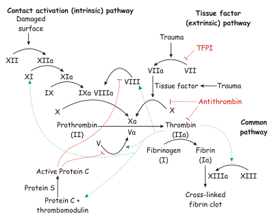
The coagulation cascade. Image source: Wikipedia, GNU Free Documentation License.
A 29 year old female has had 5 episodes of lip and tongue swelling and abdominal pain. C4 level, C1-INH level and C1-INH functional level are all normal. Which one of the following should be checked next?
(A) CBC
(B) Factor III
(C) Factor X
(D) Factor VII
(E) Factor XII
(F) Factor II
Answer: E, Factor XII
References
Angioedema, Hereditary. eMedicine Specialties > Dermatology > Allergy & Immunology.
http://emedicine.medscape.com/article/1048994-overview
Angioedema. eMedicine Specialties > Allergy and Immunology > Urticaria and Angioedema.
http://emedicine.medscape.com/article/135208-overview
Hereditary angioedema with normal C1 inhibition. Current Allergy and Asthma Reports, Volume 9, Number 4 / July, 2009.
Ratnoff OD, Margolius A (1955). Hageman trait: an asymptomatic disorder of blood coagulation. Trans. Assoc. Am. Physicians 68: 149–54. PMID 13299324.
Published: 02/23/2010
Updated: 11/23/2012
Reviewer: S. Randhawa, M.D., Assistant Professor at NSU
A 32-year-old Caucasian female was seen in the clinic for evaluation of an episode of uvular and peritonsillar edema which resolved within 12 hours of taking 40 mg of prednisone previously prescribed by her primary care physician. She has a long history of angioedema since the age of 11 with temporary improvement during pregnancy, but without full resolution of her symptoms. Her mother and her uncle also have symptoms of angiodema history.
Past medical history
Angioedema.
Medications
Prednisone 20 mg po daily and Zyrtec 10 mg po bid.
The physical examination is unremarkable.
The previous laboratory tests included CBC with differential, CMP, sedimentation rate, ANA, rheumatoid factor, TSH, and thyroid antibodies, as well as previous evaluation of the complement components, and there were no significant abnormalities.
What is the most likely diagnosis?
Hereditary angioedema is suspected since similar symptoms have occurred in other members of her family. We suspect that the patient might have type 3 hereditary angioedema.
What would you recommend as next step?
She was advised to taper the prednisone dose to 15 mg po daily for 2 weeks and then to continue prednisone 10 mg po daily for another 2 weeks.
While staying on the maintenance dose of prednisone 10 mg po daily, she was advised to start Celebrex 200 mg po daily and to make a follow up appointment in 4 weeks. Celebrex controlled symptoms in a family member (uncle) who also has angioedema.
If at the 8 week follow up, the patient had no improvement with Celebrex, then we will consider starting progesterone to control her symptoms.
Diagnosis
Hereditary angioedema type III. There is unfortunately no test for "type III" angioedema (source: AAAAI Ask the Expert). Currently, the only place in the U.S. where they ahve performed a test for “type 3 angioedema” is National Jewish Complement Lab http://buff.ly/VkoOxb
The contact information is:
National Jewish Complement Laboratory
Director: Patricia C. Giclas, PhD
Assistant Director: Ashley Frazer-Abel, PhD
Laboratory: 303.398.1541
What did we learn from this case?
Hereditary angioedema type III is typically described in women but can occur in men as well. Certain patients have symptomatic improvement during pregnancy and for them a trial of progesterone therapy may be worth a consideration. Please have in mind that progesterone will stop the menstruation in the such female patients.
Factor XII (Hageman factor)
Hereditary angioedema type III occurs in people with normal C1 inhibitor activity. In some families, mutations in the coagulation factor XII (Hageman factor) gene were detected in the affected persons. Factor XII is encoded by the F12 gene located on the tip of the long arm of the fifth chromosome. Two missense mutations have been identified in F12, the gene encoding human coagulation factor XII. These mutations are thought to be the cause of HAE type III.
Hageman factor was first discovered in 1955 when a routine preoperative blood sample of the 37-year-old railroad brakeman John Hageman was found to have prolonged clotting time in test tubes, even though he had no hemorrhagic symptoms. Hageman was then examined by Dr. Oscar Ratnoff who found that Mr. Hageman lacked a previously unidentified clotting factor. Dr. Ratnoff later found that the Hageman factor deficiency is an autosomal recessive disorder. Paradoxically, pulmonary embolism contributed to Hageman's death after an occupational accident. Since then, case series clinical studies have identified an association of thrombosis and Factor XII deficiency, though the pathophysiology of the relationship is unclear.
The coagulation cascade. Image source: Wikipedia, GNU Free Documentation License.

A 29 year old female has had 5 episodes of lip and tongue swelling and abdominal pain. C4 level, C1-INH level and C1-INH functional level are all normal. Which one of the following should be checked next?
(A) CBC
(B) Factor III
(C) Factor X
(D) Factor VII
(E) Factor XII
(F) Factor II
Answer: E, Factor XII
References
Angioedema, Hereditary. eMedicine Specialties > Dermatology > Allergy & Immunology.
http://emedicine.medscape.com/article/1048994-overview
Angioedema. eMedicine Specialties > Allergy and Immunology > Urticaria and Angioedema.
http://emedicine.medscape.com/article/135208-overview
Hereditary angioedema with normal C1 inhibition. Current Allergy and Asthma Reports, Volume 9, Number 4 / July, 2009.
Ratnoff OD, Margolius A (1955). Hageman trait: an asymptomatic disorder of blood coagulation. Trans. Assoc. Am. Physicians 68: 149–54. PMID 13299324.
Published: 02/23/2010
Updated: 11/23/2012
Angioedema
 Editor: V. Dimov, M.D., Allergist/Immunologist and Assistant Professor at University of Chicago
Information For Patients
Hives (urticaria) and angioedema
Allergic Reactions
Allergic Skin Conditions
Allergy Testing
Food Allergy
Angioedema (AE) Classification (click to enlarge the image):
Diagnosis of Chronic Urticaria and Angioedema (click to enlarge the image).
What to expect when visiting an allergy clinic
Current allergy skin tests are virtually painless. This video by Dr. Bassett, a board-certified allergist from New York City, shows what to expect when visiting an allergy clinic for diagnosis and treatment:
Information For Doctors
Angioedema: Teaching Cases
Angioedema Due to Angiotensin Converting Enzyme Inhibitors
Abdominal angioedema secondary to acquired C1 esterase functional deficiency
Hereditary Angioedema (HAE) in a Teenager: Diagnosis and Treatment
Typical laboratory workup for chronic idiopathic urticaria and angioedema
Delayed allergic reactions to omalizumab: Are patients reporting all cases? JACI, 03/2008.
A 28-Year-Old Woman With Undiagnosed Hereditary Angioedema. Medscape, 2009.
"Traumatic" Angioedema in a Patient on ACEi. Life in the Fast Lane, 2009.
Related Reading
Angioedema: Brief Review
Mind Maps: Angioedema
Mnemonics: Angioedema
Patient stories about hereditary angioedema: Marsha.
Blog articles from AllergyNotes
Published: 06/28/2007
Updated: 05/26/2012
Hereditary Angioedema (HAE) in a Teenager: Diagnosis and Treatment
Author: V. Dimov, M.D., Allergist/Immunologist and Assistant Professor, University of Chicago
Reviewer: S. Randhawa, M.D.
A 12-year-old Caucasian female is seen as a new patient by the allergy and immunology clinic for abdominal pain for 3 months. She has a family history of hereditary angioedema (HAE) diagnosed in her maternal grandfather, mother, and brother.
As a part of family screening, she had a C1 esterase inhibitor level checked 2 years ago and it was 15 mg/dL (normal 19 to 37 mg/dL). The level was repeated several months later and was was in the range of 32% to 10% of the normal.
The patient had no symptoms until 3 months ago when she started to have daily abdominal pain below the umbilicus. The pain lasts a whole day and she has night symptoms as well. There is no consistent relief provided by pain medications.
Medications
None.
Family history
Maternal grandfather with hereditary angioedema with abdominal attacks. Mother with hereditary angioedema with cutaneous attacks. Younger brother with hereditary angioedema with attacks manifesting with “throat closing” symptoms.
Physical examination
Normal.
Laboratory results
C1 esterase inhibitor level 2 years ago was 15 mg/dL (normal 19 to 37 mg/dL). C4 level was 13 mg/dL (normal 10-40 mg/dL). The C1 esterase level was 14 mg/dL (normal 16-33 mg/dL).
What is the most likely diagnosis?
This is a patient with hereditary angioedema, which affected multiple members of her family including her mother, brother, and maternal grandfather. Her hereditary angioedema manifests with abdominal symptoms.
What prophylaxis and treatment would you recommend?
She was given a prescription for a C1 inhibitor, Cinryze 1,000 units IV every four days. The rate of infusion is 1 ml/min and the infusion takes 10 minutes. She was given a prescription to discuss the administration of the medication as a prophylaxis with her primary care physician.
She is to return to our clinic in three months.
Final diagnosis
Hereditary Angioedema (HAE)
Summary
Angioedema (AE) Classification (click to enlarge the image):
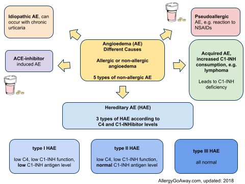
Angioedema (AE) can be allergic or non-allergic.
There are 5 types of non-allergic angioedema (AE):
- acquired AE
- hereditary AE (HAE)
- ACE-inhibitor induced AE
- idiopathic AE, can occur with chronic urticaria
- pseudoallergic AE, e.g. reaction to NSAIDs
There are 3 types of HAE that are differentiated by C4 and C1-INH levels
- type I HAE - low C4, low C1-INH function, low C1-INH antigen level
- type II HAE - low C4, low C1-INH function, normal C1-INH antigen level
- type III HAE - all normal
In most cases (85%), hereditary angioedema (HAE) is an autosomal dominant condition associated with episodic attacks of nonpitting edema. Patients with HAE have low levels of C1 inhibitor (a serine protease inhibitor). Edema is caused by unregulated generation of bradykinin.
Treatment of acute HAE attacks
- C1-INH, 20 units/kg, IV infusion
- Icatibant, 30 mg SC, bradykinin B2 receptor antagonist
- Ecallantide, 30 mg SC, kallikrein receptor antagonist
Prophylaxis of HAE attacks
- C1-INH, 1,000 units, IV infusion every 3-4 days
- attenuated androgen, e.g. danocrine 200 mg PO TID
There are 2 preparations of C1 inhibitor purified from plasma which have been used in Europe for decades (Cinryze and Berinert P). There is also a recombinant C1 inhibitor (not obtained from plasma). A kallikrein inhibitor (Ecallantide) and a bradykinin type 2 receptor antagonist (Icatibant) are in testing phases. It is likely that HAE treatment will change dramatically in near future.
.jpg)
New therapies for hereditary angioedema (HAE)
In summary, the new products for acute treatment and prophylaxis of hereditary angioedema (HAE) are:
- C1 inhibitor purified from plasma (Cinryze and Berinert P)
- recombinant C1 inhibitor
- kallikrein inhibitor (Ecallantide)
- bradykinin type 2 receptor antagonist (Icatibant)
References
What is the difference between hereditary angioedema (HAE) type I, II and III?
C1 Inhibitor Cinryze Approved by FDA for Prophylaxis Against Hereditary Angioedema Attacks
New therapies for hereditary angioedema (HAE)
Biomarkers to help diagnose abdominal attacks in angioedema due to C1-inhibitor deficiency, prevent unnecessary surgeryJournal News: Icatibant for treatment of hereditary angioedema
Hereditary Angioedema: Prophylaxis and Long-Term Management http://goo.gl/l9bY
HAE: annual drug cost alone for prophylactic C1 esterase inhibitor is $450k - nearly $5 mln for every decade of life http://goo.gl/BCVtu
New Directions in the Treatment of Angioedema. Medscape, 2012.
Related reading
35 years ago: “We probably will never know why you swell, but it’s called Angioneurotic Edema” - things have changed a lot since then. HAEA.org.
Published: 02/26/2009
Updated: 06/26/2012
Reviewer: S. Randhawa, M.D.
A 12-year-old Caucasian female is seen as a new patient by the allergy and immunology clinic for abdominal pain for 3 months. She has a family history of hereditary angioedema (HAE) diagnosed in her maternal grandfather, mother, and brother.
As a part of family screening, she had a C1 esterase inhibitor level checked 2 years ago and it was 15 mg/dL (normal 19 to 37 mg/dL). The level was repeated several months later and was was in the range of 32% to 10% of the normal.
The patient had no symptoms until 3 months ago when she started to have daily abdominal pain below the umbilicus. The pain lasts a whole day and she has night symptoms as well. There is no consistent relief provided by pain medications.
Medications
None.
Family history
Maternal grandfather with hereditary angioedema with abdominal attacks. Mother with hereditary angioedema with cutaneous attacks. Younger brother with hereditary angioedema with attacks manifesting with “throat closing” symptoms.
Physical examination
Normal.
Laboratory results
C1 esterase inhibitor level 2 years ago was 15 mg/dL (normal 19 to 37 mg/dL). C4 level was 13 mg/dL (normal 10-40 mg/dL). The C1 esterase level was 14 mg/dL (normal 16-33 mg/dL).
What is the most likely diagnosis?
This is a patient with hereditary angioedema, which affected multiple members of her family including her mother, brother, and maternal grandfather. Her hereditary angioedema manifests with abdominal symptoms.
What prophylaxis and treatment would you recommend?
She was given a prescription for a C1 inhibitor, Cinryze 1,000 units IV every four days. The rate of infusion is 1 ml/min and the infusion takes 10 minutes. She was given a prescription to discuss the administration of the medication as a prophylaxis with her primary care physician.
She is to return to our clinic in three months.
Final diagnosis
Hereditary Angioedema (HAE)
Summary
Angioedema (AE) Classification (click to enlarge the image):
Angioedema (AE) can be allergic or non-allergic.
There are 5 types of non-allergic angioedema (AE):
- acquired AE
- hereditary AE (HAE)
- ACE-inhibitor induced AE
- idiopathic AE, can occur with chronic urticaria
- pseudoallergic AE, e.g. reaction to NSAIDs
There are 3 types of HAE that are differentiated by C4 and C1-INH levels
- type I HAE - low C4, low C1-INH function, low C1-INH antigen level
- type II HAE - low C4, low C1-INH function, normal C1-INH antigen level
- type III HAE - all normal
In most cases (85%), hereditary angioedema (HAE) is an autosomal dominant condition associated with episodic attacks of nonpitting edema. Patients with HAE have low levels of C1 inhibitor (a serine protease inhibitor). Edema is caused by unregulated generation of bradykinin.
Treatment of acute HAE attacks
- C1-INH, 20 units/kg, IV infusion
- Icatibant, 30 mg SC, bradykinin B2 receptor antagonist
- Ecallantide, 30 mg SC, kallikrein receptor antagonist
Prophylaxis of HAE attacks
- C1-INH, 1,000 units, IV infusion every 3-4 days
- attenuated androgen, e.g. danocrine 200 mg PO TID
There are 2 preparations of C1 inhibitor purified from plasma which have been used in Europe for decades (Cinryze and Berinert P). There is also a recombinant C1 inhibitor (not obtained from plasma). A kallikrein inhibitor (Ecallantide) and a bradykinin type 2 receptor antagonist (Icatibant) are in testing phases. It is likely that HAE treatment will change dramatically in near future.
.jpg)
New therapies for hereditary angioedema (HAE)
In summary, the new products for acute treatment and prophylaxis of hereditary angioedema (HAE) are:
- C1 inhibitor purified from plasma (Cinryze and Berinert P)
- recombinant C1 inhibitor
- kallikrein inhibitor (Ecallantide)
- bradykinin type 2 receptor antagonist (Icatibant)
References
What is the difference between hereditary angioedema (HAE) type I, II and III?
C1 Inhibitor Cinryze Approved by FDA for Prophylaxis Against Hereditary Angioedema Attacks
New therapies for hereditary angioedema (HAE)
Biomarkers to help diagnose abdominal attacks in angioedema due to C1-inhibitor deficiency, prevent unnecessary surgeryJournal News: Icatibant for treatment of hereditary angioedema
Hereditary Angioedema: Prophylaxis and Long-Term Management http://goo.gl/l9bY
HAE: annual drug cost alone for prophylactic C1 esterase inhibitor is $450k - nearly $5 mln for every decade of life http://goo.gl/BCVtu
New Directions in the Treatment of Angioedema. Medscape, 2012.
Related reading
35 years ago: “We probably will never know why you swell, but it’s called Angioneurotic Edema” - things have changed a lot since then. HAEA.org.
Published: 02/26/2009
Updated: 06/26/2012
Abdominal angioedema secondary to acquired C1 esterase functional deficiency
Author: V. Dimov, M.D., Allergist/Immunologist and Assistant Professor at University of Chicago
Reviewer: S. Randhawa, M.D., Allergist/Immunologist and Assistant Professor at NSU
A 58-year-old Caucasian male with past medical history of hypertension (on ACE-inhibor and HCTZ) and diverticulosis was referred to our clinic for work-up of angioedema.
Past medical history (PMH)
Hypertension and diverticulosis.
Two months ago, he developed severe abdominal pain after returning from a trip to Ecuador. He was hospitalized and had an EGD which showed erosions, swelling and edema in the area of the esophagus and stomach. He was placed on PPI. Pancreatitis, cholecystitis and hepatitis were ruled out. Hydrochlorothiazide (HCTZ) and Advil were stopped. He was discharged home only to be readmitted 10 days later with abdominal pain and small bowel obstruction (SBO). Altace was stopped and SBO resolved. He was discharged home and hospitalized again 2 weeks later with similar symptoms of abdominal pain and small bowel obstruction. A CT scan of the abdomen showed dilated small bowel loops with edema in the area of the duodenum and small bowel. Symptoms resolved and he was discharged home. He has never had similar symptoms of abdominal pain in the past.
During laboratory workup, C1 esterase inhibitor level was found to be normal, but the functional activity was 29% of the normal level during his last hospitalization (3 weeks prior to the clinic visit). The test was repeated a week ago: the C1 esterase inhibitor level was again normal and the functional capacity was 87% of the normal levels.
The patient was referred to us for work-up of suspected abdominal angioedema secondary to functional C1 esterase inhibitor deficiency.
Medications
Norvasc 5 mg po daily, Protonix 40 mg po daily, Toprol XL 100 mg daily, Colace 100 mg po daily.
Social history
Negative for smoking, no pets. Alcohol: He used to drink a 6-pack of beer per week, but he stopped drinking two months ago after his symptoms started.
Family history
Negative for angioedema, childhood asthma.
Physical examination
VSS
Normal, no rash.
Nose: Normal.
Lymph nodes: Not palpable.
Respiratory system: Clear to auscultation bilaterally.
Cardiovascular system: Clear S1, S2. Abdomen is soft, non-tender, non-distended, no masses. Neurological exam: Normal strength.
Extremities: No cyanosis, clubbing, or edema.
What is the most likely diagnosis?
Abdominal angioedema secondary to acquired C1 esterase functional deficiency. The C1 esterase functional deficiency is probably secondary an infection such as Helicobacter pylori acquired in Ecuador.
What laboratory tests would you order?
Liver enzymes, ANA, CBC with manual differential, hepatitis B core antibody, hepatitis B surface antigen and hepatitis C antibodies for workup of remote hepatitis in conjunction with his travel to Ecuador.
We checked PSA level and specific tests for angioedema work-up including C1Q, C4, C2, CH50, C1 esterase inhibitor qualitative and C1 esterase inhibitor quantitative levels.
What happened next?
The patient was due to have a repeat EGD with biopsy to rule out eosinophilic esophagitis and colonoscopy next week. He has no symptoms, signs or laboratory findings of a lymphoproliferative disorder or any other malignancy.
All the tests came back normal and he was diagnosed with transient C1 esterase functional deficiency. He had no symptoms at his most recent follow-up 2 months after the initial visit.
Final diagnosis
Abdominal angioedema secondary to acquired C1 esterase functional deficiency. Our patient has acquired angioedema (AAE) type II (see below for explanation).
What did we learn from this case?
Acquired C1 esterase inhibitor deficiency is a rare condition which is associated with autoimmune or low-grade lymphoproliferative disorders. The angioedema may precede the lymphoma by many years. Optimal management requires that both angioedema and the underlying lymphoma be recognized and treated.
Adults (after the 4th decade) or elderly patients are most commonly affected.
It often presents with recurrent angioedema and low serum levels of C4 with normal levels of C3. Low levels of C1q and low C1 esterase inhibitor activity confirm the diagnosis.
There are 2 forms of acquired angioedema (AAE): type I and II.
AAE type I is a very rare syndrome associated with lymphoproliferative disorder, autoimmune disease or paraproteinemia. Complement-activating process acts to increase consumption of C1 esterase inhibitor.
In AAE type II, an autoantibody is produced against the C1 esterase inhibitor. The antibodies adhere to the C1 esterase molecule and cause a conformational change leading to decreased function or enhanced metabolism. AAE is differentiated from HAE by decreased C1q, C1r, and C1s levels and decreased functional activity of C1 esterase inhibitor.
Summary
Angioedema (AE) Classification (click to enlarge the image):
Angioedema (AE) can be allergic or non-allergic. There are 5 types of non-allergic angioedema (AE):
- acquired AE
- hereditary AE (HAE)
- ACE-inhibitor induced AE
- idiopathic AE, can occur with chronic urticaria
- pseudoallergic AE, e.g. reaction to NSAIDs
There are 3 types of HAE that are differentiated by C4 and C1-INH levels
- type I HAE - low C4, low C1-INH function, low C1-INH antigen level
- type II HAE - low C4, low C1-INH function, normal C1-INH antigen level
- type III HAE - all normal
Treatment of acute HAE attacks
- C1-INH, 20 units/kg, IV infusion
- Icatibant, 30 mg SC, bradykinin B2 receptor antagonist
- Ecallantide, 30 mg SC, kallikrein receptor antagonist
Prophylaxis of HAE attacks
- C1-INH, 1,000 units, IV infusion every 3-4 days
- attenuated androgen, e.g. danocrine 200 mg PO TID
References
Acquired C1 Esterase Inhibitor Deficiency. Svetomir N. Markovic, MD, PhD; David J. Inwards, MD; Evangelos A. Frigas, MD; and Robert P. Phyliky, MD. Ann of Int Med, 18 January 2000 | Volume 132 Issue 2 | Pages 144-150. Full text PDF.
Acquired C1-esterase inhibitor deficiency: Three case reports and commentary on the syndrome. TK Lipscombe, DI Orton, AG Bird 1 JD Wilkinson. Australasian Journal of Dermatology, Volume 37 Issue 3, Pages 145 - 148, 2007.
C1 Esterase Inhibitor Deficiency. 5-Min Clinical Consult. Unbound Medicine, Inc.
New Directions in the Treatment of Angioedema. Medscape, 2012.
Related reading
35 years ago: “We probably will never know why you swell, but it’s called Angioneurotic Edema” - things have changed a lot since then. HAEA.org.
Published: 12/17/2008
Updated: 06/17/2012
Reviewer: S. Randhawa, M.D., Allergist/Immunologist and Assistant Professor at NSU
A 58-year-old Caucasian male with past medical history of hypertension (on ACE-inhibor and HCTZ) and diverticulosis was referred to our clinic for work-up of angioedema.
Past medical history (PMH)
Hypertension and diverticulosis.
Two months ago, he developed severe abdominal pain after returning from a trip to Ecuador. He was hospitalized and had an EGD which showed erosions, swelling and edema in the area of the esophagus and stomach. He was placed on PPI. Pancreatitis, cholecystitis and hepatitis were ruled out. Hydrochlorothiazide (HCTZ) and Advil were stopped. He was discharged home only to be readmitted 10 days later with abdominal pain and small bowel obstruction (SBO). Altace was stopped and SBO resolved. He was discharged home and hospitalized again 2 weeks later with similar symptoms of abdominal pain and small bowel obstruction. A CT scan of the abdomen showed dilated small bowel loops with edema in the area of the duodenum and small bowel. Symptoms resolved and he was discharged home. He has never had similar symptoms of abdominal pain in the past.
During laboratory workup, C1 esterase inhibitor level was found to be normal, but the functional activity was 29% of the normal level during his last hospitalization (3 weeks prior to the clinic visit). The test was repeated a week ago: the C1 esterase inhibitor level was again normal and the functional capacity was 87% of the normal levels.
The patient was referred to us for work-up of suspected abdominal angioedema secondary to functional C1 esterase inhibitor deficiency.
Medications
Norvasc 5 mg po daily, Protonix 40 mg po daily, Toprol XL 100 mg daily, Colace 100 mg po daily.
Social history
Negative for smoking, no pets. Alcohol: He used to drink a 6-pack of beer per week, but he stopped drinking two months ago after his symptoms started.
Family history
Negative for angioedema, childhood asthma.
Physical examination
VSS
Normal, no rash.
Nose: Normal.
Lymph nodes: Not palpable.
Respiratory system: Clear to auscultation bilaterally.
Cardiovascular system: Clear S1, S2. Abdomen is soft, non-tender, non-distended, no masses. Neurological exam: Normal strength.
Extremities: No cyanosis, clubbing, or edema.
What is the most likely diagnosis?
Abdominal angioedema secondary to acquired C1 esterase functional deficiency. The C1 esterase functional deficiency is probably secondary an infection such as Helicobacter pylori acquired in Ecuador.
What laboratory tests would you order?
Liver enzymes, ANA, CBC with manual differential, hepatitis B core antibody, hepatitis B surface antigen and hepatitis C antibodies for workup of remote hepatitis in conjunction with his travel to Ecuador.
We checked PSA level and specific tests for angioedema work-up including C1Q, C4, C2, CH50, C1 esterase inhibitor qualitative and C1 esterase inhibitor quantitative levels.
What happened next?
The patient was due to have a repeat EGD with biopsy to rule out eosinophilic esophagitis and colonoscopy next week. He has no symptoms, signs or laboratory findings of a lymphoproliferative disorder or any other malignancy.
All the tests came back normal and he was diagnosed with transient C1 esterase functional deficiency. He had no symptoms at his most recent follow-up 2 months after the initial visit.
Final diagnosis
Abdominal angioedema secondary to acquired C1 esterase functional deficiency. Our patient has acquired angioedema (AAE) type II (see below for explanation).
What did we learn from this case?
Acquired C1 esterase inhibitor deficiency is a rare condition which is associated with autoimmune or low-grade lymphoproliferative disorders. The angioedema may precede the lymphoma by many years. Optimal management requires that both angioedema and the underlying lymphoma be recognized and treated.
Adults (after the 4th decade) or elderly patients are most commonly affected.
It often presents with recurrent angioedema and low serum levels of C4 with normal levels of C3. Low levels of C1q and low C1 esterase inhibitor activity confirm the diagnosis.
There are 2 forms of acquired angioedema (AAE): type I and II.
AAE type I is a very rare syndrome associated with lymphoproliferative disorder, autoimmune disease or paraproteinemia. Complement-activating process acts to increase consumption of C1 esterase inhibitor.
In AAE type II, an autoantibody is produced against the C1 esterase inhibitor. The antibodies adhere to the C1 esterase molecule and cause a conformational change leading to decreased function or enhanced metabolism. AAE is differentiated from HAE by decreased C1q, C1r, and C1s levels and decreased functional activity of C1 esterase inhibitor.
Summary
Angioedema (AE) Classification (click to enlarge the image):
Angioedema (AE) can be allergic or non-allergic. There are 5 types of non-allergic angioedema (AE):
- acquired AE
- hereditary AE (HAE)
- ACE-inhibitor induced AE
- idiopathic AE, can occur with chronic urticaria
- pseudoallergic AE, e.g. reaction to NSAIDs
There are 3 types of HAE that are differentiated by C4 and C1-INH levels
- type I HAE - low C4, low C1-INH function, low C1-INH antigen level
- type II HAE - low C4, low C1-INH function, normal C1-INH antigen level
- type III HAE - all normal
Treatment of acute HAE attacks
- C1-INH, 20 units/kg, IV infusion
- Icatibant, 30 mg SC, bradykinin B2 receptor antagonist
- Ecallantide, 30 mg SC, kallikrein receptor antagonist
Prophylaxis of HAE attacks
- C1-INH, 1,000 units, IV infusion every 3-4 days
- attenuated androgen, e.g. danocrine 200 mg PO TID
References
Acquired C1 Esterase Inhibitor Deficiency. Svetomir N. Markovic, MD, PhD; David J. Inwards, MD; Evangelos A. Frigas, MD; and Robert P. Phyliky, MD. Ann of Int Med, 18 January 2000 | Volume 132 Issue 2 | Pages 144-150. Full text PDF.
Acquired C1-esterase inhibitor deficiency: Three case reports and commentary on the syndrome. TK Lipscombe, DI Orton, AG Bird 1 JD Wilkinson. Australasian Journal of Dermatology, Volume 37 Issue 3, Pages 145 - 148, 2007.
C1 Esterase Inhibitor Deficiency. 5-Min Clinical Consult. Unbound Medicine, Inc.
New Directions in the Treatment of Angioedema. Medscape, 2012.
Related reading
35 years ago: “We probably will never know why you swell, but it’s called Angioneurotic Edema” - things have changed a lot since then. HAEA.org.
Published: 12/17/2008
Updated: 06/17/2012
Angioedema: Brief Review
Author: V. Dimov, M.D., Allergist/Immunologist and Assistant Professor, University of Chicago
Reviewer: S. Randhawa, M.D., Assistant Professor at NSU
History
Dr Heinrich Quincke first described the clinical picture of angioedema in 1882, hence the eponym Quincke's edema. Sir William Osler remarked in 1888 that some cases may have a hereditary basis; he coined the term hereditary angio-neurotic edema.
Pathophysiology
Bradykinin is the important mediator behind the symptoms of angioedema. Bradykinin is a potent vasodilator, leading to rapid accumulation of fluid in the interstitium. This is most obvious in the face, where the skin has relatively little supporting connective tissue, and edema develops easily.
Various mechanisms that interfere with bradykinin production or degradation can lead to angioedema:
1. ACE inhibitors block ACE, the enzyme that among other actions, degrades bradykinin.
2. In hereditary angioedema, bradykinin formation is caused by continuous activation of the complement system due to a deficiency in one of its prime inhibitors, C1-esterase inhibitor (C1INH), and continuous production of kallikrein, another process inhibited by C1INH. This serine protease inhibitor normally inhibits the conversion of C1 to C1r and C1s, which - in turn - activate other proteins of the complement system.
Etiology
Angioedema (AE) Classification (click to enlarge the image):
Angioedema (AE) can be allergic or non-allergic. There are 5 types of non-allergic angioedema (AE):
- acquired AE
- hereditary AE (HAE)
- ACE-inhibitor induced AE
- idiopathic AE, can occur with chronic urticaria
- pseudoallergic AE, e.g. reaction to NSAIDs
There are 3 types of HAE that are differentiated by C4 and C1-INH levels
- type I HAE - low C4, low C1-INH function, low C1-INH antigen level
- type II HAE - low C4, low C1-INH function, normal C1-INH antigen level
- type III HAE - all normal
Angioedema is divided in 2 categories: acquired and hereditary.
Acquired Angioedema
A
Acquired
Angioedema
ACEi-related
Hereditary
Angioedema
Autosomal dominant - 11 chromosome
Androgens for prophylaxis
Angioedema involves swelling of the deep dermal and subcutaneous/submucosal tissues. Approximately 50% of patients have both urticaria and angioedema. Angioedema is non-pitting and non-hot.

Angioedema: a 24-hour photo diary by a patient posted on Flickr. The patient took pictures of herself and uploaded them to the photo sharing website Flickr under a Creative Commons license. She had the impression her symptoms were due to urticaria but since the process affects the subcutaneous tissues (note the upper lip edema), the more likely diagnosis is angioedema and urticaria.
When you see a patient with angioedema, the first question is: "are you on ACE inhibitor?" ARBs are well tolerated as an alternative therapy with a reported cross-reactivity of less than 5-10%.


ACE-inhibitor-induced angioedema affecting the upper lip
Acquired angioedema (AAE) rarely starts after 50 years of age.
Acquired angioedema is divided into:
- Type I seen in lymphoproliferative disoders characterized by massive amounts of immune complexes
- Type II - autoantibodies to C1 inhibitor
Both type I and II have low C1q level AND low C4/C2 levels. In contrast, in HAE the C1q level is normal.

C1 protein, showing subunits C1r, C1s, and the C1q tails. Image source: Wikipedia. Patients with acquired angioedema have low C1q levels AND low C4/C2 levels. In contrast, in hereditary angioedema (HAE) the C1q level is normal.

Classical and alternative complement pathways. Image source: Wikipedia.
Hereditary Angioedema (HAE)
Hereditary angioedema (HAE) is an autosomal dominant condition associated with episodic attacks of nonpitting edema. Patients with HAE have low levels of C1 inhibitor (a serine protease inhibitor). Edema is caused by unregulated generation of bradykinin. In HAE the C1q level is normal which differentiates from acquired angioedema type I and II.
1
C1 inhibitor is low
C1 q level is normal
11 chromosome
11 years is the mean age of onset
11 years before the diagnosis is made, on average
Oral contraceptives can unmask HAE -- estrogens unmask the disease, androgens are used for treatment.
Barium swallow can show bowel wall edema seen as "coin stacking" on X-ray.
Diagnosis
Diagnostic workup:
C1q, C4, C2
C1-esterase inhibitor - qualitative and quantitative
CH50
In HAE and AAE, C4 levels are low during angioedema episodes but may be normal between attackes.
To differentiate between AAE and HAE, the C1q level should be measured.
The hallmarks of AAE are low C1q, C2, C4, and C1-INH levels.
In HAE, C1q levels are usually normal or only slightly decreased (rarely less than 50% of the normal values).
Treatment of HAE
Treatment of acute HAE attacks
- C1-INH, 20 units/kg, IV infusion
- Icatibant, 30 mg SC, bradykinin B2 receptor antagonist
- Ecallantide, 30 mg SC, kallikrein receptor antagonist
Prophylaxis of HAE attacks
- C1-INH, 1,000 units, IV infusion every 3-4 days
- attenuated androgen, e.g. danocrine 200 mg PO TID
Effective chronic therapy for HAE has been available for decades -- androgens or plasmin inhibitors. Until recently, there was no therapy for acute attacks available in the U.S.
How do male steroids (androgens) work?
Androgens upregulate the gene.
A follow-up study of the long-term use of stanozolol showed that 21 patients with HAE benefited from stanozolol therapy up to 2 mg daily for more than 25 years. Treatment-related symptoms, predominantly hirsutism, menstrual cycle disorders, and weight gain, developed in 10 patients, but these were easily controlled with dose reduction.
In recent years, 5 pharmaceutical companies have developed drugs which stop acute attacks of HAE or can be used for prophylaxis.
There are 2 preparations of C1 inhibitor purified from plasma which have been used in Europe for decades (Cinryze and Berinert P). There is also a recombinant C1 inhibitor (not obtained from plasma). A kallikrein inhibitor (Ecallantide) and a bradykinin type 2 receptor antagonist (Icatibant) are in testing phases. It is likely that HAE treatment will change dramatically in near future.
New therapies for hereditary angioedema (HAE)
In summary, the new products for acute treatment and prophylaxis of hereditary angioedema (HAE) are:
- C1 inhibitor purified from plasma (Cinryze and Berinert P)
- recombinant C1 inhibitor
- kallikrein inhibitor (Ecallantide)
- bradykinin type 2 receptor antagonist (Icatibant), can be given SQ
R
Recombinant human C1 inhibitor from mammary secretions of trangenic rabbit
Rabbit
Rhucin
C1 inhibitor concentrates
Farkas et al. reported the use of a human C1 inhibitor concentrate therapy for 468 acute attacks in 61 patients with HAE, including 22 children, with efficacy in more than 90% of cases and no reported adverse effects. C1 inhibitor therapy is the best approach from a physiological perspective since it replaces the missing factor. However, the C1 inhibitor has to be administered intravenously which precludes home therapy. Recombinant human C1 inhibitor avoids the infection transmission risks but is difficult to produce.
Kallikrein inhibitors
Kallikrein inhibitors are also effective in reducing HAE symptoms. Angioedema pathogenesis involves the activation of kallikrein to generate bradykinin. A placebo-controlled trial of 48 patients with HAE treated with ecallantide, a kallikrein inhibitor, showed a reduction of HAE attack symptoms in 72% of patients.
Bradykinin type 2 receptor antagonists
A bradykinin type 2 receptor antagonist (Icatibant) has a distinctive advantage over the C1 inhibitor concentrate therapy from the fact that it can be administered subcutaneously. Patients could potentially administer icatibant at home, similar to insulin therapy for diabetes.
References
International consensus on hereditary and acquired angioedema. Annals, 2012.
New therapies for hereditary angioedema: Disease outlook changes dramatically. Frank et al. JACI, Volume 121, Issue 1, Pages 272-280 (January 2008).
New Directions in the Treatment of Angioedema. Medscape, 2012.
Advances in basic and clinical immunology in 2007. Journal of Allergy and Clinical Immunology - Volume 122, Issue 1 (July 2008).
Hereditary Angioedema. NEJM, Volume 359:1027-1036, September 4, 2008.
Angioedema. Maurice Reid, MD. eMedicine.
Angioedema. Nedra R Dodds, MD. eMedicine.
Related reading
Angioedema Due to Angiotensin Converting Enzyme Inhibitors. Allergy Cases, 01/2008.
Audio: New Therapies on Horizon for Angioedema Attacks. AAAAI, 03/2008.
Angioedema, from Wikipedia, the free encyclopedia.
A 28-Year-Old Woman With Undiagnosed Hereditary Angioedema. Medscape, 2009.
"Traumatic" Angioedema in a Patient on ACEi. Life in the Fast Lane, 2009.
35 years ago: “We probably will never know why you swell, but it’s called Angioneurotic Edema” - things have changed a lot since then. HAEA.org.
Published: 07/12/2008
Updated: 11/25/2012
Reviewer: S. Randhawa, M.D., Assistant Professor at NSU
History
Dr Heinrich Quincke first described the clinical picture of angioedema in 1882, hence the eponym Quincke's edema. Sir William Osler remarked in 1888 that some cases may have a hereditary basis; he coined the term hereditary angio-neurotic edema.
Pathophysiology
Bradykinin is the important mediator behind the symptoms of angioedema. Bradykinin is a potent vasodilator, leading to rapid accumulation of fluid in the interstitium. This is most obvious in the face, where the skin has relatively little supporting connective tissue, and edema develops easily.
Various mechanisms that interfere with bradykinin production or degradation can lead to angioedema:
1. ACE inhibitors block ACE, the enzyme that among other actions, degrades bradykinin.
2. In hereditary angioedema, bradykinin formation is caused by continuous activation of the complement system due to a deficiency in one of its prime inhibitors, C1-esterase inhibitor (C1INH), and continuous production of kallikrein, another process inhibited by C1INH. This serine protease inhibitor normally inhibits the conversion of C1 to C1r and C1s, which - in turn - activate other proteins of the complement system.
Etiology
Angioedema (AE) Classification (click to enlarge the image):
Angioedema (AE) can be allergic or non-allergic. There are 5 types of non-allergic angioedema (AE):
- acquired AE
- hereditary AE (HAE)
- ACE-inhibitor induced AE
- idiopathic AE, can occur with chronic urticaria
- pseudoallergic AE, e.g. reaction to NSAIDs
There are 3 types of HAE that are differentiated by C4 and C1-INH levels
- type I HAE - low C4, low C1-INH function, low C1-INH antigen level
- type II HAE - low C4, low C1-INH function, normal C1-INH antigen level
- type III HAE - all normal
Angioedema is divided in 2 categories: acquired and hereditary.
Acquired Angioedema
A
Acquired
Angioedema
ACEi-related
Hereditary
Angioedema
Autosomal dominant - 11 chromosome
Androgens for prophylaxis
Angioedema involves swelling of the deep dermal and subcutaneous/submucosal tissues. Approximately 50% of patients have both urticaria and angioedema. Angioedema is non-pitting and non-hot.

Angioedema: a 24-hour photo diary by a patient posted on Flickr. The patient took pictures of herself and uploaded them to the photo sharing website Flickr under a Creative Commons license. She had the impression her symptoms were due to urticaria but since the process affects the subcutaneous tissues (note the upper lip edema), the more likely diagnosis is angioedema and urticaria.
When you see a patient with angioedema, the first question is: "are you on ACE inhibitor?" ARBs are well tolerated as an alternative therapy with a reported cross-reactivity of less than 5-10%.

ACE-inhibitor-induced angioedema affecting the upper lip
Acquired angioedema (AAE) rarely starts after 50 years of age.
Acquired angioedema is divided into:
- Type I seen in lymphoproliferative disoders characterized by massive amounts of immune complexes
- Type II - autoantibodies to C1 inhibitor
Both type I and II have low C1q level AND low C4/C2 levels. In contrast, in HAE the C1q level is normal.

C1 protein, showing subunits C1r, C1s, and the C1q tails. Image source: Wikipedia. Patients with acquired angioedema have low C1q levels AND low C4/C2 levels. In contrast, in hereditary angioedema (HAE) the C1q level is normal.


Classical and alternative complement pathways. Image source: Wikipedia.
Hereditary Angioedema (HAE)
Hereditary angioedema (HAE) is an autosomal dominant condition associated with episodic attacks of nonpitting edema. Patients with HAE have low levels of C1 inhibitor (a serine protease inhibitor). Edema is caused by unregulated generation of bradykinin. In HAE the C1q level is normal which differentiates from acquired angioedema type I and II.
1
C1 inhibitor is low
C1 q level is normal
11 chromosome
11 years is the mean age of onset
11 years before the diagnosis is made, on average
Oral contraceptives can unmask HAE -- estrogens unmask the disease, androgens are used for treatment.
Barium swallow can show bowel wall edema seen as "coin stacking" on X-ray.
Diagnosis
Diagnostic workup:
C1q, C4, C2
C1-esterase inhibitor - qualitative and quantitative
CH50
In HAE and AAE, C4 levels are low during angioedema episodes but may be normal between attackes.
To differentiate between AAE and HAE, the C1q level should be measured.
The hallmarks of AAE are low C1q, C2, C4, and C1-INH levels.
In HAE, C1q levels are usually normal or only slightly decreased (rarely less than 50% of the normal values).
Treatment of HAE
Treatment of acute HAE attacks
- C1-INH, 20 units/kg, IV infusion
- Icatibant, 30 mg SC, bradykinin B2 receptor antagonist
- Ecallantide, 30 mg SC, kallikrein receptor antagonist
Prophylaxis of HAE attacks
- C1-INH, 1,000 units, IV infusion every 3-4 days
- attenuated androgen, e.g. danocrine 200 mg PO TID
Effective chronic therapy for HAE has been available for decades -- androgens or plasmin inhibitors. Until recently, there was no therapy for acute attacks available in the U.S.
How do male steroids (androgens) work?
Androgens upregulate the gene.
A follow-up study of the long-term use of stanozolol showed that 21 patients with HAE benefited from stanozolol therapy up to 2 mg daily for more than 25 years. Treatment-related symptoms, predominantly hirsutism, menstrual cycle disorders, and weight gain, developed in 10 patients, but these were easily controlled with dose reduction.
In recent years, 5 pharmaceutical companies have developed drugs which stop acute attacks of HAE or can be used for prophylaxis.
There are 2 preparations of C1 inhibitor purified from plasma which have been used in Europe for decades (Cinryze and Berinert P). There is also a recombinant C1 inhibitor (not obtained from plasma). A kallikrein inhibitor (Ecallantide) and a bradykinin type 2 receptor antagonist (Icatibant) are in testing phases. It is likely that HAE treatment will change dramatically in near future.
New therapies for hereditary angioedema (HAE)
In summary, the new products for acute treatment and prophylaxis of hereditary angioedema (HAE) are:
- C1 inhibitor purified from plasma (Cinryze and Berinert P)
- recombinant C1 inhibitor
- kallikrein inhibitor (Ecallantide)
- bradykinin type 2 receptor antagonist (Icatibant), can be given SQ
R
Recombinant human C1 inhibitor from mammary secretions of trangenic rabbit
Rabbit
Rhucin
C1 inhibitor concentrates
Farkas et al. reported the use of a human C1 inhibitor concentrate therapy for 468 acute attacks in 61 patients with HAE, including 22 children, with efficacy in more than 90% of cases and no reported adverse effects. C1 inhibitor therapy is the best approach from a physiological perspective since it replaces the missing factor. However, the C1 inhibitor has to be administered intravenously which precludes home therapy. Recombinant human C1 inhibitor avoids the infection transmission risks but is difficult to produce.
Kallikrein inhibitors
Kallikrein inhibitors are also effective in reducing HAE symptoms. Angioedema pathogenesis involves the activation of kallikrein to generate bradykinin. A placebo-controlled trial of 48 patients with HAE treated with ecallantide, a kallikrein inhibitor, showed a reduction of HAE attack symptoms in 72% of patients.
Bradykinin type 2 receptor antagonists
A bradykinin type 2 receptor antagonist (Icatibant) has a distinctive advantage over the C1 inhibitor concentrate therapy from the fact that it can be administered subcutaneously. Patients could potentially administer icatibant at home, similar to insulin therapy for diabetes.
References
International consensus on hereditary and acquired angioedema. Annals, 2012.
New therapies for hereditary angioedema: Disease outlook changes dramatically. Frank et al. JACI, Volume 121, Issue 1, Pages 272-280 (January 2008).
New Directions in the Treatment of Angioedema. Medscape, 2012.
Hereditary angioedema, Supplement of Annals of Allergy, Asthma and Immunology, 01/2008.
Hereditary angioedema: a decade of human C1-inhibitor concentrate therapy. Farkas et al. J Allergy Clin Immunol 120. 914-917.2007.Advances in basic and clinical immunology in 2007. Journal of Allergy and Clinical Immunology - Volume 122, Issue 1 (July 2008).
Hereditary Angioedema. NEJM, Volume 359:1027-1036, September 4, 2008.
Angioedema. Maurice Reid, MD. eMedicine.
Angioedema. Nedra R Dodds, MD. eMedicine.
Related reading
Angioedema Due to Angiotensin Converting Enzyme Inhibitors. Allergy Cases, 01/2008.
Audio: New Therapies on Horizon for Angioedema Attacks. AAAAI, 03/2008.
Angioedema, from Wikipedia, the free encyclopedia.
A 28-Year-Old Woman With Undiagnosed Hereditary Angioedema. Medscape, 2009.
"Traumatic" Angioedema in a Patient on ACEi. Life in the Fast Lane, 2009.
35 years ago: “We probably will never know why you swell, but it’s called Angioneurotic Edema” - things have changed a lot since then. HAEA.org.
Published: 07/12/2008
Updated: 11/25/2012
Angioedema Due to Angiotensin Converting Enzyme Inhibitors
Author: V. Dimov, M.D., Allergist/Immunologist and Assistant Professor at University of Chicago
Reviewer: S. Randhawa, M.D., Allergist/Immunologist and Assistant Professor at NSU
A 41-year-old African American female (AAF) came to the emergency room (ER) with an upper lip swelling which she noted when she woke up in the morning. She denied shortness of breath, change in her voice or wheezing. She had been taking Lotrel (amlodipine/benazepril) for hypertension (HTN) for 6 months. She had one previous episode of angioedema affecting the the upper lip 7 years ago while she was taking Zestril (lisinopril). The previous episode did not involve any airway compromise and was treated as an outpatient.
Past medical history (PMH)
Hypertension (HTN), diabetes type 2 (DM2), obesity.
Medications
Lotrel (amlodipine/benazepril), metformin.
Family medical history (FMH)
No history of serious allergic reaction in family.
Physical examination
Vital signs stable (VSS), in no apparent distress (NAD).
HEENT: upper lip swelling, no skin rash, no tongue or eyelid swelling.
Chest: CTA (B).
CVS: Clear S1S2.
Abdomen: Soft, NT, ND, +BS.
Extremities: no c/c/e.
ACE-inhibitor-induced angioedema affecting the upper lip (click to enlarge the images).
Laboratory results
The complete blood count (CBC) and basic metabolic panel (BMP) were normal.
What is the most likely diagnosis?
Angioedema due to ACEi without airway compromise, second episode.
What diagnostic tests would you suggest?
C1q, C4, C2 levels
C1-esterase inhibitor - qualitative and quantitative
CH50
Other laboratory tests that can be considered:
CBC+DIFF
CMP
ANA
ESR
IgE
What is the most appropriate treatment?
Stop ACEi and start amlodipine (Norvasc) as a blood pressure (BP) medication.
Solu-Medrol (methylprednisolone) 40 mg IV q 6 hr, when better, switch to oral steroids.
Benadryl (diphenhydramine) 25 mg po q 6 hr (H1-blocker).
Pepcid (famotidine) 20 mg po bid (H2-blocker).
Continuous monitoring of SpO2.
What happened?
The patient was seen by an ENT specialist who did not find any laryngeal edema and she was admitted for a 23-hour observation.
She was given Solu-Medrol (methylprednisolone) 120 mg IV x 1, and then 40 mg IV q 6 hr. The H1- and H2-blockers were continued.
What happened next?
The upper lip swelling has completely resolved by next morning. The patient had no further complaints and was discharged home with oral prednisone taper for 7 days and Benadryl po q 6 hr x 3 days, then prn for itching, rash or swelling. She was advised to avoid driving while taking Benadryl and to follow-up with her primary care physician (PCP) in 3-5 days to check the pending laboratory tests done on admission.
Final diagnosis
Angiodema due to Angiotensin Converting Enzyme Inhibitors (ACEi).
Summary
Angioedema (AE) Classification (click to enlarge the image):
Angioedema (AE) can be allergic or non-allergic. There are 5 types of non-allergic angioedema (AE):
- acquired AE
- hereditary AE (HAE)
- ACE-inhibitor induced AE
- idiopathic AE, can occur with chronic urticaria
- pseudoallergic AE, e.g. reaction to NSAIDs
ACE inhibitors are the most common cause of drug-induced angioedema. Several reports have also linked angiotensin II receptor blockers (ARBs), such as losartan and valsartan, with the development of angioedema but the risk is much lower. ACE-inhibitor induced angiodema typically resolves within 24 to 48 hours.
ACE-inhibitor induced angioedema is an example of idiosyncratic reaction, from Greek, "a peculiar temperament."
Classification of adverse reactions to drugs: "SOAP III" mnemonic (click to enlarge the image):
Adverse drug reactions (ADRs) affect 10–20% of hospitalized patients and 25% of outpatients.
Rule of 10s in ADR
10% of patients develop ADR
10% of these are due to allergy
10% of these lead to anaphylaxis
10% of these lead to death
SOAP III:
Side effect
Overdose
Allergy
Pseudoallergy
Interaction
Intolerance
Idiosyncrasy
In a study of 42,000 patients treated with antihypertensive medications, angioedema occurred in 0.13% of (53 people). The distribution was as follows:
- 70% were receiving lisinopril (an ACEI)
- 15% received chlorthalidone (a diuretic)
- 9% received doxazosin (an alpha blocker)
- 6% received amlodipine (a Ca++ channel blocker)
What is the cross-reactivity risk when prescribing ARB to a patient with ACE-inhibitor-related angioedema?
Less than 5 %.
A literature review of ACEi/ARB angioedema cross-reactivity, shows incidence of 3 to 8%. In a risk-benefit assessment, ARBs should be used cautiously in patients with a history of ACE inhibitor-induced angioedema
Can you prescribe ARB to a patient with ACE-inhibitor-related angioedema?
Yes, but only for populations that have demonstrated a clear benefit from angiotensin II antagonism, for example, patients with CHF and CKD.
The above recommendation has been adopted by the National Kidney Foundation guidelines and the American College of Cardiology and American Heart Association (ACC/AHA) consensus guidelines. Given the strong potential for harm with drug-induced angioedema, however, close monitoring is necessary to ensure that repeat angioedema does not occur with ARB.
ACE inhibitor-related cough
Cough associated with ACE inhibitors was first reported with captopril in 1985. Early reviews reported a frequency of 1-2% but recent reviews found it to be as high as 15-39%. Cough related to ACE inhibitors usually resolves within 2 weeks of stopping the medication but the median time is 26 days.
References
Incidence of angioedema with different antihypertension treatments. JACI, Beyond Our Pages, Volume 119, Issue 5, Pages 1287-1288 (May 2007). Primary source: Diller et al. J Clin Hypertension 2006;8:649-56.
New therapies for hereditary angioedema (HAE). Allergy Notes, 01/2008.
Hereditary angioedema, Supplement of Annals of Allergy, Asthma and Immunology, 01/2008.
Cross-Reactivity of ACE Inhibitor–Induced Angioedema with ARBs. U.S. Pharmacist. Vol. No: 32:2 Posted: 2/20/2007.
Valsartan-Induced Angioedema. The Annals of Pharmacotherapy: Vol. 37, No. 7, pp. 1024-1027, 2003.
Adverse Reactions to Drugs: A Short Review
Treatment of ACE Inhibitor-Induced Cough. Medscape, 1999.
Cough and Angioedema From Angiotensin-Converting Enzyme Inhibitors: New Insights Into Mechanisms and Management. Medscape, 2004.
ACE inhibitor- versus angiotensin II blocker-induced cough and angioedema. The Annals of Pharmacotherapy, 1998.
Angioedema. Maurice Reid, MD. eMedicine.
Angioedema. Nedra R Dodds, MD. eMedicine.
Multiple choice questions
Chapter 57: Drug Allergy. Allergy and Immunology Review Corner: Chapter 57 of Pediatric Allergy: Principles & Practices, edited by Donald Y.M. Leung, et al.
Related reading
Visceral angioedema due to angiotensin-converting enzyme inhibitor therapy - diagnosed with abdominal CT. CCJM, 2011.
The first ACE inhibitor (captopril) was developed from viper venom by a Brazilian post-doc. The FASEB Journal, 2003;17:788-789.
Angioedema due to ACE inhibitor. NEJM, 07/2011.
Angioedema due to the renin inhibitor aliskiren. CCJM, 2011.
Published: 06/23/2007
Updated: 06/21/2012
The photographed patient gave a written permission for her photograph be taken and used for medical education.
Comments from Twitter
Dr John Weiner @AllergyNet: Still occasionally unrecognized in EDs
Reviewer: S. Randhawa, M.D., Allergist/Immunologist and Assistant Professor at NSU
A 41-year-old African American female (AAF) came to the emergency room (ER) with an upper lip swelling which she noted when she woke up in the morning. She denied shortness of breath, change in her voice or wheezing. She had been taking Lotrel (amlodipine/benazepril) for hypertension (HTN) for 6 months. She had one previous episode of angioedema affecting the the upper lip 7 years ago while she was taking Zestril (lisinopril). The previous episode did not involve any airway compromise and was treated as an outpatient.
Past medical history (PMH)
Hypertension (HTN), diabetes type 2 (DM2), obesity.
Medications
Lotrel (amlodipine/benazepril), metformin.
Family medical history (FMH)
No history of serious allergic reaction in family.
Physical examination
Vital signs stable (VSS), in no apparent distress (NAD).
HEENT: upper lip swelling, no skin rash, no tongue or eyelid swelling.
Chest: CTA (B).
CVS: Clear S1S2.
Abdomen: Soft, NT, ND, +BS.
Extremities: no c/c/e.
ACE-inhibitor-induced angioedema affecting the upper lip (click to enlarge the images).
Laboratory results
The complete blood count (CBC) and basic metabolic panel (BMP) were normal.
What is the most likely diagnosis?
Angioedema due to ACEi without airway compromise, second episode.
What diagnostic tests would you suggest?
C1q, C4, C2 levels
C1-esterase inhibitor - qualitative and quantitative
CH50
Other laboratory tests that can be considered:
CBC+DIFF
CMP
ANA
ESR
IgE
What is the most appropriate treatment?
Stop ACEi and start amlodipine (Norvasc) as a blood pressure (BP) medication.
Solu-Medrol (methylprednisolone) 40 mg IV q 6 hr, when better, switch to oral steroids.
Benadryl (diphenhydramine) 25 mg po q 6 hr (H1-blocker).
Pepcid (famotidine) 20 mg po bid (H2-blocker).
Continuous monitoring of SpO2.
What happened?
The patient was seen by an ENT specialist who did not find any laryngeal edema and she was admitted for a 23-hour observation.
She was given Solu-Medrol (methylprednisolone) 120 mg IV x 1, and then 40 mg IV q 6 hr. The H1- and H2-blockers were continued.
What happened next?
The upper lip swelling has completely resolved by next morning. The patient had no further complaints and was discharged home with oral prednisone taper for 7 days and Benadryl po q 6 hr x 3 days, then prn for itching, rash or swelling. She was advised to avoid driving while taking Benadryl and to follow-up with her primary care physician (PCP) in 3-5 days to check the pending laboratory tests done on admission.
Final diagnosis
Angiodema due to Angiotensin Converting Enzyme Inhibitors (ACEi).
Summary
Angioedema (AE) Classification (click to enlarge the image):
Angioedema (AE) can be allergic or non-allergic. There are 5 types of non-allergic angioedema (AE):
- acquired AE
- hereditary AE (HAE)
- ACE-inhibitor induced AE
- idiopathic AE, can occur with chronic urticaria
- pseudoallergic AE, e.g. reaction to NSAIDs
ACE inhibitors are the most common cause of drug-induced angioedema. Several reports have also linked angiotensin II receptor blockers (ARBs), such as losartan and valsartan, with the development of angioedema but the risk is much lower. ACE-inhibitor induced angiodema typically resolves within 24 to 48 hours.
ACE-inhibitor induced angioedema is an example of idiosyncratic reaction, from Greek, "a peculiar temperament."
Classification of adverse reactions to drugs: "SOAP III" mnemonic (click to enlarge the image):
Adverse drug reactions (ADRs) affect 10–20% of hospitalized patients and 25% of outpatients.
Rule of 10s in ADR
10% of patients develop ADR
10% of these are due to allergy
10% of these lead to anaphylaxis
10% of these lead to death
SOAP III:
Side effect
Overdose
Allergy
Pseudoallergy
Interaction
Intolerance
Idiosyncrasy
In a study of 42,000 patients treated with antihypertensive medications, angioedema occurred in 0.13% of (53 people). The distribution was as follows:
- 70% were receiving lisinopril (an ACEI)
- 15% received chlorthalidone (a diuretic)
- 9% received doxazosin (an alpha blocker)
- 6% received amlodipine (a Ca++ channel blocker)
What is the cross-reactivity risk when prescribing ARB to a patient with ACE-inhibitor-related angioedema?
Less than 5 %.
A literature review of ACEi/ARB angioedema cross-reactivity, shows incidence of 3 to 8%. In a risk-benefit assessment, ARBs should be used cautiously in patients with a history of ACE inhibitor-induced angioedema
Can you prescribe ARB to a patient with ACE-inhibitor-related angioedema?
Yes, but only for populations that have demonstrated a clear benefit from angiotensin II antagonism, for example, patients with CHF and CKD.
The above recommendation has been adopted by the National Kidney Foundation guidelines and the American College of Cardiology and American Heart Association (ACC/AHA) consensus guidelines. Given the strong potential for harm with drug-induced angioedema, however, close monitoring is necessary to ensure that repeat angioedema does not occur with ARB.
ACE inhibitor-related cough
Cough associated with ACE inhibitors was first reported with captopril in 1985. Early reviews reported a frequency of 1-2% but recent reviews found it to be as high as 15-39%. Cough related to ACE inhibitors usually resolves within 2 weeks of stopping the medication but the median time is 26 days.
References
Incidence of angioedema with different antihypertension treatments. JACI, Beyond Our Pages, Volume 119, Issue 5, Pages 1287-1288 (May 2007). Primary source: Diller et al. J Clin Hypertension 2006;8:649-56.
New therapies for hereditary angioedema (HAE). Allergy Notes, 01/2008.
Hereditary angioedema, Supplement of Annals of Allergy, Asthma and Immunology, 01/2008.
Cross-Reactivity of ACE Inhibitor–Induced Angioedema with ARBs. U.S. Pharmacist. Vol. No: 32:2 Posted: 2/20/2007.
Valsartan-Induced Angioedema. The Annals of Pharmacotherapy: Vol. 37, No. 7, pp. 1024-1027, 2003.
Adverse Reactions to Drugs: A Short Review
Treatment of ACE Inhibitor-Induced Cough. Medscape, 1999.
Cough and Angioedema From Angiotensin-Converting Enzyme Inhibitors: New Insights Into Mechanisms and Management. Medscape, 2004.
ACE inhibitor- versus angiotensin II blocker-induced cough and angioedema. The Annals of Pharmacotherapy, 1998.
Angioedema. Maurice Reid, MD. eMedicine.
Angioedema. Nedra R Dodds, MD. eMedicine.
Multiple choice questions
Chapter 57: Drug Allergy. Allergy and Immunology Review Corner: Chapter 57 of Pediatric Allergy: Principles & Practices, edited by Donald Y.M. Leung, et al.
Related reading
Visceral angioedema due to angiotensin-converting enzyme inhibitor therapy - diagnosed with abdominal CT. CCJM, 2011.
The first ACE inhibitor (captopril) was developed from viper venom by a Brazilian post-doc. The FASEB Journal, 2003;17:788-789.
Angioedema due to ACE inhibitor. NEJM, 07/2011.
Angioedema due to the renin inhibitor aliskiren. CCJM, 2011.
Published: 06/23/2007
Updated: 06/21/2012
The photographed patient gave a written permission for her photograph be taken and used for medical education.
Comments from Twitter
Dr John Weiner @AllergyNet: Still occasionally unrecognized in EDs
Mnemonics: Angioedema
Author: V. Dimov, M.D., Allergist/Immunologist and Assistant Professor at University of Chicago
Reviewer: S. Randhawa, M.D., Allergist/Immunologist and Assistant Professor at NSU
A
Acquired
Angioedema
ACEi-related
Hereditary
Angioedema
Autosomal dominant
Androgens for prophylaxis
HAE mnemonic
1
C1 inhibitor is low
C1 q level is normal
11 chromosome
11 years is the mean age of onset
11 years before the diagnosis is made, on average
Treatment of HAE
R
Recombinant human C1 inhibitor from mammary secretions of trangenic rabbit
Rabbit
Rhucin
Angioedema (AE) can be allergic or non-allergic.
There are 5 types of non-allergic angioedema (AE):
- acquired AE
- hereditary AE (HAE)
- ACE-inhibitor induced AE
- idiopathic AE, can occur with chronic urticaria
- pseudoallergic AE, e.g. reaction to NSAIDs
There are 3 types of HAE that are differentiated by C4 and C1-INH levels
- type I HAE - low C4, low C1-INH function, low C1-INH antigen level
- type II HAE - low C4, low C1-INH function, normal C1-INH antigen level
- type III HAE - all normal
Treatment of acute HAE attacks
- C1-INH, 20 units/kg, IV infusion
- Icatibant, 30 mg SC, bradykinin B2 receptor antagonist
- Ecallantide, 30 mg SC, kallikrein receptor antagonist
Prophylaxis of HAE attacks
- C1-INH, 1,000 units, IV infusion every 3-4 days
- attenuated androgen, e.g. danocrine 200 mg PO TID
Angioedema (AE) Classification (click to enlarge the image):
References
New Directions in the Treatment of Angioedema. Medscape, 2012.
International consensus on hereditary and acquired angioedema. Annals, 2012.
Published: 02/07/2008
Updated: 12/12/2012
Reviewer: S. Randhawa, M.D., Allergist/Immunologist and Assistant Professor at NSU
A
Acquired
Angioedema
ACEi-related
Hereditary
Angioedema
Autosomal dominant
Androgens for prophylaxis
HAE mnemonic
1
C1 inhibitor is low
C1 q level is normal
11 chromosome
11 years is the mean age of onset
11 years before the diagnosis is made, on average
Treatment of HAE
R
Recombinant human C1 inhibitor from mammary secretions of trangenic rabbit
Rabbit
Rhucin
Angioedema (AE) can be allergic or non-allergic.
There are 5 types of non-allergic angioedema (AE):
- acquired AE
- hereditary AE (HAE)
- ACE-inhibitor induced AE
- idiopathic AE, can occur with chronic urticaria
- pseudoallergic AE, e.g. reaction to NSAIDs
There are 3 types of HAE that are differentiated by C4 and C1-INH levels
- type I HAE - low C4, low C1-INH function, low C1-INH antigen level
- type II HAE - low C4, low C1-INH function, normal C1-INH antigen level
- type III HAE - all normal
Treatment of acute HAE attacks
- C1-INH, 20 units/kg, IV infusion
- Icatibant, 30 mg SC, bradykinin B2 receptor antagonist
- Ecallantide, 30 mg SC, kallikrein receptor antagonist
Prophylaxis of HAE attacks
- C1-INH, 1,000 units, IV infusion every 3-4 days
- attenuated androgen, e.g. danocrine 200 mg PO TID
Angioedema (AE) Classification (click to enlarge the image):
References
New Directions in the Treatment of Angioedema. Medscape, 2012.
International consensus on hereditary and acquired angioedema. Annals, 2012.
Published: 02/07/2008
Updated: 12/12/2012
Mind Maps: Angioedema
Author: V. Dimov, M.D., Allergist/Immunologist and Assistant Professor at University of Chicago
Reviewer: S. Randhawa, M.D., Allergist/Immunologist and Assistant Professor at NSU
Angioedema (AE) Classification (click to enlarge the image):
New therapies for hereditary angioedema (HAE)
References
International consensus on hereditary and acquired angioedema. Annals, 2012.
Published: 01/24/2008
Updated: 11/26/2012
Reviewer: S. Randhawa, M.D., Allergist/Immunologist and Assistant Professor at NSU
Angioedema (AE) Classification (click to enlarge the image):
New therapies for hereditary angioedema (HAE)
References
International consensus on hereditary and acquired angioedema. Annals, 2012.
Published: 01/24/2008
Updated: 11/26/2012
Subscribe to:
Posts (Atom)