Author: V. Dimov, M.D., Allergist/Immunologist at Cleveland Clinic
Reviewer: S. Randhawa, M.D., Allergist/Immunologist and Assistant Professor at NSU
A 60-year-old male is admitted to the hospital with septic arthritis of the left knee. He has a remote history of penicillin allergy. At age 7, he was given a penicillin antibiotic, and immediately there was shortness of breath, throat closing and symptoms of anaphylaxis. Epineprine was administered, and he has been warned by his parents all his life not to take antibiotics.
He responded well to the initial antibiotic treatment with Vancomycin and rifampicin. Blood cultures showed MSSA sensitive to cefazolin. The ID consultant would like to discharge patient home with a PICC lines on 4-week antibiotic treatment.
How to administer cefazolin in a patient with history of penicillin allergy?
1. Skin test for penicillin with Pre-Pen and penicillin G.
2. If the skin test is negative, administer cefazolin via graded dose challenge in a monitored setting (ICU). Observe the patient for 24 hours after the test dose.
Historically, soon after cephalosporins were introduced, there were reports of cephalosoprin anaphylaxis in patients who also had experienced penicillin anaphylaxis. During the initial clinical trials with first-generation cephalosporins and cefamandole, 8.1% of patients with a history of allergy to penicillin had a possible allergy to a cephalosporin, versus 4.5% of patients with no such history . Therefore, the standard teaching is that patients who have had possible penicillin anaphylaxis should not be treated with cephalosporins.
This dogma has been questioned. There is increasing evidence that, in most allergic reactions to cephalosporins, it is the side chain rather than the beta-lactam ring that is the antigen. Older cephalosporins (cephalothin, cephaloridine, and cefamandole) have a side chain similar to that of penicillin and were often contaminated with penicillin. These 2 facts may account for some of the early reports of cross-reactivity between penicillins and cephalosporins.
There is evidence that, among patients with a history of penicillin allergy, the rate of allergic reaction to any other antibiotic is 3 times the rate among control subjects. In the general population, the risk of serious allergic reactions to cephalosporins appears to be below 0.02%.
References and related reading
AInotes - PCN Allergy http://buff.ly/1uD0CsY
Practical Aspects of Choosing an Antibiotic for Patients with a Reported Allergy to an Antibiotic http://buff.ly/1xkxxIt
Published: 07/12/2010
Updated: 03/15/2014
Showing posts with label Drug Allergy. Show all posts
Showing posts with label Drug Allergy. Show all posts

Delayed reaction to oral amoxicillin
Author: V. Dimov, M.D., Allergist/Immunologist and Assistant Professor at University of Chicago
Reviewer: S. Randhawa, M.D., Allergist/Immunologist and Assistant Professor at NSU
A 12-year-old female is at the allergy clinic for evaluation of suspected drug allergy to amoxicillin. Two years ago, she was treated with Augmentin for acute sinusitis. After 7 days of taking the antibiotic, she developed hives. No angioedema or systemic symptoms. No allergy to other antibiotics or medications.
What is the most likely diagnosis?
One episode of acute urticaria with differential diagnosis including:
- idiopathic urticaria
- viral-induced urticaria
- delayed reaction to amoxicillin
What tests would you recommend?
Percutaneous skin testing with negative and positive control, and amoxicillin 1:1, 125 mg/5 ml. The test was negative for amoxicillin.
ImmunoCAP sIgE for PCN and amoxicillin was ordered and it was reported as negative 10 days later.
What would be the next step in management?
Regarding Delayed reaction to oral amoxicillin/clavulanic acid, skin test with amoxicillin and sIgE were negative. The risk for IgE-mediated reaction is low.
There are 2 diagnostic options:
1. Perform skin and intradermal test with Pre-Pen and penicillin G, followed by graded dose oral challenge with amoxicillin.
2. Perform graded dose oral challenge with amoxicillin.
Considering her history of delayed reaction and low risk, option 2 was recommended, a graded dose oral challenge with amoxicillin in the allergy clinic with observation for anaphylaxis.
Dosing protocol for Amoxicillin graded dose challenge:
Step 1: 0.1% of the dose of 500 mg (1/100th)
20 min later
Step 2: 10% of the dose
20 min later
Step 3: 30% of the dose
20 min later
Step 4: The remaining 60% of the dose.
Observe for 1 hour after the last dose.
What happened next?
The challenge was negative. The patient does not have an IgE-mediated allergy to amoxicillin.
For delayed or nonimmediate types of reactions, challenge procedures are less standardized. In general, the time between doses should be long enough that delayed symptoms have time to develop before the next higher dose is administered. Some protocols take 7-14 days. An example of a challenge procedure for delayed reactions would involve administering doses at weekly intervals, starting with 1/100 of a usual dose on day 0, 1/10 of a dose on day 7, and a standard dose on day 14. However, other studies have documented that some delayed reactions only develop after several days at a full therapeutic dose, or in the presence of a concomitant viral infection. Thus, there may be certain drug reactions that cannot be easily elicited with any challenge protocol.
Summary
Although allergy to antibiotics is commonly claimed, true allergy to these drugs is often absent http://buff.ly/1tIbcTl
Adverse drug reactions affect up to 10% of people. When drug reactions resembling allergy happen, they are called drug hypersensitivity reactions (DHRs). Drug hypersensitivity reactions may be allergic or nonallergic. Drug allergies are drug hypersensitivity reactions caused by the immune system.
Clinical presentation in hypersensitivity reactions to beta-lactams is remembered by the mnemonic MAUS:
Maculopapular exanthema, 19.1%
Anaphylaxis without shock, 19.1%
Urticaria, 36.7%
Shock, anaphylactic shock, 17.6%
Anaphylactic shock and anaphylaxis occurs within 1 hour of beta-lactam administration. Exanthema occurs after 24 hours. Urticaria can occur at any time.
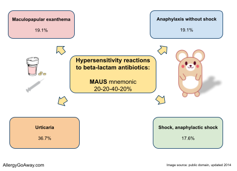
Hypersensitivity reactions to beta-lactam antibiotics: MAUS mnemonic 20-20-40-20%. Click here to enlarge the image.
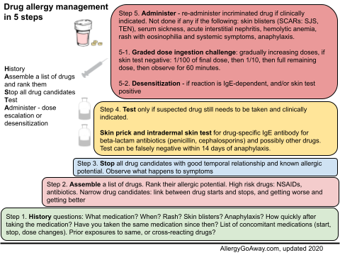
Drug allergy management in 5 steps (click to enlarge the image).
Gell and Goombs classification of hypersensitivity reactions: ACID
Anaphylaxis, angioedema, asthma, type I
Cytotoxic, antibody-mediated, type II, e.g AIHA, ITP, Graves'
Immune complex disease (CIC), type III, e.g. GN, serum sickness, drug fever
Delayed, cell-mediated, type IV, e.g. contact dermatitis
Type I, IgE-mediated drug reactions may involve anaphylaxis or urticaria. Type I reactions occur early or late in a course of therapy and can persist for weeks or months after drug withdrawal.
Type II cytolytic reactions are to rapidly haptenating drugs such as penicillin.
Type III, drug-specific immune complexes result from high-dose, prolonged therapy. They produce drug fever, serum sickness, and cutaneous vasculitis.
Type IV, contact dermatitis occurs with topically applied drugs. Highly sensitizing drugs such as penicillins are no longer provided in a topical form.
What is serum sickness?
From a historical perspective, the term serum sickness means a self-limited immune complex disease caused by exposure to foreign animal proteins or haptens.
Some proteins and large polypeptide drugs (e.g. insulin) can directly stimulate antibody production. However, most drugs act as haptens, binding to proteins, and then stimulating an allergic reaction.
Gell and Coombs classification of drug hypersensitivity was established before T cell subsets were known.
T cell-meditated immunopathology has been subclassified as types IVa-IVd reactions
This subclassification considers the cytokine production and type of cells:
- monocytes (type IVa)
- eosinophils (type IVb)
- cytotoxic activity of both CD4 and CD8 T cells (type IVc)
- neutrophils (type IVd)
Type IVa (Th1)
Th1 cells activate macrophages by secreting large amounts of interferon-gamma. Th1 cells drive the production of complement-fixing antibodies involved in type II and III reactions (IgG1, IgG3).
TH1 are co-stimulatory for TNF, IL-12 and CD8 T cell responses. In vivo correlate is monocyte activation - in PPD or granuloma formation in sarcoidosis. Th1 cells activate CD8 cells, which might explain the common combination of IVa and IVc reactions (e.g., in contact dermatitis).
Type IVb (Th2)
Th2 cells secrete IL-4, IL13, and IL-5, which promote B cell production of IgE and IgG4, mast cells and eosinophils. There is a link to type I reactions, as Th2 cells boost IgE production by IL-4/IL-13 secretion.
Type IVc
T cells can act as cytotoxic cells. They emigrate to the tissue and kill tissue cells like hepatocytes or keratinocytes in a perforin/granzyme B and Fas ligand dependent manner.
Cytotoxic T cells play an important role in:
- maculopapular and bullous skin diseases
- neutrophilic inflammations (acute generalized exanthematous pustulosis, AGEP)
- contact dermatitis
Type IVc reactions are important in bullous skin reactions like Stevens–Johnson syndrome (SJS) and toxic epidermal necrolysis (TEN), where activated CD8 T cells kill keratinocytes.
Type IVd
T cells coordinate sterile neutrophilic inflammation of the skin, in particular AGEP. In this drug-induced disease, T cells recruit neutrophils via CXCL8 release and prevent their apoptosis via GM-CSF. Such T cell reactions are also found in Behçet disease and pustular psoriasis.
Mnemonic
T cell-meditated drug hypersensitivity subclassified as types IVa-IVd:
IVa - 1 = Th1
IVb - 2 = Th2
IVc = cytotoxic CD8 T cells
IVd
References:
Clinical presentation and time course in hypersensitivity reactions to β-lactams. P. J. Bousquet, V. Kvedariene, H.-B. Co-Minh, P. Martins, M. Rongier, B. Arnoux, P. Demoly (2007). Allergy 62 (8), 872–876.
Drug Allergy. Middleton's Allergy: Principles and Practice, Mosby; 7 edition (November 19, 2008).
Clinical review: ABC of allergies. Adverse reactions to drugs. BMJ 1998;316:1511-1514.
Severe Cutaneous Adverse Reactions to Drugs. Current Opinion in Allergy and Clinical Immunology. Faith L. Chia; Khai Pang Leong. Published on Medscape, 08/2007.
References: 04-12-2013
Updated: 05-12-2104
Reviewer: S. Randhawa, M.D., Allergist/Immunologist and Assistant Professor at NSU
A 12-year-old female is at the allergy clinic for evaluation of suspected drug allergy to amoxicillin. Two years ago, she was treated with Augmentin for acute sinusitis. After 7 days of taking the antibiotic, she developed hives. No angioedema or systemic symptoms. No allergy to other antibiotics or medications.
What is the most likely diagnosis?
One episode of acute urticaria with differential diagnosis including:
- idiopathic urticaria
- viral-induced urticaria
- delayed reaction to amoxicillin
What tests would you recommend?
Percutaneous skin testing with negative and positive control, and amoxicillin 1:1, 125 mg/5 ml. The test was negative for amoxicillin.
ImmunoCAP sIgE for PCN and amoxicillin was ordered and it was reported as negative 10 days later.
What would be the next step in management?
Regarding Delayed reaction to oral amoxicillin/clavulanic acid, skin test with amoxicillin and sIgE were negative. The risk for IgE-mediated reaction is low.
There are 2 diagnostic options:
1. Perform skin and intradermal test with Pre-Pen and penicillin G, followed by graded dose oral challenge with amoxicillin.
2. Perform graded dose oral challenge with amoxicillin.
Considering her history of delayed reaction and low risk, option 2 was recommended, a graded dose oral challenge with amoxicillin in the allergy clinic with observation for anaphylaxis.
Dosing protocol for Amoxicillin graded dose challenge:
Step 1: 0.1% of the dose of 500 mg (1/100th)
20 min later
Step 2: 10% of the dose
20 min later
Step 3: 30% of the dose
20 min later
Step 4: The remaining 60% of the dose.
Observe for 1 hour after the last dose.
What happened next?
The challenge was negative. The patient does not have an IgE-mediated allergy to amoxicillin.
For delayed or nonimmediate types of reactions, challenge procedures are less standardized. In general, the time between doses should be long enough that delayed symptoms have time to develop before the next higher dose is administered. Some protocols take 7-14 days. An example of a challenge procedure for delayed reactions would involve administering doses at weekly intervals, starting with 1/100 of a usual dose on day 0, 1/10 of a dose on day 7, and a standard dose on day 14. However, other studies have documented that some delayed reactions only develop after several days at a full therapeutic dose, or in the presence of a concomitant viral infection. Thus, there may be certain drug reactions that cannot be easily elicited with any challenge protocol.
Summary
Although allergy to antibiotics is commonly claimed, true allergy to these drugs is often absent http://buff.ly/1tIbcTl
Adverse drug reactions affect up to 10% of people. When drug reactions resembling allergy happen, they are called drug hypersensitivity reactions (DHRs). Drug hypersensitivity reactions may be allergic or nonallergic. Drug allergies are drug hypersensitivity reactions caused by the immune system.
Clinical presentation in hypersensitivity reactions to beta-lactams is remembered by the mnemonic MAUS:
Maculopapular exanthema, 19.1%
Anaphylaxis without shock, 19.1%
Urticaria, 36.7%
Shock, anaphylactic shock, 17.6%
Anaphylactic shock and anaphylaxis occurs within 1 hour of beta-lactam administration. Exanthema occurs after 24 hours. Urticaria can occur at any time.
Hypersensitivity reactions to beta-lactam antibiotics: MAUS mnemonic 20-20-40-20%. Click here to enlarge the image.
Drug allergy management in 5 steps (click to enlarge the image).
Gell and Goombs classification of hypersensitivity reactions: ACID
Anaphylaxis, angioedema, asthma, type I
Cytotoxic, antibody-mediated, type II, e.g AIHA, ITP, Graves'
Immune complex disease (CIC), type III, e.g. GN, serum sickness, drug fever
Delayed, cell-mediated, type IV, e.g. contact dermatitis
Type I, IgE-mediated drug reactions may involve anaphylaxis or urticaria. Type I reactions occur early or late in a course of therapy and can persist for weeks or months after drug withdrawal.
Type II cytolytic reactions are to rapidly haptenating drugs such as penicillin.
Type III, drug-specific immune complexes result from high-dose, prolonged therapy. They produce drug fever, serum sickness, and cutaneous vasculitis.
Type IV, contact dermatitis occurs with topically applied drugs. Highly sensitizing drugs such as penicillins are no longer provided in a topical form.
What is serum sickness?
From a historical perspective, the term serum sickness means a self-limited immune complex disease caused by exposure to foreign animal proteins or haptens.
Some proteins and large polypeptide drugs (e.g. insulin) can directly stimulate antibody production. However, most drugs act as haptens, binding to proteins, and then stimulating an allergic reaction.
Gell and Coombs classification of drug hypersensitivity was established before T cell subsets were known.
T cell-meditated immunopathology has been subclassified as types IVa-IVd reactions
This subclassification considers the cytokine production and type of cells:
- monocytes (type IVa)
- eosinophils (type IVb)
- cytotoxic activity of both CD4 and CD8 T cells (type IVc)
- neutrophils (type IVd)
Type IVa (Th1)
Th1 cells activate macrophages by secreting large amounts of interferon-gamma. Th1 cells drive the production of complement-fixing antibodies involved in type II and III reactions (IgG1, IgG3).
TH1 are co-stimulatory for TNF, IL-12 and CD8 T cell responses. In vivo correlate is monocyte activation - in PPD or granuloma formation in sarcoidosis. Th1 cells activate CD8 cells, which might explain the common combination of IVa and IVc reactions (e.g., in contact dermatitis).
Type IVb (Th2)
Th2 cells secrete IL-4, IL13, and IL-5, which promote B cell production of IgE and IgG4, mast cells and eosinophils. There is a link to type I reactions, as Th2 cells boost IgE production by IL-4/IL-13 secretion.
Type IVc
T cells can act as cytotoxic cells. They emigrate to the tissue and kill tissue cells like hepatocytes or keratinocytes in a perforin/granzyme B and Fas ligand dependent manner.
Cytotoxic T cells play an important role in:
- maculopapular and bullous skin diseases
- neutrophilic inflammations (acute generalized exanthematous pustulosis, AGEP)
- contact dermatitis
Type IVc reactions are important in bullous skin reactions like Stevens–Johnson syndrome (SJS) and toxic epidermal necrolysis (TEN), where activated CD8 T cells kill keratinocytes.
Type IVd
T cells coordinate sterile neutrophilic inflammation of the skin, in particular AGEP. In this drug-induced disease, T cells recruit neutrophils via CXCL8 release and prevent their apoptosis via GM-CSF. Such T cell reactions are also found in Behçet disease and pustular psoriasis.
Mnemonic
T cell-meditated drug hypersensitivity subclassified as types IVa-IVd:
IVa - 1 = Th1
IVb - 2 = Th2
IVc = cytotoxic CD8 T cells
IVd
References:
Clinical presentation and time course in hypersensitivity reactions to β-lactams. P. J. Bousquet, V. Kvedariene, H.-B. Co-Minh, P. Martins, M. Rongier, B. Arnoux, P. Demoly (2007). Allergy 62 (8), 872–876.
Drug Allergy. Middleton's Allergy: Principles and Practice, Mosby; 7 edition (November 19, 2008).
Clinical review: ABC of allergies. Adverse reactions to drugs. BMJ 1998;316:1511-1514.
Severe Cutaneous Adverse Reactions to Drugs. Current Opinion in Allergy and Clinical Immunology. Faith L. Chia; Khai Pang Leong. Published on Medscape, 08/2007.
References: 04-12-2013
Updated: 05-12-2104
Acute urticaria (hives) due to allergy to terbinafine
Author: V. Dimov, M.D., Allergist/Immunologist and Assistant Professor at University of Chicago
Reviewer: S. Randhawa, M.D., Allergist/Immunologist and Assistant Professor at NSU, Fort Lauderdale
A 25-year-old female was referred for evaluation of urticaria and lip angioedema after being treated with terbinafine (Lamisil) for 14 days for ringworm. One day after the last dose of terbinafine, she developed hives that progressed to affect her whole body. No systemic symptoms. She developed lip swelling 2 days later and the hives lasted a total of 3 days. They resolved with cetiririzine and oral steroid.
Past Medical History, Past Surgical History: Negative.
Current Medications: None.
Family Medical History and Social History: Not contributory.
Review Of Systems: 12/14 systems were reviewed, negative apart from history of present illness above.
Physical Examination: Unremarkable, no hives.
What is the most likely diagnosis?
Acute urticaria (hives) and lip angioedema (swelling) likely due to adverse reaction to terbinafine (Lamisil). The reaction can happen soon after stopping the medication, or after taking it for weeks. There is no evidence of chronic urticaria, blistering or systemic symptoms. No hives or angioedema now. Food allergy is unlikely with her history, but we can do a skin test for food and aiborne allergens for completeness.
What management would you suggest?
Take Claritin or Zyrtec 10 mg po daily for 2 weeks. Do not take terbinafine (Lamisil) or related medications in the future. An allergy to terbinafine (Lamisil) was added to her allergies list in EMR.
A graded dose drug challenge can be considered in the future if she must take terbinafine and there are no alternative options.
Summary
Terbinafine is an allylamine antifungal agent widely used to treat dermatophyte onychomycosis and dermatomycoses. Br J Dermatol. included 10 case reports of severe cutaneous adverse reactions associated with terbinafine therapy which required discontinuation of the antifungal agent: erythema multiforme, erythroderma, severe urticaria, pityriasis rosea and worsening of pre-existing psoriasis.
References:
Cutaneous adverse effects associated with terbinafine therapy: 10 case reports and a review of the literature. Gupta AK, Lynde CW, Lauzon GJ, Mehlmauer MA, Braddock SW, Miller CA, Del Rosso JQ, Shear NH. Br J Dermatol. 1998 Mar;138(3):529-32.
Association between terbinafine and arthralgia, fever and urticaria: symptoms or syndrome?
van Puijenbroek EP, Egberts AC, Meyboom RH, Leufkens HG. Pharmacoepidemiol Drug Saf. 2001 Mar-Apr;10(2):135-42.
Published: 08-20-2013
Updated: 09-17-2013
Reviewer: S. Randhawa, M.D., Allergist/Immunologist and Assistant Professor at NSU, Fort Lauderdale
A 25-year-old female was referred for evaluation of urticaria and lip angioedema after being treated with terbinafine (Lamisil) for 14 days for ringworm. One day after the last dose of terbinafine, she developed hives that progressed to affect her whole body. No systemic symptoms. She developed lip swelling 2 days later and the hives lasted a total of 3 days. They resolved with cetiririzine and oral steroid.
Past Medical History, Past Surgical History: Negative.
Current Medications: None.
Family Medical History and Social History: Not contributory.
Review Of Systems: 12/14 systems were reviewed, negative apart from history of present illness above.
Physical Examination: Unremarkable, no hives.
What is the most likely diagnosis?
Acute urticaria (hives) and lip angioedema (swelling) likely due to adverse reaction to terbinafine (Lamisil). The reaction can happen soon after stopping the medication, or after taking it for weeks. There is no evidence of chronic urticaria, blistering or systemic symptoms. No hives or angioedema now. Food allergy is unlikely with her history, but we can do a skin test for food and aiborne allergens for completeness.
What management would you suggest?
Take Claritin or Zyrtec 10 mg po daily for 2 weeks. Do not take terbinafine (Lamisil) or related medications in the future. An allergy to terbinafine (Lamisil) was added to her allergies list in EMR.
A graded dose drug challenge can be considered in the future if she must take terbinafine and there are no alternative options.
Summary
Terbinafine is an allylamine antifungal agent widely used to treat dermatophyte onychomycosis and dermatomycoses. Br J Dermatol. included 10 case reports of severe cutaneous adverse reactions associated with terbinafine therapy which required discontinuation of the antifungal agent: erythema multiforme, erythroderma, severe urticaria, pityriasis rosea and worsening of pre-existing psoriasis.
References:
Cutaneous adverse effects associated with terbinafine therapy: 10 case reports and a review of the literature. Gupta AK, Lynde CW, Lauzon GJ, Mehlmauer MA, Braddock SW, Miller CA, Del Rosso JQ, Shear NH. Br J Dermatol. 1998 Mar;138(3):529-32.
Association between terbinafine and arthralgia, fever and urticaria: symptoms or syndrome?
van Puijenbroek EP, Egberts AC, Meyboom RH, Leufkens HG. Pharmacoepidemiol Drug Saf. 2001 Mar-Apr;10(2):135-42.
Published: 08-20-2013
Updated: 09-17-2013
Evaluation of adverse reaction to DTaP vaccine
Author: V. Dimov, M.D., Allergist/Immunologist and Assistant Professor at University of Chicago
Reviewer: S. Randhawa, M.D., Allergist/Immunologist and Assistant Professor at NSU
A 6-month-old boy is in the allergy clinic for evaluation of adverse reaction to DTaP vaccine. One hour after the first dose of the DTaP vaccine, he became restless and had hives on his abdomen that stayed for 2 days and resolved with cetirizine. The family is very worried because his mother had a systemic reaction as a young child to what she thinks was tetanus vaccine (or DTP). She was 3-year-old at the time, had hives, passed out and required hospitalization.
The boy has no history of asthma, allergic rhinitis, food allergy, or atopic dermatitis.
Physical examination was normal.
What is the most likely diagnosis?
- idiopathic acute urticaria
- adverse reaction to DTaP vaccine
What diagnostic method would you suggest?
Skin test and graded drug challenge with DTaP vaccine.
What happened?
The skin test and the graded dose challenge with DTaP vaccine were negative. There was no evidence of IgE-mediated drug allergic reaction to DTaP. The risk of delayed, non-IgE mediated reaction cannot be ruled out.
Ideally, skin test for each of the individual components of DTP should be performed, followed by challenge in the allergy clinic. However, since the separate vaccines are no longer available, then a test with the DTaP is indicated following the modified protocol below:
1. Skin test with positive control, negative skin control, 1:100, 1:10, 1:1 concentration of DTP. Observe for 15 min. If negative, proceed with:
2. Intradermal test with 1:100 concentration of DTP. Observe for 15 min. If negative, proceed with:
3. Inject 10% of the DTP dose as indicated. Observe for 30 min. If negative, proceed with:
4. Inject 90% of the dose. Observe for 30 min.
All 4 steps were completed and were negative for signs or symptoms of IgE-mediated reaction.
The plan is based on the reference below:
If the skin test (prick and intradermal skin test) result is negative, the chance that the patient has IgE antibody to any vaccine constituent is negligible, and the vaccine can be administered in the usual manner. Nonetheless, in a patient with a history suggestive of an anaphylactic reaction, it is prudent to administer the vaccine under observation, with epinephrine and other treatments available. Thus if the skin test results to the vaccine and its ingredients are negative, particularly at the intradermal level (with the vaccine diluted 1:100), then it is unlikely that the patient has IgE antibody to any component of the vaccine, and they can be given the vaccine in the usual manner but observed for at least 30 minutes afterward. If vaccine or vaccine component skin test results are positive, the vaccine might still be administered, if necessary, in graded doses. If the full vaccine dose is normally a volume of 0.5 mL, the patient is first given 0.05 mL of a 1:10 dilution and then given full-strength vaccine (at 15-minute intervals) at doses of 0.05, 0.1, 0.15, and finally 0.2 mL, for a cumulative dose of 0.5 mL. A simplified protocol can be used, with administration of 10% of the dose (followed by 30 minute observation), followed by 90% of the dose (followed by 30 minute observation).
This procedure in a patient who is presumed to be allergic to the vaccine being administered needs to be performed under direct medical supervision, with emergency medications and equipment immediately available to promptly treat an anaphylactic reaction should it occur. Such challenges can be performed in an office or hospital setting with or without an intravenous line in place, depending on the severity of the original reaction to the vaccine and the patient’s medical condition.
Source: J Allergy Clin Immunol. 2012 Jul;130(1):25-43. doi: 10.1016/j.jaci.2012.04.003. Epub 2012 May 16. Adverse reactions to vaccines practice parameter 2012 update. Kelso JM, Greenhawt MJ, Li JT, Nicklas RA, Bernstein DI, Blessing-Moore J, Cox L, Khan D, Lang DM, Oppenheimer J, Portnoy JM, Randolph CR, Schuller DE, Spector SL, Tilles SA, Wallace D.
What happened next?
There was no evidence of IgE-mediated drug allergic reaction to DTaP. The risk of delayed, non-IgE mediated reaction cannot be ruled out because the mechanism of those reactions is different. The patient can proceed with the rest of the immunization schedule as per the current CDC guidelines.
Overdiagnosis of vaccine allergy is a major public health problem http://buff.ly/1tI9y44
References
J Allergy Clin Immunol. 2012 Jul;130(1):25-43. doi: 10.1016/j.jaci.2012.04.003. Epub 2012 May 16. Adverse reactions to vaccines practice parameter 2012 update. Kelso JM, Greenhawt MJ, Li JT, Nicklas RA, Bernstein DI, Blessing-Moore J, Cox L, Khan D, Lang DM, Oppenheimer J, Portnoy JM, Randolph CR, Schuller DE, Spector SL, Tilles SA, Wallace D.
Published: 03/12/2012
Updated: 05/22/2013
Reviewer: S. Randhawa, M.D., Allergist/Immunologist and Assistant Professor at NSU
A 6-month-old boy is in the allergy clinic for evaluation of adverse reaction to DTaP vaccine. One hour after the first dose of the DTaP vaccine, he became restless and had hives on his abdomen that stayed for 2 days and resolved with cetirizine. The family is very worried because his mother had a systemic reaction as a young child to what she thinks was tetanus vaccine (or DTP). She was 3-year-old at the time, had hives, passed out and required hospitalization.
The boy has no history of asthma, allergic rhinitis, food allergy, or atopic dermatitis.
Physical examination was normal.
What is the most likely diagnosis?
- idiopathic acute urticaria
- adverse reaction to DTaP vaccine
What diagnostic method would you suggest?
Skin test and graded drug challenge with DTaP vaccine.
What happened?
The skin test and the graded dose challenge with DTaP vaccine were negative. There was no evidence of IgE-mediated drug allergic reaction to DTaP. The risk of delayed, non-IgE mediated reaction cannot be ruled out.
Ideally, skin test for each of the individual components of DTP should be performed, followed by challenge in the allergy clinic. However, since the separate vaccines are no longer available, then a test with the DTaP is indicated following the modified protocol below:
1. Skin test with positive control, negative skin control, 1:100, 1:10, 1:1 concentration of DTP. Observe for 15 min. If negative, proceed with:
2. Intradermal test with 1:100 concentration of DTP. Observe for 15 min. If negative, proceed with:
3. Inject 10% of the DTP dose as indicated. Observe for 30 min. If negative, proceed with:
4. Inject 90% of the dose. Observe for 30 min.
All 4 steps were completed and were negative for signs or symptoms of IgE-mediated reaction.
The plan is based on the reference below:
If the skin test (prick and intradermal skin test) result is negative, the chance that the patient has IgE antibody to any vaccine constituent is negligible, and the vaccine can be administered in the usual manner. Nonetheless, in a patient with a history suggestive of an anaphylactic reaction, it is prudent to administer the vaccine under observation, with epinephrine and other treatments available. Thus if the skin test results to the vaccine and its ingredients are negative, particularly at the intradermal level (with the vaccine diluted 1:100), then it is unlikely that the patient has IgE antibody to any component of the vaccine, and they can be given the vaccine in the usual manner but observed for at least 30 minutes afterward. If vaccine or vaccine component skin test results are positive, the vaccine might still be administered, if necessary, in graded doses. If the full vaccine dose is normally a volume of 0.5 mL, the patient is first given 0.05 mL of a 1:10 dilution and then given full-strength vaccine (at 15-minute intervals) at doses of 0.05, 0.1, 0.15, and finally 0.2 mL, for a cumulative dose of 0.5 mL. A simplified protocol can be used, with administration of 10% of the dose (followed by 30 minute observation), followed by 90% of the dose (followed by 30 minute observation).
This procedure in a patient who is presumed to be allergic to the vaccine being administered needs to be performed under direct medical supervision, with emergency medications and equipment immediately available to promptly treat an anaphylactic reaction should it occur. Such challenges can be performed in an office or hospital setting with or without an intravenous line in place, depending on the severity of the original reaction to the vaccine and the patient’s medical condition.
Source: J Allergy Clin Immunol. 2012 Jul;130(1):25-43. doi: 10.1016/j.jaci.2012.04.003. Epub 2012 May 16. Adverse reactions to vaccines practice parameter 2012 update. Kelso JM, Greenhawt MJ, Li JT, Nicklas RA, Bernstein DI, Blessing-Moore J, Cox L, Khan D, Lang DM, Oppenheimer J, Portnoy JM, Randolph CR, Schuller DE, Spector SL, Tilles SA, Wallace D.
What happened next?
There was no evidence of IgE-mediated drug allergic reaction to DTaP. The risk of delayed, non-IgE mediated reaction cannot be ruled out because the mechanism of those reactions is different. The patient can proceed with the rest of the immunization schedule as per the current CDC guidelines.
Overdiagnosis of vaccine allergy is a major public health problem http://buff.ly/1tI9y44
References
J Allergy Clin Immunol. 2012 Jul;130(1):25-43. doi: 10.1016/j.jaci.2012.04.003. Epub 2012 May 16. Adverse reactions to vaccines practice parameter 2012 update. Kelso JM, Greenhawt MJ, Li JT, Nicklas RA, Bernstein DI, Blessing-Moore J, Cox L, Khan D, Lang DM, Oppenheimer J, Portnoy JM, Randolph CR, Schuller DE, Spector SL, Tilles SA, Wallace D.
Published: 03/12/2012
Updated: 05/22/2013
Protocol for desensitization to rosuvastatin
Statins decrease cholesterol levels by inhibiting 3-hydroxy-3-methyl-glutaryl–coenzyme A reductase levels. Statins reduce morbidity and mortality in patients with a previous myocardial infarction (MI).
Use of statins has been associated with myositis and skin reactions, such as toxic epidermal necrolysis, urticarial vasculitis, drug reaction with eosinophilia and systemic symptoms syndrome, and chronic urticaria. However, anaphylaxis to statins and desensitization have been rarely reported.
A letter to the editor published in JACI, reported successful rapid oral desensitization to rosuvastatin in a 37-year-old man. He had a reported anaphylaxis to multiple statin agents.
He had a life-threatening reaction to 2 different statins that was consistent with an IgE-mediated immediate hypersensitivity mechanis. He was not challenged with a full-dose statin agent because of the significant risks.
Rosuvastatin was chosen as the agent for rapid oral desensitization because he had not previously received this statin. Rosuvastatin contains a sulfonamide group, but the patient had no prior history of adverse reactions to sulfonamides.
The patient was admitted to the cardiac intensive care unit (ICU) and an arterial line was placed. In preparation for the desensitization, beta-blocker was held for 72 hours before the procedure should epinephrine need to be administered. He was pretreated with montelukast and zileuton for 48 hours before the procedure, and on the morning of the desensitization, he received aspirin, ranitidine, and odansetron. H1 blockers were held before desensitization over concerns that this might mask an early IgE-mediated reaction.
He was started on a dose of 0.01 mg of rosuvastatin, which was increased as outlined in Table 1.
Table 1. Oral rosuvastatin desensitization protocol.
Time -- Dose (mg) -- Concentration (mg/mL) -- mL
0:00 0.01 0.01 1
0:30 0.02 0.01 2
1:00 0.04 0.01 4
1:30 0.08 0.01 8
2:00 0.1 0.1 1
2:30 0.2 0.1 2
3:00 0.4 0.1 4
3:30 0.8 0.1 8
4:00 1 1 1
4:30 2 1 2
5:00 4 1 4
5:30 8 1 8
6:00 10 10-mg tablet 10-mg tablet
Solutions were made by dissolving a 10-mg tablet of rosuvastatin in 10 mL of saline, then diluting to the 0.1 and 0.01 mg/mL concentrations. Doses were given every 30 minutes; if any reaction occurs, the dose can be repeated or decreased based on severity.
After receiving the 0.1-mg dose, the patient reported pruritus but had no urticaria. Blood pressure was stable. He was treated with 50 mg of diphenhydramine and 100 mg of hydrocortisone, the 0.1-mg dose was repeated, and the protocol was continued and successfully completed with a final oral dose of 10 mg. The patient stayed overnight in ICU and was discharged the next day.
The authors claim that this is the first published desensitization protocol to statin agents in a patient with a history of anaphylaxis to these agents. The protocol enabled a high-risk cardiac patient to undergo successful desensitization in a carefully monitored intensive care setting.
References:
Successful desensitization to rosuvastatin in a patient with a history of anaphylaxis to multiple statins. The Journal of Allergy and Clinical Immunology, Volume 131, Issue 1 , Pages 234-236, January 2013.
http://www.jacionline.org/article/S0091-6749(12)01620-X/fulltext
Use of statins has been associated with myositis and skin reactions, such as toxic epidermal necrolysis, urticarial vasculitis, drug reaction with eosinophilia and systemic symptoms syndrome, and chronic urticaria. However, anaphylaxis to statins and desensitization have been rarely reported.
A letter to the editor published in JACI, reported successful rapid oral desensitization to rosuvastatin in a 37-year-old man. He had a reported anaphylaxis to multiple statin agents.
He had a life-threatening reaction to 2 different statins that was consistent with an IgE-mediated immediate hypersensitivity mechanis. He was not challenged with a full-dose statin agent because of the significant risks.
Rosuvastatin was chosen as the agent for rapid oral desensitization because he had not previously received this statin. Rosuvastatin contains a sulfonamide group, but the patient had no prior history of adverse reactions to sulfonamides.
The patient was admitted to the cardiac intensive care unit (ICU) and an arterial line was placed. In preparation for the desensitization, beta-blocker was held for 72 hours before the procedure should epinephrine need to be administered. He was pretreated with montelukast and zileuton for 48 hours before the procedure, and on the morning of the desensitization, he received aspirin, ranitidine, and odansetron. H1 blockers were held before desensitization over concerns that this might mask an early IgE-mediated reaction.
He was started on a dose of 0.01 mg of rosuvastatin, which was increased as outlined in Table 1.
Table 1. Oral rosuvastatin desensitization protocol.
Time -- Dose (mg) -- Concentration (mg/mL) -- mL
0:00 0.01 0.01 1
0:30 0.02 0.01 2
1:00 0.04 0.01 4
1:30 0.08 0.01 8
2:00 0.1 0.1 1
2:30 0.2 0.1 2
3:00 0.4 0.1 4
3:30 0.8 0.1 8
4:00 1 1 1
4:30 2 1 2
5:00 4 1 4
5:30 8 1 8
6:00 10 10-mg tablet 10-mg tablet
Solutions were made by dissolving a 10-mg tablet of rosuvastatin in 10 mL of saline, then diluting to the 0.1 and 0.01 mg/mL concentrations. Doses were given every 30 minutes; if any reaction occurs, the dose can be repeated or decreased based on severity.
After receiving the 0.1-mg dose, the patient reported pruritus but had no urticaria. Blood pressure was stable. He was treated with 50 mg of diphenhydramine and 100 mg of hydrocortisone, the 0.1-mg dose was repeated, and the protocol was continued and successfully completed with a final oral dose of 10 mg. The patient stayed overnight in ICU and was discharged the next day.
The authors claim that this is the first published desensitization protocol to statin agents in a patient with a history of anaphylaxis to these agents. The protocol enabled a high-risk cardiac patient to undergo successful desensitization in a carefully monitored intensive care setting.
References:
Successful desensitization to rosuvastatin in a patient with a history of anaphylaxis to multiple statins. The Journal of Allergy and Clinical Immunology, Volume 131, Issue 1 , Pages 234-236, January 2013.
http://www.jacionline.org/article/S0091-6749(12)01620-X/fulltext
Hypersensitivity reactions to sulfonamides - the term “sulfa allergy” is misleading
From a JACI 2012 review:
Chemical structure and cross-reactivity in sulfonamide allergy
Sulfonamides are drugs carrying the SO2-NH2 group. This group is present in many different drugs. Of allergenic relevance are antibacterial sulfonamides (sulfamethoxazole [SMX], sulfadoxine, and sulfapyridine), which are derivatives of sulfanilamides.
Non-sulfanilamide drugs, such as glibenclamide, furosemide, and celecoxib, are not stimulatory and tolerated by patients allergic to sulfanilamides. The term “sulfa allergy” is therefore misleading.
Immunology of sulfanilamide allergies
The mechanism of sulfanilamide hypersensitivity reactions involves:
- frequent T cell–mediated reactions. They are different types and of varying severity - rash (maculopapular exanthem), DRESS, Stevens-Johnson syndrome (SJS), toxic epidermal necrolysis (TEN)
- rare IgE reactions - IgE-mediated urticaria and anaphylaxis
- occasional IgG antibody–mediated reactions - hemolytic anemia
One medication can cause reactiobs via different mechanisms. For example, sulfamethoxazole [SMX] can elicit an IgE response, a T cell–mediated response, or both to modified proteins, which can result in different clinical pictures.
SMX can also directly stimulate T cells. This is a typical example of the pharmacologic interaction with immune receptor (p-i) concept. This means that SMX directly binds to the HLA receptor (p-i HLA), T-cell receptor (p-i TCR), or both and thereby elicits T-cell stimulation.
Symptoms of sulfonamide hypersensitivities
Under treatment with sulfonamide antimicrobial agents, 2% of the general population have adverse drug reactions suggestive of an allergic mechanism.
A special problem is the rather high occurrence of a sulfanilamide-related side effect in patients with HIV. Cotrimoxazole was widely in HIV-positive patients and the prevalence of rashes is higher than in the general population.
Two sulfonilamide (amprenavir und fosamprenavir) are used as protease inhibitors in HIV therapy. They induce a high degree of rashes (19-29%), which in 1-3% require to stop therapy.
Sulfanilamides and DRESS
Sulfanilamide allergies include potentially life-threatening reactions, such as SJS/TEN and DRESS. This latter syndrome is also called drug (induced) hypersensitivity syndrome (DHS or DiHS), because not all patients have eosinophilia. DRESS appears typically after a 2-10-week drug exposure.
Patients with DRESS have skin rash, fever, lymph node swelling, hepatitis, or involvement of other organs. Many patients have facial swelling; some have signs of a capillary leak syndrome. More than 70% have marked eosinophilia (often higher than 1 G/L). The lethality is 5-10% and mainly caused by liver failure.
There are some peculiar features that make DRESS a vexing disease:
- recurrent symptoms long after having stopped drug treatment is common. It is often related to reactivation of herpesviruses, in particular human herpesvirus 6, EBV, or CMV.
- intolerance of other drugs/chemicals during the active phase of DRESS: different “innocuous” drugs, such as acetaminophen, can cause flare-ups. Occasionally, even a permanent second drug allergy to a further compound can develop (ie, multiple drug hypersensitivity syndrome).
Diagnosis of sulfonamide hypersensitivity
In the acute phase of an anaphylactic reaction, increased serum tryptase levels (1-4 hours after anaphylaxis) support the diagnosis of an acute allergy. However, these tests do not pinpoint the relevant drug.
An allergy workup is normally recommended 1-6 months after the reaction and it might comprise of skin and in vitro tests.
- The sensitivity of these tests is low, but the specificity is good, which makes a positive result valuable.
Intradermal skin tests might be helpful in both immediate and nonimmediate reactions. SMX at a concentration of 80 mg/mL has been shown to be nonirritating in intradermal tests. However, the sensitivity of intradermally applied SMX in different skin manifestations is not known.
In addition, intradermal skin tests have a small risk for eliciting systemic allergic reactions (mostly mild and transient).
- Patch tests and LTTs are used in Europe in patients with nonimmediate reactions. The latter seems to have a fairly good sensitivity and specificity in lamotrigine- and carbamazepine-induced DRESS.
The risk of a patch test (10% in dimethyl sulfoxide or petrolatum) is negligible; however, its sensitivity seems to be lower than that of late (24 hours) reading of intradermal tests.
LTT seems to be more sensitive and allows also testing compounds in vitro, which are not available for in vivo tests. The test measures the drug-induced proliferation of the patient’s PBMCs during a 6-day culture. However, the LTT is still a rather complex procedure that is not available in many centers.
Treatment od sulfonamide hypersensitivity
The presumably causative drugs should immediately be withdrawn.
- In nonimmediate SMX reactions with mild rashes and no signs of mucosal or extracutaneous symptoms, the cotrimoxazole treatment can be continued or readministered after a “desensitization” protocol. Such “treating through” or “desensitization” is most often used in HIV-positive patients and is successful in 44-79% of cases. It requires monitoring for systemic involvement (fever, eosinophilia, lymphadenopathy, and hepatitis).
- In patients with severe nonimmediate reactions, the T-cell immune system is massively activated, and these patients might temporarily react to many “innocuous” drugs with a flare-up. It is common practice to reduce drug therapy in patients with DRESS as much as possible as long as activated lymphocytes are detectable in the circulation.
- For the treatment of DRESS with severe organ involvements (eg, alanine aminotransferase/aspartate aminotranse level higher than 500 U/L), corticosteroids are used.
Patients with sulfanilamide-induced SJS/TEN are best referred in specialized (eg, burn) centers.
Table 1. Classification of sulfonamides
Sulfanilamides (sometimes refered to as sulflonyl-arylamines)
Antibacterial agents:
Sulfadiazine
Sulfamethoxazole
Sulfisoxazole
Sulfapyridine (in sulfasalazine)
Protease inhibitors:
Amprenavir
Fosamprenavir
----------
Non-sufanilamides
COX-2 inhibitors:
Celecoxib
Etoricoxib
Carbonic anhydrase inhibitors:
Acetazolamide
Brinzolamide
Dorzolamide
Methazolamide
Diuretics:
Bumetanide
Chlorthalidone
Chlorothiazide
Diazoxide
Furosemide
Hydrochlorothiazide
Indapamide
Metolazone
Torasemide
Sulfonylureas:
Chlorpropamide
Glibenclamide
Glimepiride
Glipizide
Glyburide
Tolbutamide
Tolazamide
Other non-sufanilamides:
Ibutilide
Sotalol
Topiramate
Zonisamide
References:
Allergy to sulfonamides. Benno Schnyder, MD, Werner J. Pichler. The Journal of Allergy and Clinical Immunology, Volume 131, Issue 1 , Pages 256-257.e5, January 2013.
http://www.jacionline.org/article/S0091-6749(12)01610-7/fulltext
Chemical structure and cross-reactivity in sulfonamide allergy
Sulfonamides are drugs carrying the SO2-NH2 group. This group is present in many different drugs. Of allergenic relevance are antibacterial sulfonamides (sulfamethoxazole [SMX], sulfadoxine, and sulfapyridine), which are derivatives of sulfanilamides.
Non-sulfanilamide drugs, such as glibenclamide, furosemide, and celecoxib, are not stimulatory and tolerated by patients allergic to sulfanilamides. The term “sulfa allergy” is therefore misleading.
Immunology of sulfanilamide allergies
The mechanism of sulfanilamide hypersensitivity reactions involves:
- frequent T cell–mediated reactions. They are different types and of varying severity - rash (maculopapular exanthem), DRESS, Stevens-Johnson syndrome (SJS), toxic epidermal necrolysis (TEN)
- rare IgE reactions - IgE-mediated urticaria and anaphylaxis
- occasional IgG antibody–mediated reactions - hemolytic anemia
One medication can cause reactiobs via different mechanisms. For example, sulfamethoxazole [SMX] can elicit an IgE response, a T cell–mediated response, or both to modified proteins, which can result in different clinical pictures.
SMX can also directly stimulate T cells. This is a typical example of the pharmacologic interaction with immune receptor (p-i) concept. This means that SMX directly binds to the HLA receptor (p-i HLA), T-cell receptor (p-i TCR), or both and thereby elicits T-cell stimulation.
Symptoms of sulfonamide hypersensitivities
Under treatment with sulfonamide antimicrobial agents, 2% of the general population have adverse drug reactions suggestive of an allergic mechanism.
A special problem is the rather high occurrence of a sulfanilamide-related side effect in patients with HIV. Cotrimoxazole was widely in HIV-positive patients and the prevalence of rashes is higher than in the general population.
Two sulfonilamide (amprenavir und fosamprenavir) are used as protease inhibitors in HIV therapy. They induce a high degree of rashes (19-29%), which in 1-3% require to stop therapy.
Sulfanilamides and DRESS
Sulfanilamide allergies include potentially life-threatening reactions, such as SJS/TEN and DRESS. This latter syndrome is also called drug (induced) hypersensitivity syndrome (DHS or DiHS), because not all patients have eosinophilia. DRESS appears typically after a 2-10-week drug exposure.
Patients with DRESS have skin rash, fever, lymph node swelling, hepatitis, or involvement of other organs. Many patients have facial swelling; some have signs of a capillary leak syndrome. More than 70% have marked eosinophilia (often higher than 1 G/L). The lethality is 5-10% and mainly caused by liver failure.
There are some peculiar features that make DRESS a vexing disease:
- recurrent symptoms long after having stopped drug treatment is common. It is often related to reactivation of herpesviruses, in particular human herpesvirus 6, EBV, or CMV.
- intolerance of other drugs/chemicals during the active phase of DRESS: different “innocuous” drugs, such as acetaminophen, can cause flare-ups. Occasionally, even a permanent second drug allergy to a further compound can develop (ie, multiple drug hypersensitivity syndrome).
Diagnosis of sulfonamide hypersensitivity
In the acute phase of an anaphylactic reaction, increased serum tryptase levels (1-4 hours after anaphylaxis) support the diagnosis of an acute allergy. However, these tests do not pinpoint the relevant drug.
An allergy workup is normally recommended 1-6 months after the reaction and it might comprise of skin and in vitro tests.
- The sensitivity of these tests is low, but the specificity is good, which makes a positive result valuable.
Intradermal skin tests might be helpful in both immediate and nonimmediate reactions. SMX at a concentration of 80 mg/mL has been shown to be nonirritating in intradermal tests. However, the sensitivity of intradermally applied SMX in different skin manifestations is not known.
In addition, intradermal skin tests have a small risk for eliciting systemic allergic reactions (mostly mild and transient).
- Patch tests and LTTs are used in Europe in patients with nonimmediate reactions. The latter seems to have a fairly good sensitivity and specificity in lamotrigine- and carbamazepine-induced DRESS.
The risk of a patch test (10% in dimethyl sulfoxide or petrolatum) is negligible; however, its sensitivity seems to be lower than that of late (24 hours) reading of intradermal tests.
LTT seems to be more sensitive and allows also testing compounds in vitro, which are not available for in vivo tests. The test measures the drug-induced proliferation of the patient’s PBMCs during a 6-day culture. However, the LTT is still a rather complex procedure that is not available in many centers.
Treatment od sulfonamide hypersensitivity
The presumably causative drugs should immediately be withdrawn.
- In nonimmediate SMX reactions with mild rashes and no signs of mucosal or extracutaneous symptoms, the cotrimoxazole treatment can be continued or readministered after a “desensitization” protocol. Such “treating through” or “desensitization” is most often used in HIV-positive patients and is successful in 44-79% of cases. It requires monitoring for systemic involvement (fever, eosinophilia, lymphadenopathy, and hepatitis).
- In patients with severe nonimmediate reactions, the T-cell immune system is massively activated, and these patients might temporarily react to many “innocuous” drugs with a flare-up. It is common practice to reduce drug therapy in patients with DRESS as much as possible as long as activated lymphocytes are detectable in the circulation.
- For the treatment of DRESS with severe organ involvements (eg, alanine aminotransferase/aspartate aminotranse level higher than 500 U/L), corticosteroids are used.
Patients with sulfanilamide-induced SJS/TEN are best referred in specialized (eg, burn) centers.
Table 1. Classification of sulfonamides
Sulfanilamides (sometimes refered to as sulflonyl-arylamines)
Antibacterial agents:
Sulfadiazine
Sulfamethoxazole
Sulfisoxazole
Sulfapyridine (in sulfasalazine)
Protease inhibitors:
Amprenavir
Fosamprenavir
----------
Non-sufanilamides
COX-2 inhibitors:
Celecoxib
Etoricoxib
Carbonic anhydrase inhibitors:
Acetazolamide
Brinzolamide
Dorzolamide
Methazolamide
Diuretics:
Bumetanide
Chlorthalidone
Chlorothiazide
Diazoxide
Furosemide
Hydrochlorothiazide
Indapamide
Metolazone
Torasemide
Sulfonylureas:
Chlorpropamide
Glibenclamide
Glimepiride
Glipizide
Glyburide
Tolbutamide
Tolazamide
Other non-sufanilamides:
Ibutilide
Sotalol
Topiramate
Zonisamide
References:
Allergy to sulfonamides. Benno Schnyder, MD, Werner J. Pichler. The Journal of Allergy and Clinical Immunology, Volume 131, Issue 1 , Pages 256-257.e5, January 2013.
http://www.jacionline.org/article/S0091-6749(12)01610-7/fulltext
How to rule out suspected allergy to Diphtheria Toxoid and Acellular Pertussis (Tdap) vaccine?
Author: V. Dimov, M.D., Allergist/Immunologist and Assistant Professor at University of Chicago
Reviewer: S. Randhawa, M.D., Allergist/Immunologist and Assistant Professor at NSU
A 3-year-old girl is seen in the allergy clinic with a referral for a suspected allergy to the Diphtheria Toxoid and Acellular Pertussis (Tdap) vaccine. At age 2 months, after the first administration of Tdap vaccine, she became irritable approximately 2 hours after that, and she was screaming for more than 4 hours. The same behavior was repeated for 3-4 days. It was a traumatic experience for both the parents and the baby who required an ER visit. Since then, she was only administered the Td component, without the acellular pertussis component. She is otherwise a healthy girl, without symptoms of allergic disease.
Past Medical History: negative.
Medications: None. NKDA.
Physical Examination is unremarkable.
What is the most likely cause of her symptoms?
The described episode with screaming for more than 4 hours may occur in approximately 1 in 1000 of the immunizations for pertussis. An allergic reaction occurs in approximately 1 in a million doses. Her symptoms at age 2 months were not suggestive of an allergic reaction.
How to rule out allergic reaction to Tdap in this patient?
A graded dose challenge with Tdap vaccine was performed. The challenge followed the established protocol with monitoring of the vital signs and serial physical examinations every 15 minutes. She received 10% of the Tdap vaccine. After 30-minute observation, the remaining 90% of the vaccine was administered. She was observed in the clinic for another 30 minutes after the second dose. There were no signs of adverse effects to the vaccine.
Final diagnosis
History of adverse reaction to Tdap vaccine. Negative graded dose challenge with Tdap vaccine.
What would you suggest during the future administrations of the Tdap vaccine?
The graded dose challenge with Tdap vaccine was negative today and future doses can be administered in the pediatrician’s office with a 30-minute observation period after the administration of the vaccine.
What is the risk for encephalopathy with Tdap vaccine?
Acute encephalopathy occurs extremely rarely with whole-cell pertussis (DTP) vaccines (ratio of 0-10.5 cases to one million doses administered). Neither anaphylaxis nor encephalopathy occurred during clinical trials that involved administration of 26,000 doses of ACEL-IMUNE vaccine. Source: CDC.
Overdiagnosis of vaccine allergy is a major public health problem http://buff.ly/1tI9y44
Vaccine Adverse Reactions - Algorithms:
References
Management of an adult with a possible allergic reaction to DPT vaccine - advice from AAAAI Ask the Expert, 2012.
Published: 11/28/2011
Updated: 05/08/2012
Reviewer: S. Randhawa, M.D., Allergist/Immunologist and Assistant Professor at NSU
A 3-year-old girl is seen in the allergy clinic with a referral for a suspected allergy to the Diphtheria Toxoid and Acellular Pertussis (Tdap) vaccine. At age 2 months, after the first administration of Tdap vaccine, she became irritable approximately 2 hours after that, and she was screaming for more than 4 hours. The same behavior was repeated for 3-4 days. It was a traumatic experience for both the parents and the baby who required an ER visit. Since then, she was only administered the Td component, without the acellular pertussis component. She is otherwise a healthy girl, without symptoms of allergic disease.
Past Medical History: negative.
Medications: None. NKDA.
Physical Examination is unremarkable.
What is the most likely cause of her symptoms?
The described episode with screaming for more than 4 hours may occur in approximately 1 in 1000 of the immunizations for pertussis. An allergic reaction occurs in approximately 1 in a million doses. Her symptoms at age 2 months were not suggestive of an allergic reaction.
How to rule out allergic reaction to Tdap in this patient?
A graded dose challenge with Tdap vaccine was performed. The challenge followed the established protocol with monitoring of the vital signs and serial physical examinations every 15 minutes. She received 10% of the Tdap vaccine. After 30-minute observation, the remaining 90% of the vaccine was administered. She was observed in the clinic for another 30 minutes after the second dose. There were no signs of adverse effects to the vaccine.
Final diagnosis
History of adverse reaction to Tdap vaccine. Negative graded dose challenge with Tdap vaccine.
What would you suggest during the future administrations of the Tdap vaccine?
The graded dose challenge with Tdap vaccine was negative today and future doses can be administered in the pediatrician’s office with a 30-minute observation period after the administration of the vaccine.
What is the risk for encephalopathy with Tdap vaccine?
Acute encephalopathy occurs extremely rarely with whole-cell pertussis (DTP) vaccines (ratio of 0-10.5 cases to one million doses administered). Neither anaphylaxis nor encephalopathy occurred during clinical trials that involved administration of 26,000 doses of ACEL-IMUNE vaccine. Source: CDC.
Overdiagnosis of vaccine allergy is a major public health problem http://buff.ly/1tI9y44
Vaccine Adverse Reactions - Algorithms:
References
Management of an adult with a possible allergic reaction to DPT vaccine - advice from AAAAI Ask the Expert, 2012.
Published: 11/28/2011
Updated: 05/08/2012
Adverse reaction with local anesthetic - how to rule out allergy?
Author: V. Dimov, M.D., Allergist/Immunologist and Assistant Professor at University of Chicago
Reviewer: S. Randhawa, M.D., Allergist/Immunologist and Assistant Professor at NSU
A 42-year-old female nurse is at the allergy clinic for evaluation of suspected latex and drug allergy to lidocaine. On several occasions she reported hives, respiratory symptoms, predominantly cough, and facial swelling with exposure to latex in different forms. Three months ago, she had skin lesions removed with local anesthesia with lidocaine. Within 15-20 minutes, she developed a runny nose, hives, and itchy eyes. She has no history of asthma, allergic rhinitis, food allergy or atopic dermatitis.
Medications: None. She has no known drug allergies.
Physical examination: unremarkable for signs of allergic disease.
What is the most likely diagnosis?
This is a patient with suspected allergy to latex with several reactions that occurred with exposure to different forms of latex. She generally avoids latex and she has no symptoms of oral allergy syndrome such as problems with consumption of tomato, kiwi, and other fruits or vegetables known to cross-react with latex.
She has a history of suspected allergic reaction to local anesthetics. However, this could be related to cross-contamination with latex during the administration of the local anesthetic or the use of gloves. For example, if the office is not latex free, then the cross contamination of the dentist’s or the dermatologist's glove can occur between the latex and nonlatex gloves. Also, during the administration of the anesthetic itself, the rubber cap of the anesthetic solution, if it contains latex, can contaminate the needle used for injection of the anesthetic and this again could be the reason for the reaction; not the anesthetic itself. IgE-mediated reactions to local anesthetics are very rare. Most reactions are related to nonallergic factors such as vasovagal response, anxiety, toxic reactions related to dysrhythmias, or to epinephrine effect, and again, cross contamination with latex needs to be ruled out.
What is the suggested plan for evaluation and treatment?
Regarding her suspected latex allergy, the general recommendation is to proceed with skin prick test with dry powder glove and a saline solution in which the latex glove has been soaked in for 1 hour. However, her history is significant and we may not need to do the skin prick test evaluation for latex allergy. Serum IgE testing is available for latex allergy; however, it is not very reliable with sensitivity in the range of 36%, and specificity in the range of 95% to 98%. If the serum IgE test for latex is positive then latex allergy is likely. However, if the serum Ig testing is negative then this does not rule out latex allergy and we need to proceed with skin prick testing, and a challenge if the skin test is negative.
Regarding the adverse reaction to local anesthetic, the likelihood of IgE mediated allergy is low, and we can confirm the absence of allergy with the well-established protocol as outlined by the drug sensitivity parameter in the 2010 edition of the Annals of Allergy, Asthma and Immunology.
The typical protocol is as follows: Skin prick test is first performed with negative control, positive control, and undiluted anesthetic. If the skin test is negative then successive subcutaneous injections are done with 0.1 mL of 1:100 dilution, 0.1 mL of 1:10 dilution, and 0.1 mL of full strength solution.
The injections are given at 15-minute intervals and they can be given either subcutaneously or intradermally. If reactions are not encountered with 0.1 mL of the full strength solution, then I would proceed with 0.5 to 1 mL of the anesthetic that is injected subcutaneously.
With this protocol there have been no serious allergic reactions reported after administration of the local anesthetic, if the skin test results and test dosing of the incremental challenge are negative.
Generally we prefer to do the test with the local anesthetic only, without epinephrine or preservatives such as parabens or sulfites. With this protocol there is no need to do intradermal testing and it is well established that the testing can be done with subcutaneous injections.
What happened?
The patient will bring the local anesthetic that the dentist is planning to use during the future planned work for dental cavity repair and then we will proceed with skin prick and incremental challenge with the local anesthetic. We can also work on the diagnosis of latex allergy but this cannot be done on the same day.
A followup was scheduled in 2 weeks for skin prick and incremental challenge with local anesthetic.
There is a difference between graded dose challenge and rapid desensitization. Minimum requirements for rapid desensitization: 1-on-1 RN, CPR/ACLS, crash cart, Epi at bedside, anesthesia/code team, allergist 3 minutes from bedside.
Neuromuscular blocking agents, antibiotics, and latex are the most common causes of anesthesia-related reactions http://buff.ly/1kVa1Xk
References
Drug Allergy: An Updated Practice Parameter. Ann Allergy, Asthma & Immunol. 2010; 105 (PDF).
Hypersensitivity reactions in anesthesia setting: Neuromuscular blocking agents (NMBAs) are most frequently incriminated http://goo.gl/Qa8EI
Related reading
Local Anesthetic Allergy - ENT blog http://buff.ly/19gfoix
Published: 02/06/2012
Reviewed: 07/10/2012
Reviewer: S. Randhawa, M.D., Allergist/Immunologist and Assistant Professor at NSU
A 42-year-old female nurse is at the allergy clinic for evaluation of suspected latex and drug allergy to lidocaine. On several occasions she reported hives, respiratory symptoms, predominantly cough, and facial swelling with exposure to latex in different forms. Three months ago, she had skin lesions removed with local anesthesia with lidocaine. Within 15-20 minutes, she developed a runny nose, hives, and itchy eyes. She has no history of asthma, allergic rhinitis, food allergy or atopic dermatitis.
Medications: None. She has no known drug allergies.
Physical examination: unremarkable for signs of allergic disease.
What is the most likely diagnosis?
This is a patient with suspected allergy to latex with several reactions that occurred with exposure to different forms of latex. She generally avoids latex and she has no symptoms of oral allergy syndrome such as problems with consumption of tomato, kiwi, and other fruits or vegetables known to cross-react with latex.
She has a history of suspected allergic reaction to local anesthetics. However, this could be related to cross-contamination with latex during the administration of the local anesthetic or the use of gloves. For example, if the office is not latex free, then the cross contamination of the dentist’s or the dermatologist's glove can occur between the latex and nonlatex gloves. Also, during the administration of the anesthetic itself, the rubber cap of the anesthetic solution, if it contains latex, can contaminate the needle used for injection of the anesthetic and this again could be the reason for the reaction; not the anesthetic itself. IgE-mediated reactions to local anesthetics are very rare. Most reactions are related to nonallergic factors such as vasovagal response, anxiety, toxic reactions related to dysrhythmias, or to epinephrine effect, and again, cross contamination with latex needs to be ruled out.
What is the suggested plan for evaluation and treatment?
Regarding her suspected latex allergy, the general recommendation is to proceed with skin prick test with dry powder glove and a saline solution in which the latex glove has been soaked in for 1 hour. However, her history is significant and we may not need to do the skin prick test evaluation for latex allergy. Serum IgE testing is available for latex allergy; however, it is not very reliable with sensitivity in the range of 36%, and specificity in the range of 95% to 98%. If the serum IgE test for latex is positive then latex allergy is likely. However, if the serum Ig testing is negative then this does not rule out latex allergy and we need to proceed with skin prick testing, and a challenge if the skin test is negative.
Regarding the adverse reaction to local anesthetic, the likelihood of IgE mediated allergy is low, and we can confirm the absence of allergy with the well-established protocol as outlined by the drug sensitivity parameter in the 2010 edition of the Annals of Allergy, Asthma and Immunology.
The typical protocol is as follows: Skin prick test is first performed with negative control, positive control, and undiluted anesthetic. If the skin test is negative then successive subcutaneous injections are done with 0.1 mL of 1:100 dilution, 0.1 mL of 1:10 dilution, and 0.1 mL of full strength solution.
The injections are given at 15-minute intervals and they can be given either subcutaneously or intradermally. If reactions are not encountered with 0.1 mL of the full strength solution, then I would proceed with 0.5 to 1 mL of the anesthetic that is injected subcutaneously.
With this protocol there have been no serious allergic reactions reported after administration of the local anesthetic, if the skin test results and test dosing of the incremental challenge are negative.
Generally we prefer to do the test with the local anesthetic only, without epinephrine or preservatives such as parabens or sulfites. With this protocol there is no need to do intradermal testing and it is well established that the testing can be done with subcutaneous injections.
What happened?
The patient will bring the local anesthetic that the dentist is planning to use during the future planned work for dental cavity repair and then we will proceed with skin prick and incremental challenge with the local anesthetic. We can also work on the diagnosis of latex allergy but this cannot be done on the same day.
A followup was scheduled in 2 weeks for skin prick and incremental challenge with local anesthetic.
There is a difference between graded dose challenge and rapid desensitization. Minimum requirements for rapid desensitization: 1-on-1 RN, CPR/ACLS, crash cart, Epi at bedside, anesthesia/code team, allergist 3 minutes from bedside.
Neuromuscular blocking agents, antibiotics, and latex are the most common causes of anesthesia-related reactions http://buff.ly/1kVa1Xk
References
Drug Allergy: An Updated Practice Parameter. Ann Allergy, Asthma & Immunol. 2010; 105 (PDF).
Hypersensitivity reactions in anesthesia setting: Neuromuscular blocking agents (NMBAs) are most frequently incriminated http://goo.gl/Qa8EI
Related reading
Local Anesthetic Allergy - ENT blog http://buff.ly/19gfoix
Published: 02/06/2012
Reviewed: 07/10/2012
Suspected allergic reaction to adalimumab (Humira): what to do?
Author: V. Dimov, M.D., Allergist/Immunologist and Assistant Professor at University of Chicago
Reviewer: S. Randhawa, M.D., Allergist/Immunologist and Assistant Professor at NSU
A 25-year-old woman with a history of Crohn’s disease developed local swelling, diziness and shortness of breath 3 hours after her second infusion with adalimumab (Humira). She wants to know if this an allergic reaction.
Past medical history
Chron’s disease
Medications
Adalimumab (Humira), acetaminophen as needed, multivitamins
Physical examination
Stable vital signs, unremarkable for allergic disease
What is the most likely diagnosis?
Delayed reaction, non-IgE mediated reaction to adalimumab (Humira) is on the differential diagnosis list.
What would you suggest?
Administer adalimumab (Humira) with premedication (see below), under careful observation. If the reaction occurs again, consider a switch to another TNF inhibitor.
Brief overview of TNF inhibitors and related adverse reactions
Etanercept (Enbrel) is a fusion protein. It consists of the TNF receptor fused with the Fc component of IgG. It binds to TNF alpha, thus inhibiting its activity. The same action can be achieved by two other biological modifiers - infliximab (Remicade) and adalimumab (Humira). Both are monoclonal antibodies. They block the activity of TNF alpha by interfering with its binding to the TNF alpha receptor. Adalimumab is a humanized monoclonal antibody (mAb), differing from infliximab, which is not humanized.
Infliximab is known to be more immunogenic than adalimumab. IgG anti-TNF are produced more frequently in patients treated with infliximab than with adalimumab and are responsible for treatment failures. Infliximab is also responsible for induction of IgE, and IgE-mediated responses, more frequently than etanercept and adalimumab.
In a 2012 review, anaphylaxis was observed in 2% of patients treated with etanercept and in no patient receiving adalimumab (http://www.annallergy.org/article/S1081-1206(11)00899-4/fulltext). The reactions to etanercept and adalimumab were mainly local and mild. Of interest, a previous history of atopy had no predictive value on the development of any type of hypersensitivity reactions to these agents.
Acute and delayed infusion reactions can be characterized as mild, moderate. 90% infusion reactions that occur with infliximab are acute.
Acute infusion reactions
Acute infusion reactions sometimes represent true allergies - IgE-mediated type I reactions (anaphylactic reactions) that include hypotension, bronchospasm, wheezing, and urticaria.
True anaphylactic reactions occur in some patients treated with infliximab. However, the great majority of acute infusion reactions that occur with infliximab treatment are characterized by nonspecific symptoms and are classified as anaphylactoid reactions (nonallergic reactions). These reactions are not mediated by IgE - and the skin test is negative.
Delayed infusion reactions
Delayed infusion reactions resemble serum sickness in the timing of their onset. They consist of skin rash, diffuse joint pains, myalgias, and fatigue, sometimes accompanied by fever. Delayed reactions may represent mild type III reactions (immune complex-mediated reactions). The skin test is negative in delayed infusion reactions.
In suspected allergic reactions to TNF inhibitors, there are 3 options:
1. Use another TNF inhibitor, for example, if the patient has a reaction to infliximab, the physican can switch to either etanercept or adalimumab.
2. Desensitization to the suspected agent. Prior to desensitization, skin prick and intradermal tests can be done. If they are positive, IgE sensitization is more likely. However, negative skin test does not rule out an IgE-mediated reaction. Serum IgEs are not well validated at this time.
The detection of IgE anti-infliximab has been reported, but the assay has not yet been validated. Skin tests can identify mast cell–sensitizing specific IgE.
In a recent study, in all cases of urticaria and in 5 of anaphylaxis (5 out of 8 cases), the skin tests were positive for infliximab, indicating an IgE-mediated response (http://www.annallergy.org/article/S1081-1206(11)00899-4/fulltext). The 3 patients with anaphylaxis and negative skin tests did not experience urticaria and developed the reaction at the first administration of the drug, suggesting a non–IgE-mediated anaphylaxis. All of the patients with positive skin tests to infliximab experienced clinical symptoms after the second infusion, suggesting a previous sensitization to this agent.
3. Pretreatment protocol is often the most practical approach.
Pretreatment protocol and preventive strategies
- Premedication with diphenhydramine (25- 50 mg, one dose) and acetaminophen (650 mg, one dose) 90 minutes prior to infusion. Alternatively, patients can be given a second-generation, non-sedating antihistamine (e.g., cetirizine or loratadine 10 mg daily) for 5 days prior to the infusion.
- Use of a test dose of the TNF inhibitor, for example, with infliximab, the test dose begins at a rate of 10 mL/hour, followed by an increase of the infusion rate as tolerated every 15 minutes until the usual rate of 125 mL/hour is reached.
- For patients with a history of anaphylaxis after infliximab, prednisone (50 mg PO every 8 hours) should be given over the 24 hours prior to infliximab infusion, in addition to diphenhydramine and acetaminophen.
Summary
Most acute infusion reactions that occur with infliximab are not mediated by IgE and are not anaphylactic (not allergic reactions). Less information is available about adalimumab (Humira) which seems less immunogenic than infliximab.
The approach to treatment is graded according to whether the reaction is mild, moderate, or severe. Many reactions respond to stopping the infusion temporarily and providing hydration, diphenhydramine, and acetaminophen.
Delayed infusion reactions to infliximab can usually be treated with the combination of acetaminophen (650 mg, four times daily) and an antihistamine, either diphenhydramine (50 mg daily or twice daily) or a second-generation antihistamine (e.g., cetirizine or loratadine 10 mg daily).
Skin testing can be done in the allergic clinic with skin prick and intradermal test but it would only be partially helpful if the patient has an acute IgE-mediated reaction (true allergy). Serum IgEs are not well validated at this time.
This table from UpToDate is very helpful in distinguishing the different reactions and planning their management: UpToDate, 2012 edition.
Classification of adverse reactions to drugs: "SOAP III" mnemonic (click to enlarge the image):

Adverse drug reactions (ADRs) affect 10–20% of hospitalized patients and 25% of outpatients.
Rule of 10s in ADR
10% of patients develop ADR
10% of these are due to allergy
10% of these lead to anaphylaxis
10% of these lead to death
References
Hypersensitivity reactions during treatment with infliximab, etanercept, and adalimumab. Ilaria Puxeddu, MD, PhD, Lucia Giori, MD, Valeria Rocchi, MD, PhD, Laura Bazzichi, MD, Stefano Bombardieri, MD, Antonio Tavoni, MD, Paola Migliorini, MD, Isabella Del Corso, MD. Annals of Allergy, Asthma & Immunology, Volume 108, Issue 2 , Pages 123-124, February 2012.
Allergy to monoclonal antibodies: cutting-edge desensitization methods - Expert Reviews, 2012.
Tumor necrosis factor-alpha inhibitors: An overview of adverse effects. John H Stone, et al. UpToDate, version 19.3: January 2012.
mAb: If it ends in -mumab, it's a fully human protein, -zumab is humanized (10% mouse), -ximab is chimeric (33% mouse). ACP blog, 2012.
Published: 02/06/2012
Reviewed: 04/10/2012
Comments from Twitter:
Chelsea Air @chelseair: Have heard in relation to TNF inhibitors that keeping the infusion rate at a slower pace is helpful with allergy reaction
Reviewer: S. Randhawa, M.D., Allergist/Immunologist and Assistant Professor at NSU
A 25-year-old woman with a history of Crohn’s disease developed local swelling, diziness and shortness of breath 3 hours after her second infusion with adalimumab (Humira). She wants to know if this an allergic reaction.
Past medical history
Chron’s disease
Medications
Adalimumab (Humira), acetaminophen as needed, multivitamins
Physical examination
Stable vital signs, unremarkable for allergic disease
What is the most likely diagnosis?
Delayed reaction, non-IgE mediated reaction to adalimumab (Humira) is on the differential diagnosis list.
What would you suggest?
Administer adalimumab (Humira) with premedication (see below), under careful observation. If the reaction occurs again, consider a switch to another TNF inhibitor.
Brief overview of TNF inhibitors and related adverse reactions
Etanercept (Enbrel) is a fusion protein. It consists of the TNF receptor fused with the Fc component of IgG. It binds to TNF alpha, thus inhibiting its activity. The same action can be achieved by two other biological modifiers - infliximab (Remicade) and adalimumab (Humira). Both are monoclonal antibodies. They block the activity of TNF alpha by interfering with its binding to the TNF alpha receptor. Adalimumab is a humanized monoclonal antibody (mAb), differing from infliximab, which is not humanized.
Infliximab is known to be more immunogenic than adalimumab. IgG anti-TNF are produced more frequently in patients treated with infliximab than with adalimumab and are responsible for treatment failures. Infliximab is also responsible for induction of IgE, and IgE-mediated responses, more frequently than etanercept and adalimumab.
In a 2012 review, anaphylaxis was observed in 2% of patients treated with etanercept and in no patient receiving adalimumab (http://www.annallergy.org/article/S1081-1206(11)00899-4/fulltext). The reactions to etanercept and adalimumab were mainly local and mild. Of interest, a previous history of atopy had no predictive value on the development of any type of hypersensitivity reactions to these agents.
Acute and delayed infusion reactions can be characterized as mild, moderate. 90% infusion reactions that occur with infliximab are acute.
Acute infusion reactions
Acute infusion reactions sometimes represent true allergies - IgE-mediated type I reactions (anaphylactic reactions) that include hypotension, bronchospasm, wheezing, and urticaria.
True anaphylactic reactions occur in some patients treated with infliximab. However, the great majority of acute infusion reactions that occur with infliximab treatment are characterized by nonspecific symptoms and are classified as anaphylactoid reactions (nonallergic reactions). These reactions are not mediated by IgE - and the skin test is negative.
Delayed infusion reactions
Delayed infusion reactions resemble serum sickness in the timing of their onset. They consist of skin rash, diffuse joint pains, myalgias, and fatigue, sometimes accompanied by fever. Delayed reactions may represent mild type III reactions (immune complex-mediated reactions). The skin test is negative in delayed infusion reactions.
In suspected allergic reactions to TNF inhibitors, there are 3 options:
1. Use another TNF inhibitor, for example, if the patient has a reaction to infliximab, the physican can switch to either etanercept or adalimumab.
2. Desensitization to the suspected agent. Prior to desensitization, skin prick and intradermal tests can be done. If they are positive, IgE sensitization is more likely. However, negative skin test does not rule out an IgE-mediated reaction. Serum IgEs are not well validated at this time.
The detection of IgE anti-infliximab has been reported, but the assay has not yet been validated. Skin tests can identify mast cell–sensitizing specific IgE.
In a recent study, in all cases of urticaria and in 5 of anaphylaxis (5 out of 8 cases), the skin tests were positive for infliximab, indicating an IgE-mediated response (http://www.annallergy.org/article/S1081-1206(11)00899-4/fulltext). The 3 patients with anaphylaxis and negative skin tests did not experience urticaria and developed the reaction at the first administration of the drug, suggesting a non–IgE-mediated anaphylaxis. All of the patients with positive skin tests to infliximab experienced clinical symptoms after the second infusion, suggesting a previous sensitization to this agent.
3. Pretreatment protocol is often the most practical approach.
Pretreatment protocol and preventive strategies
- Premedication with diphenhydramine (25- 50 mg, one dose) and acetaminophen (650 mg, one dose) 90 minutes prior to infusion. Alternatively, patients can be given a second-generation, non-sedating antihistamine (e.g., cetirizine or loratadine 10 mg daily) for 5 days prior to the infusion.
- Use of a test dose of the TNF inhibitor, for example, with infliximab, the test dose begins at a rate of 10 mL/hour, followed by an increase of the infusion rate as tolerated every 15 minutes until the usual rate of 125 mL/hour is reached.
- For patients with a history of anaphylaxis after infliximab, prednisone (50 mg PO every 8 hours) should be given over the 24 hours prior to infliximab infusion, in addition to diphenhydramine and acetaminophen.
Summary
Most acute infusion reactions that occur with infliximab are not mediated by IgE and are not anaphylactic (not allergic reactions). Less information is available about adalimumab (Humira) which seems less immunogenic than infliximab.
The approach to treatment is graded according to whether the reaction is mild, moderate, or severe. Many reactions respond to stopping the infusion temporarily and providing hydration, diphenhydramine, and acetaminophen.
Delayed infusion reactions to infliximab can usually be treated with the combination of acetaminophen (650 mg, four times daily) and an antihistamine, either diphenhydramine (50 mg daily or twice daily) or a second-generation antihistamine (e.g., cetirizine or loratadine 10 mg daily).
Skin testing can be done in the allergic clinic with skin prick and intradermal test but it would only be partially helpful if the patient has an acute IgE-mediated reaction (true allergy). Serum IgEs are not well validated at this time.
This table from UpToDate is very helpful in distinguishing the different reactions and planning their management: UpToDate, 2012 edition.
Classification of adverse reactions to drugs: "SOAP III" mnemonic (click to enlarge the image):
Adverse drug reactions (ADRs) affect 10–20% of hospitalized patients and 25% of outpatients.
Rule of 10s in ADR
10% of patients develop ADR
10% of these are due to allergy
10% of these lead to anaphylaxis
10% of these lead to death
References
Hypersensitivity reactions during treatment with infliximab, etanercept, and adalimumab. Ilaria Puxeddu, MD, PhD, Lucia Giori, MD, Valeria Rocchi, MD, PhD, Laura Bazzichi, MD, Stefano Bombardieri, MD, Antonio Tavoni, MD, Paola Migliorini, MD, Isabella Del Corso, MD. Annals of Allergy, Asthma & Immunology, Volume 108, Issue 2 , Pages 123-124, February 2012.
Allergy to monoclonal antibodies: cutting-edge desensitization methods - Expert Reviews, 2012.
Tumor necrosis factor-alpha inhibitors: An overview of adverse effects. John H Stone, et al. UpToDate, version 19.3: January 2012.
mAb: If it ends in -mumab, it's a fully human protein, -zumab is humanized (10% mouse), -ximab is chimeric (33% mouse). ACP blog, 2012.
Published: 02/06/2012
Reviewed: 04/10/2012
Comments from Twitter:
Chelsea Air @chelseair: Have heard in relation to TNF inhibitors that keeping the infusion rate at a slower pace is helpful with allergy reaction
Facial angioedema after dental work - how to rule out latex allergy?
Author: V. Dimov, M.D., Allergist/Immunologist and Assistant Professor at University of Chicago
Reviewer: S. Randhawa, M.D., Allergist/Immunologist, Fort Lauderdale, FL
A 7-year-old boy is in the allergy clinic for evaluation for latex allergy. Three months ago, he had dental work done on 7 teeth on the same side (on the right) and 2-3 dental caps on the other side. The procedure lasted 22 minutes. Soon after, while in recovery, he developed facial angioedema. An allergic reaction to latex was presumed because the dentist used latex gloves. In the dental office, he received two epinephrine doises, antihistamines and Solu-Medrol IV (methylprednisolone). Despite that, it took about a week for the swelling to subside.
His specific IgE testing (sIgE) for latex (ImmunoCAP) was negative. However, ImmunoCAP test is only helpful if positive (similar to sIgE for penicillin). A negative specific IgE test for latex does not rule out latex allergy and he was scheduled for skin prick test and challenge with the same. The dentist provided the same type of latex glove that he worked with during the procedure.
Past Medical History, Past Surgical History, Social History are unremarkable. Medications: none. Physical Examination: unremarkable.
What is the differential diagnosis in this case?
- Latex allergy
- Postoperative facial edema secondary to local trauma
- Allergic reaction to anesthetic or antibiotic
What test would you suggest?
Skin prick testing with latex, followed by challenge if the skin test is negative.
What happened?
He had skin prick testing with latex. It was performed with the same type of latex glove that the dentist was wearing during the 22-minute surgical procedure that was was followed by suspected angioedema.
The skin test was done with the prick-puncture technique with:
- negative/positive control
- wet glove
- wet glove solution
- dry glove
The skin prick test with latex was completely negative.
This was followed by a negative challenge test: with rubbing of the latex glove on his left hand, there was no reaction, and then the latex glove, with powder on, was rubbed on his right cheek, and again he had no reaction immediately, and then during the observation period, 40 minutes after the test. This is considered a negative skin prick and challenge test with latex.
Final diagnosis
Postoperative facial edema secondary to local trauma. There is no evidence of latex allergy, especially with negative skin prick test, specific IgE, and direct challenge with latex. The differential diagnosis includes an allergic reaction to anesthetic or antibiotic.
Summary
This is a patient with facial edema soon after dental procedure involving work on multiple teeth. It was initially attributed to latex, however, he had negative specific IgE testing for latex. Also, he had a negative skin prick test for latex today and a negative skin challenge with latex. In addition, he has a long history of exposure to latex products and they do not report any allergic reactions, including when he plays with balloons with latex powder.
There is no contraindication to using latex products or latex gloves during surgical procedures.
An approach to performing a test for immediate hypersensitivity to latex:
1. Prick test with the solution obtained from a soaked latex glove left in saline for one hour. Await 15 minutes.
2. If test 1. is negative, place a wet latex glove on the arm for 15 minutes.
3. If there is no response to test 2., prick through the wet latex glove. Await 15 minutes.
4. Simultaneously draw an ELISA for IgE-anti-latex (ImmunoCAP). Await the results of the ELISA before clearing for the surgical procedure.
Evaluation of patient with possible latex allergy. AAAAI Ask the Expert, 2012.
List of some common products that may contain latex
Rubber sink stoppers and sink mats
Rubber or rubber-grip utensils
Rubber electrical cords or water hoses
Bath mats and floor rugs that have rubber backing
Toothbrushes with rubber grips or handles
Rubber tub toys
Sanitary napkins (that contain rubber)
Condoms/diaphragms
Diapers that contain rubber
Adult undergarments that contain rubber
Waterproof bed pads containing rubber
Undergarments, socks and other clothing with elastic bands that contain rubber
Adhesives such as glue, paste, art supplies, glue pens
Older Barbie dolls and other dolls that are made of rubber
Rubber bands, mouse and keyboard cords, desktop and chair pads, rubber stamps
Mouse and wrist pads containing rubber
Keyboards and calculators with rubber keys or switches
Pens with comfort grip or any rubber coating
Remote controllers for TVs or VCRs with rubber grips or keys
Camera, telescope or binocular eye pieces
Bathing caps and elastic in bathing suits
Tests to rule out latex allergy
- Skin prick test with the liquid from the soaked latex glove, and then if that is negative, apply a wet glove to the forearm and prick through the glove. If all of the above are negative using the same gloves the patient has used in the past it would make the diagnosis of latex allergy unlikely. This should be followed by a challenge when appropriate.
- Specific IgE blood test may be helpful but the skin testing is the preferred diagnostic method. sIgE for latex allergy has much lower sensitivity than previously reported - sensitivity was 35%; specificity 98% (Ann of Allergy, Asthma, Imm, 2012). Sensitivity, specificity, NPV, and PPV of latex-specific IgE assay are 91, 72, 96 and 50%, respectively (http://goo.gl/9gX0F).
- Pre- and post- pulmonary function tests as objective confirmation of obstruction if a patients complains of shortness of breath but did not exhibit visible cutaneous manifestations
Classification of adverse reactions to drugs: "SOAP III" mnemonic (click to enlarge the image):
Adverse drug reactions (ADRs) affect 10–20% of hospitalized patients and 25% of outpatients. Neuromuscular blocking agents, antibiotics, and latex are the most common causes of anesthesia-related reactions http://buff.ly/1kVa1Xk
Rule of 10s in ADR
10% of patients develop ADR
10% of these are due to allergy
10% of these lead to anaphylaxis
10% of these lead to death
Angioedema (AE) can be allergic or non-allergic.
There are 5 types of non-allergic angioedema (AE):
- acquired AE
- hereditary AE (HAE)
- ACE-inhibitor induced AE
- idiopathic AE, can occur with chronic urticaria
- pseudoallergic AE, e.g. reaction to NSAIDs
There are 3 types of HAE that are differentiated by C4 and C1-INH levels
- type I HAE - low C4, low C1-INH function, low C1-INH antigen level
- type II HAE - low C4, low C1-INH function, normal C1-INH antigen level
- type III HAE - all normal
References
Latex Allergy. Cleveland Clinic.
Latex-free Consumer Products. American Latex Allergy Association.
Latex Allergy. The American Family Physician, 1998.
A patient information handout on latex allergy.
Diagnosis of latex allergy. AAAAI Ask the Expert, 2011.
Approach putative reactions to local anesthetics by a skin test followed by a “graded challenge” protocol goo.gl/vJfQp
Contact dermatitis to latex surgical gloves? There are limited choices for non-latex gloves: vinyl or nitrile. AAAAI, 2011. Hypersensitivity reactions due to nitrile gloves - presence of latex was the cause of the symptoms (JACI, 2012).
Published: 05/12/2010
Updated: 04/22/2012
Reviewer: S. Randhawa, M.D., Allergist/Immunologist, Fort Lauderdale, FL
A 7-year-old boy is in the allergy clinic for evaluation for latex allergy. Three months ago, he had dental work done on 7 teeth on the same side (on the right) and 2-3 dental caps on the other side. The procedure lasted 22 minutes. Soon after, while in recovery, he developed facial angioedema. An allergic reaction to latex was presumed because the dentist used latex gloves. In the dental office, he received two epinephrine doises, antihistamines and Solu-Medrol IV (methylprednisolone). Despite that, it took about a week for the swelling to subside.
His specific IgE testing (sIgE) for latex (ImmunoCAP) was negative. However, ImmunoCAP test is only helpful if positive (similar to sIgE for penicillin). A negative specific IgE test for latex does not rule out latex allergy and he was scheduled for skin prick test and challenge with the same. The dentist provided the same type of latex glove that he worked with during the procedure.
Past Medical History, Past Surgical History, Social History are unremarkable. Medications: none. Physical Examination: unremarkable.
What is the differential diagnosis in this case?
- Latex allergy
- Postoperative facial edema secondary to local trauma
- Allergic reaction to anesthetic or antibiotic
What test would you suggest?
Skin prick testing with latex, followed by challenge if the skin test is negative.
What happened?
He had skin prick testing with latex. It was performed with the same type of latex glove that the dentist was wearing during the 22-minute surgical procedure that was was followed by suspected angioedema.
The skin test was done with the prick-puncture technique with:
- negative/positive control
- wet glove
- wet glove solution
- dry glove
The skin prick test with latex was completely negative.
This was followed by a negative challenge test: with rubbing of the latex glove on his left hand, there was no reaction, and then the latex glove, with powder on, was rubbed on his right cheek, and again he had no reaction immediately, and then during the observation period, 40 minutes after the test. This is considered a negative skin prick and challenge test with latex.
Final diagnosis
Postoperative facial edema secondary to local trauma. There is no evidence of latex allergy, especially with negative skin prick test, specific IgE, and direct challenge with latex. The differential diagnosis includes an allergic reaction to anesthetic or antibiotic.
Summary
This is a patient with facial edema soon after dental procedure involving work on multiple teeth. It was initially attributed to latex, however, he had negative specific IgE testing for latex. Also, he had a negative skin prick test for latex today and a negative skin challenge with latex. In addition, he has a long history of exposure to latex products and they do not report any allergic reactions, including when he plays with balloons with latex powder.
There is no contraindication to using latex products or latex gloves during surgical procedures.
An approach to performing a test for immediate hypersensitivity to latex:
1. Prick test with the solution obtained from a soaked latex glove left in saline for one hour. Await 15 minutes.
2. If test 1. is negative, place a wet latex glove on the arm for 15 minutes.
3. If there is no response to test 2., prick through the wet latex glove. Await 15 minutes.
4. Simultaneously draw an ELISA for IgE-anti-latex (ImmunoCAP). Await the results of the ELISA before clearing for the surgical procedure.
Evaluation of patient with possible latex allergy. AAAAI Ask the Expert, 2012.
List of some common products that may contain latex
Rubber sink stoppers and sink mats
Rubber or rubber-grip utensils
Rubber electrical cords or water hoses
Bath mats and floor rugs that have rubber backing
Toothbrushes with rubber grips or handles
Rubber tub toys
Sanitary napkins (that contain rubber)
Condoms/diaphragms
Diapers that contain rubber
Adult undergarments that contain rubber
Waterproof bed pads containing rubber
Undergarments, socks and other clothing with elastic bands that contain rubber
Adhesives such as glue, paste, art supplies, glue pens
Older Barbie dolls and other dolls that are made of rubber
Rubber bands, mouse and keyboard cords, desktop and chair pads, rubber stamps
Mouse and wrist pads containing rubber
Keyboards and calculators with rubber keys or switches
Pens with comfort grip or any rubber coating
Remote controllers for TVs or VCRs with rubber grips or keys
Camera, telescope or binocular eye pieces
Bathing caps and elastic in bathing suits
Tests to rule out latex allergy
- Skin prick test with the liquid from the soaked latex glove, and then if that is negative, apply a wet glove to the forearm and prick through the glove. If all of the above are negative using the same gloves the patient has used in the past it would make the diagnosis of latex allergy unlikely. This should be followed by a challenge when appropriate.
- Specific IgE blood test may be helpful but the skin testing is the preferred diagnostic method. sIgE for latex allergy has much lower sensitivity than previously reported - sensitivity was 35%; specificity 98% (Ann of Allergy, Asthma, Imm, 2012). Sensitivity, specificity, NPV, and PPV of latex-specific IgE assay are 91, 72, 96 and 50%, respectively (http://goo.gl/9gX0F).
- Pre- and post- pulmonary function tests as objective confirmation of obstruction if a patients complains of shortness of breath but did not exhibit visible cutaneous manifestations
Classification of adverse reactions to drugs: "SOAP III" mnemonic (click to enlarge the image):
Adverse drug reactions (ADRs) affect 10–20% of hospitalized patients and 25% of outpatients. Neuromuscular blocking agents, antibiotics, and latex are the most common causes of anesthesia-related reactions http://buff.ly/1kVa1Xk
Rule of 10s in ADR
10% of patients develop ADR
10% of these are due to allergy
10% of these lead to anaphylaxis
10% of these lead to death
Angioedema (AE) can be allergic or non-allergic.
There are 5 types of non-allergic angioedema (AE):
- acquired AE
- hereditary AE (HAE)
- ACE-inhibitor induced AE
- idiopathic AE, can occur with chronic urticaria
- pseudoallergic AE, e.g. reaction to NSAIDs
There are 3 types of HAE that are differentiated by C4 and C1-INH levels
- type I HAE - low C4, low C1-INH function, low C1-INH antigen level
- type II HAE - low C4, low C1-INH function, normal C1-INH antigen level
- type III HAE - all normal
References
Latex Allergy. Cleveland Clinic.
Latex-free Consumer Products. American Latex Allergy Association.
Latex Allergy. The American Family Physician, 1998.
A patient information handout on latex allergy.
Diagnosis of latex allergy. AAAAI Ask the Expert, 2011.
Approach putative reactions to local anesthetics by a skin test followed by a “graded challenge” protocol goo.gl/vJfQp
Contact dermatitis to latex surgical gloves? There are limited choices for non-latex gloves: vinyl or nitrile. AAAAI, 2011. Hypersensitivity reactions due to nitrile gloves - presence of latex was the cause of the symptoms (JACI, 2012).
Published: 05/12/2010
Updated: 04/22/2012
"Allergic reaction" to albuterol disproved by a negative albuterol challenge
Author: V. Dimov, M.D., Allergist/Immunologist, Cleveland Clinic Florida
Reviewer: S. Randhawa, M.D., Allergist/Immunologist and Assistant Professor at NSU
A 6-month-old boy, who has a history of bronchiolitis, developed urticaria after the first dose of treatment with albuterol syrup 2 weeks ago. The symptoms have since resolved with the administration of diphenhydramine (Benadryl (TM). He has no history of food allergies and no history of asthma. He has some occasional sneezing and congestion which started recently. He has no history of eczema.
He is not on any medications. Family history is positive for seasonal allergies in his mother.
On physical examination, this is a well-developed, well-nourished male in no apparent distress. The physical examination is normal.
What would you suggest in this sutation?
Please write your thoughts in the comment section below.
What happened?
He had a drug challenge test with nebulized albuterol. He received 1 dose and we observed him for 30 minutes after that, without any evidence of urticaria or any drug allergic reaction.
What is the most likely diagnosis?
This is a child with a history of bronchiolitis with suspected drug allergic reaction to albuterol syrup. He had urticaria which was most likely related to a viral infection. He had a negative drug challenge with nebulized albuterol which makes a drug allergic reaction to albuterol syrup considerably less likely.
What is the next step?
We suggested the use of nebulized albuterol in the future, if needed for respiratory symptoms, instead of albuterol syrup. Considering that he had a history of sneezing, nasal congestion and visible mold in the house, we can perform skin prick testing for environmental allergens, including dust mite, cat, dog, cockroach and mold when he is approximately 9-12 months of age, or earlier. The testing for environmental allergens is one of the major criteria in the modified asthma predictive index (mAPI) and it would be helpful to determine his risk of developing asthma later in life.
Classification of adverse reactions to drugs using the "SOAP III" mnemonic (click to enlarge the image):
Adverse drug reactions (ADRs) affect 10–20% of hospitalized patients and 25% of outpatients.
Rule of 10s in ADR
10% of patients develop ADR
10% of these are due to allergy
10% of these lead to anaphylaxis
10% of these lead to death
There is a difference between graded dose challenge and rapid desensitization. Minimum requirements for rapid desensitization: 1-on-1 RN, CPR/ACLS, crash cart, Epi at bedside, anesthesia/code team, allergist 3 minutes from bedside.
ALBUTEROL INHALATION CHALLENGE TEST
|
Dose Number |
Time |
Dose (mg) |
Pulse |
BP |
RR |
Reaction |
|
1 |
|
90 mcg, 1spray |
|
|
|
|
|
2 |
|
180 mcg, 2 sprays |
|
|
|
|
|
Post Test |
|
|
|
|
|
|
Available formulation: Albuterol MDI (90 mcg/actuation spray).
Pharmacy instructions for labeling: see table above.
Nursing instructions for administration and monitoring
1) Obtain vital signs (BP, Pulse and Respiration Rate) before each dose and before discharge from clinic
2) Time intervals are as follows:
a) after dose 1 : 30 minutes
b) after dose 2 : 60 minutes
3) Notify the doctor if the patient develops any symptoms and document above
Published: 11/12/2010
Updated: 01/15/2021
Subscribe to:
Posts (Atom)