Author: V. Dimov, M.D., Allergist/Immunologist and Assistant Professor at University of Chicago Reviewer: S. Randhawa, M.D., Allergist/Immunologist and Assistant Professor at LSU (Shreveport) Department of Allergy and Immunology
CD4 T cells make 60% of circulating T cells. CD4 T cells are cytokine-secreting helper cells. They recognize antigens only through MHC II -- the antigen has to be ingested and degraded by antigen-presenting cells (APC) and presented on the cell surface via MHC II molecules.
Schematic representation of MHC class II. Image source: Wikipedia.
MHC Class II Processing. This video describes the assembly of MHC Class II molecules. This video is from: Janeway's Immunobiology, 7th Edition, Murphy, Travers, & Walport. Source: Garland Science.
The CD4 molecule is a protein of the immunoglobulin superfamily. The HIV virus binds to CD4 via its gp120 and therefore is has a special predilection for infecting CD4 cells.
CD4/CD8 ratio is a sensitive marker to predict HIV infection in infants. Shearer et al. analyzed HIV-exposed infants and found that a CD4/CD8 ratio of 1.8 or less at 3 months of age was more sensitive than CD4+ T-cell counts to predict HIV infection. This finding may be useful to help HIV diagnosis in poorer countries without access to virology laboratories.
Th1 and Th2 Cells
CD4 helpers are divided into Th1 and Th2 subsets. The differentiation into Th1 or Th2 is driven by cytokines (IL2, IL4) which upregulate STAT molecules (signal transducers and activators of transcription).
Th1
Th1 cells participate in cell-mediated immunity (delayed type hypersensitivity reactions and killing of intracellular pathogens), autoimmune diseases (e.g. RA, MS).
Th2
Th2 cells participate in humoral immunity via activation of B cells. They play an important role in allergic inflammation (e.g. asthma, allergic rhinitis) via activation of mast cells andeosinophils. Th1/Th2 bias is a term which describes whether Th1 or Th2 response is predominant.
Th I Infection Intracellular pathogens Immune diseases, i.e. autoimmune diseases Inflammatory bowel disease (IBD)
Th2 Antibodies, i.e. humoral immunity Allergy Asthma Allergic rhinitis
- stimulating a TH2-type immune response - decreasing "microbial pressure," thus shifting the balance between TH1 and TH2 immunity
Number of CD4 T cells /mcL is different in children and adults
Adults, older than 18 years More than 500
Children - depends on age
0-1 year More than 1500
1-5 year More than 1000
6-12 year More than 500
The hygiene hypothesis
Decreased atopic sensitization associated with living in a farm was explored by studying bacteria found in cowsheds. Acinetobacter lwoffii and Lactococcus lactis shifted the immune response toward the secretion of TH1 cytokines in a murine model. One begins to wonder if biotherapy with bacterial extracts would be in the future of studies of allergy prevention. T helper cells (click to enlarge the image).
Regulatory T cells - 6 groups have been described as of year 2010 (click to enlarge the image).
Author: V. Dimov, M.D., Allergist/Immunologist and Assistant Professor at University of Chicago
Reviewer: S. Randhawa, M.D., Allergist/Immunologist and Assistant Professor at LSU (Shreveport) Department of Allergy and Immunology
These cells were formerly known as suppressor T cells. The "classic" T regulatory cells (T-regs) express CD4, CD25 and FOXP3.
FOXP3 (forkhead box P3) functions as the master regulator in the development and function of regulatory T cells. FOX (forkhead box) proteins are a family of transcription factors that play important roles in regulating the expression of genes involved in cell growth, proliferation, differentiation, and longevity. The fork head domain is a type of protein domain which is often found in transcription factors and whose purpose is to bind DNA.
CD25 is interleukin-2 receptor alpha chain (IL-2Ra). The IL-2 receptor (IL-2R) was the first interleukin receptor to be described and characterized. Daclizumab (Zenapax) is a humanized monoclonal antibody to the alpha subunit of the IL-2 receptor of T cells (anti-IL2Ra). It is used to prevent rejection in organ transplantation, especially in kidney transplants.
Regulatory T cells - 5 groups have been described as of year 2010:
- CD4+CD25+ regulatory T cells ("classic" T regs)
- TR1 cells, CD4 cells that secrete IL-10
- Th3 cells, a subset of CD4+ cells that secrete TGF-b
- CD8+ suppressor T cells
- γ/δ T cells
Regulatory T cells - 6 groups have been described as of year 2010 (click to enlarge the image).
Author: V. Dimov, M.D., Allergist/Immunologist and Assistant Professor at University of Chicago
Reviewer: S. Randhawa, M.D., Allergist/Immunologist and Assistant Professor at NSU
Src Kinases
Src family kinases are named after the transforming gene of Rous sarcoma virus, the first animal tumor virus identified.
Src (pronounced "sarc" as it is short for sarcoma) is a family of proto-oncogenic tyrosine kinases discovered by J. Michael Bishop and Harold Varmus, for which they won the 1989 Nobel Prize. The discovery of Src family proteins has been instrumental to the understanding of cancer as a disease where normally healthy cellular signalling has gone awry.
The Src family includes 9 members:
SrcA subfamily
- Src
- Yes
- Fyn
- Fgr
SrcB subfamily
- Lck
- Hck
- Blk
- Lyn
Frk is in its own subfamily
Phosphorylation events in T cell activation:
Lck (Src kinases Lyn, Fyn, Blk in B cells) ZAP (Syk in B cells)
PLC LAT (SLP/Btk in B cells) Grb2
SOS
RAS, MAPK
NAN (NFAT, AP-1, NFkB)
C-src tyrosine kinase
C-src tyrosine kinase, also known as CSK, includes an SH2 domain, an SH3 domain, and a tyrosine kinase domain. C-src also acts on the LYN and FYN kinases.
LYN
LYN is V-yes-1 Yamaguchi sarcoma viral related oncogene homolog. LYN is a member of the Src family of protein tyrosine kinases, which is mainly expressed in hematopoietic cells and in neural tissues. LYN is associated with cell surface receptor proteins, including the B cell antigen receptor (BCR), CD40, or CD19.
Lck
Lck (or leukocyte-specific protein tyrosine kinase) is a protein that is found inside lymphocytes. Lck is a member of the Src family of tyrosine kinases.
SYK
SYK (Spleen tyrosine kinase) is an enyzme encoded by the SYK gene. SYK and ZAP-70 are members of the Syk family of tyrosine kinases. Please make a distinction between the Src family of tyrosine kinases (9 members, listed above) and the Syk family of tyrosine kinases (SYK and ZAP-70).
Within B and T cells respectively, SYK and ZAP-70 transmit signals from the B-Cell receptor and T-Cell receptor.
B-cells have SYK
T-cells have ZAP
LAT
LAT (Linker of Activated T cells) is a protein phosphorylated by ZAP70/SYK protein tyrosine kinases following activation of the T-cell antigen receptor (TCR) signal transduction pathway.
T and B Cells - Naive and Memory Cell Markers (click to enlarge the image).
T cell activation involves all of the following steps EXCEPT:
A. RAS-MAPK pathway
B. activation of Syk
C. activation of NFAT
D. activation of Protein Kinase C
Answer: B. Syk is part of the B cell activation. ZAP-70 is its equivalent in T cells.
References
Src (gene). Wikipedia. LYN. Wikipedia. Lck. Wikipedia. SYK. Wikipedia. LAT, Linker of activated T cells. Wikipedia.
Related reading
Discovery of T-cell Receptors (video)
National Jewish Health scientists John Kappler, PhD, and Philippa Marrack, PhD, discuss their discovery of the T-cell receptor - the protein found on the surface of T Cells that helps detect specific antigens, or foreign invaders, in the body:
Author: V. Dimov, M.D., Allergist/Immunologist and Assistant Professor at University of Chicago Reviewer: S. Randhawa, M.D., Allergist/Immunologist and Assistant Professor at LSU (Shreveport) Department of Allergy and Immunology
There is no T cell response without costimulation. If T cells recognize the antigen but are not co-stimulated, they go in a state of anergy or nonresponsiveness.
Two co-stimulators produce negative response instead of positive response (activation). The negative co-stimulators are CTLA-4 and PD-1 on T cells. They are connected to ITIMs.
Pairs of co-stimulators and their ligands:
Left - antigen presenting cells (APC) -- right - T cells
CD 80 (B7-1 in mouse) -- CD 28 CD 86 (B7-2 in mouse) -- CTLA-4 (negative response) ICOS-L -- ICOS (class switch) OX40L -- OX40 CD40 -- CD40L (class switch) PD-L1/PD-L2 -- PD-1 (negative response, PD - Programmed Death)
CD152 is cytotoxic T-lymphocyte antigen-4 (CTLA-4). CTLA-4 binds to CD80 and CD86 receptors with a higher affinity than CD28, and inhibits T cell activation.
Abatacept (Orencia) is CTLA4-human IgG1 fusion protein against B7-1 (CD80) and B7-2 (CD86). It is used for treatment of rheumatoid arthritis and juvenile rheumatoid arthritis.
Author: V. Dimov, M.D., Allergist/Immunologist and Assistant Professor at University of Chicago
Reviewer: S. Randhawa, M.D., Allergist/Immunologist and Assistant Professor at NSU
Advanced T-cell testing includes:
- Lymphocyte proliferative assays (LPA) to evaluate response to mitogens, Ag, and allogenic cells
- Lymphocyte mediated cytotoxicity – NK and ADCC activity
- Production of cytokines, TH2/TH1 and functional response to cytokines
- Signal transduction studies
- Northern blot analysis for mRNA
- T-cell receptor excision circles (TRECs)
Formation of TRECs
During their passage through the thymus, T-cell precursors rearrange their TCR genes. There are excisions of segments of DNA, the ends are ligated to form small circles called T-cell receptor excision circles (TRECs).
T-cell receptor excision circles (TRECs) can be used as a routine newborn screening protocol for SCID. DNA was extracted from DBSs NBS cards, and real-time quantitative PCR determined the number of TRECs (dried blood spots (DBSs), newborn screening (NBS) cards). No TRECs were detected in either the SCID or naive T-cell-depleted samples.
It is unknown what role the relatively bulky extracellular region of CD45 plays during cell interactions, but CD45 has various isoforms that change in size depending on the Th cell activation and maturation status. For example, CD45 shortens in length following Th activation (CD45RA+ to CD45RO+).
Thymus-derived naive CD45RA+ T cells carry TRECs. TREC in PBMC correlate with the percentage expression of CD45RA+.
The thymus is crucial in establishing a normal, diverse T-cell receptor (TCR) repertoire. TCRalpha/beta diversity is generated through rearrangements of the TCR alpha and TCR beta chain genes. The TCR delta chain locus lies within the TCR alpha chain locus and its excision forms the first step in TCR alpha chain gene rearrangement. The intervening excised DNA is circularized by the formation of a "signal joint" forming a DNA episome, termed a T-cell receptor excision circle (TREC).
70% of T cells emerging from the thymus contain one or two TRECs secondary to whether one or both TCRalpha loci genes were rearranged.
The thymus contributes naïve T (CD45RA+) cells with TRECs to the peripheral immune system, but memory T cells (CD45RO+) contain few if any detectable TRECs.
TRECs can serve as specific phenotypic marker for recent thymic emigrants. Real-time quantitative PCR can determine absolute TREC numbers and diagnose T- SCID.
T-cell receptor excision circles (TRECs) as routine newborn screening protocol for severe combined immunodeficiency (SCID)
Severe combined immunodeficiency (SCID) is characterized by the absence of functional T cells and B cells. Without early diagnosis and treatment, infants with SCID die from severe infections within the first year of life.
The researches tried to determined the feasibility of detecting SCID T(-)B(-)NK(+) in newborns by quantitating T-cell receptor excision circles (TRECs) from dried blood spots (DBSs) on newborn screening (NBS) cards (See what a TREC is here).
DNA was extracted from DBSs on deidentified NBS cards, and real-time quantitative PCR (RT-qPCR) was used to determine the number of TRECs.
No TRECs were detected in either the SCID or naive T-cell-depleted samples.
The authors concluded that the use of RT-qPCR to quantitate TRECs from DNA extracted from newborn DBSs is a highly sensitive and specific screening test for SCID. This assay is currently being used in Wisconsin for routine screening infants for SCID.
HIV and TRECs
HIV-1 infection decreases levels of TRECs. Successful highly active antiretroviral therapy (HAART) increases level of TRECs.
T and B Cells - Naive and Memory Cell Markers (click to enlarge the image).
What CD marker correlates well with T cell receptor excision circles (TRECs)?
(A) CD19
(B) CD45RO
(C) CD27
(D) CD45RA
(E) CD18
(F) CD15
A: D. Naive T lymphocytes express large CD45 isoforms and are usually positive for CD45RA. CD45RA cells contain TRECs. Activated and memory T lymphocytes express the shortest CD45 isoform, CD45RO. CD45RA is large and it shortens as the cell matures.
Receptors for IL-2, IL-4, IL-7, IL-9, IL-15, and IL-21 contain γ chain, which is affected in X-linked SCID.
Severe combined immunodeficiency (SCID) - 4 groups according to T/B/NK cells (click to enlarge the image).
In SCID, the younger the age of the patient at the time of transplantation, the better the prognosis. There is a 95% survival rate in an infant who undergoes a transplant before 3 months of age. After six months, the survival rate decreases dramatically, to 50%.
References
Population-based screening for SCID in neonates: The winner is T-cell receptor excision circles. JACI, 2012. See the TRECs figure here: http://goo.gl/dAXHv Development of a routine newborn screening protocol for severe combined immunodeficiency. Baker MW, Grossman WJ, Laessig RH, Hoffman GL, Brokopp CD, Kurtycz DF, Cogley MF, Litsheim TJ, Katcher ML, Routes JM. J Allergy Clin Immunol. 2009 May 29.
The state of Wisconsin approach to newborn screening for SCID: 5 infants with SCID detected in 3 years. JACI, 2012.
Author: V. Dimov, M.D., Fellow, Creighton University Division of Allergy & Immunology Reviewer: S. Randhawa, M.D., Fellow, LSU (Shreveport) Department of Allergy & Immunology
CD45, leukocyte common antigen (LCA), is a type I transmembrane protein present on all hemopoietic cells, except erythrocytes. CD45 is defective in T-B+NK+ SCID.
Type of cell
CD markers
stem cells
CD34+,CD31-
all leukocyte groups
CD45+
Granulocyte
CD45+,CD15+
Monocyte
CD45+,CD14+
T lymphocyte
CD45+,CD3+
T helper cell
CD45+,CD3+,CD4+
Cytotoxic T cell
CD45+,CD3+,CD8+
B lymphocyte
CD45+,CD19+ or CD45+,CD20+
Thrombocyte
CD45+,CD61+
Natural killer cell
CD16+,CD56+,CD3-
Protein tyrosine phosphatase, receptor type, C also known as PTPRC is an enzyme also known as CD45 antigen. CD45 was originally called leukocyte common antigen (LCA).
CD45 is a protein tyrosine phosphatase (PTP) and an essential regulator of T- and B-cell antigen receptor signaling. CD45 suppresses JAK kinases, and thus functions as a regulator of cytokine receptor signaling. CD45 activates various Src family kinases required for the antigen receptor signaling.
Key steps of the JAK-STAT pathway. Image source: Wikpedia, public domain.
Type I cytokine receptors are mediated through JAK/STAT and bind: IL-2, 3, 4, 5, 6, 7, 9, 10, 12, 13, 15, GM-CSF.
The first event following the TCR ligation (binding) is the activation of Src kinases - autoinhibitory loop.
CD45 phosphorylation in TCR signaling is of early and key importance. CD45 and ZAP70 defects cause SCID. ZAP-70 deficiency is a form of SCID with no CD8 cells and no T cell function, but with normal B and NK cells.
Mnemonic
J JAK3 defect causes a form of SCID. Janus "Just fine" B cells (T-/B+/NK-)
This video describes the cellular signaling that takes place with the T cell receptor. This video is from: Janeway's Immunobiology, 7th Edition Murphy, Travers, & Walport. Source: Garland Science.
CD45 is a highly glycosylated and a very long protein.
Naive T lymphocytes express large CD45 isoforms and are usually positive for CD45RA. CD45RA cells contain TRECs. Activated and memory T lymphocytes express the shortest CD45 isoform, CD45RO. CD45RA is large and it shortens as the cell matures.
The cytoplasmic domain of CD45 is one of the largest known and it has an intrinsic phosphatase activity that removes an inhibitory phosphate group on a tyrosine kinase called Lck (in T cells) or Lyn/Fyn/Lck (in B cells) and activates it.
Memory T cells
- Central memory TCM cells express L-selectin and the chemokine receptor CCR7, they secrete IL-2, but not IFNγ or IL-4.
- Effector memory TEM cells produce IFNγ and IL-4.
CD markers for central vs. effector memory T cells:
Central memory T cells: CD45RO+, CCR7+, CD62L+
Effector memory T cells: CD45RO+, CCR7-, CD62L-
Memory B cells have CD27.
CD45 is expressed in lymphomas, B-cell CLL, hairy cell leukemia, and acute nonlymphocytic leukemia.
T and B Cells - Naive and Memory Cell Markers (click to enlarge the image).
CD44
CD44 CD45 - leukocyte common antigen, present on all hemopoietic cells except erythrocytes
CD44 is a cell-surface glycoprotein involved in cell-cell interactions, cell adhesion and migration. PMN adhesion to the endothelium is mediated through CD44.
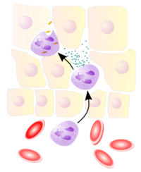
Neutrophils extravasate from blood vessels to the site of tissue injury or infection during the innate immune response. Image source: Wikipedia, GNU Free Documentation License.
CD44 expression is an indicative marker for effector-memory T-cells.
CD44 CD99 Both participate in leukocyte migration.
What CD marker correlates well with T cell receptor excision circles (TRECs)?
(A) CD19
(B) CD45RO
(C) CD27
(D) CD45RA
(E) CD18
(F) CD15
A: D. Naive T lymphocytes express large CD45 isoforms and are usually positive for CD45RA. CD45RA cells contain TRECs. Activated and memory T lymphocytes express the shortest CD45 isoform, CD45RO. CD45RA is large and it shortens as the cell matures.
Severe combined immunodeficiency (SCID) - 4 groups according to T/B/NK cells (click to enlarge the image).
Author: V. Dimov, M.D., Allergist/Immunologist and Assistant Professor at University of Chicago
Reviewer: S. Randhawa, M.D., Allergist/Immunologist and Assistant Professor at NSU
T cells are derived from pluripotent hematopoietic stem cells (CD34+) which migrate from the bone marrow to the thymus.
B cells can bind free antigen but T cells cannot. They can recognize an antigen only when it is bound and presented by antigen-presenting cells (APC) via MHC molecules.
Antigen presentation stimulates T cells to become either "cytotoxic" CD8+ cells or "helper" CD4+ cells. Image source: Wikipedia.
In this video, a T cell becomes activated when it interacts with a dedritic cell. This video is from: Janeway's Immunobiology, 7th Edition Murphy, Travers, & Walport. Source: Garland Science.
Antigen
An antigen is a molecule that is recognized by the immune system - [anti]body [gen]erator.
Superantigen is an antigen that activates a large number of polyclonal T lymphocytes. Superantigens bind to the Vβ chain of the T cell receptor (TCR) bypassing the need for MCH.
Different antigens according to their structure
- Proteins are excellent immunogens, they are "classic" antigens and are T-cell-dependent antigens. Vaccines which are protein based antigens include diphtheria and tetanus.
- Polysaccharides are T-cell-independent antigens. The "old" unconjugated meningococcal and 23-valent pneumococcal vaccines are examples of T-cell independent antigens. Updated recommendations for immunization include the use of quadrivalent (serogroups A, C, Y, and W-135) meningococcal conjugate vaccines (Medscape, 2011).
- Nucleic acids - DNA and RNA
- Lipids - MHC-like CD1 molecules bind lipid antigens that are recognized by natural killer T lymphocytes (NKT cells) and γδ T lymphocytes. NKT cells express NK cell and T lymphocytes markers. NKT cells recognize lipids in the context of CD-1.
MHC Genome
Genes that encode MHC molecules are located on the short arm of chromosome 6. The β2 microglobulin chain (part of MCH I) is encoded on chromosome 15.
Bare lymphocyte syndromes include MHC class I and MHC class II deficiencies. These are primary immune deficiency disorders (PIDD) due to a lack of expression of either MHC I or MHC II. MHC class I deficiency leads to CD8 lymphopenia. MHC class II deficiency leads to CD4 lymphopenia.
Thymic education
The thymus plays a major role in T cells development and selecting which of the T cells entering the thymus will leave "alive." This process is called thymic education. The extreme selectivity of the thymus education is underlined by the fact that 95% of entering stem cells are eliminated within the thymus, never to become circulating T cells.
B cells undergo only negative selection, whereas T cells undergo both positive and negative selection.
Intrathymic T Cell Differentiation. Image source: Wikipedia.
After pre-T cells enter the thymus, they first attempt to rearrange their T cell receptor (TCR) genes. This process is mediated by the same genes used by B cells to rearrange the DNA for immunoglobulins: recombination activating genes (RAG1, RAG2).
Gene Recombination. This video shows how gene recombination affects immunoglobulins. This video is from: Janeway's Immunobiology, 7th Edition Murphy, Travers, & Walport. Source: Garland Science.
The two chains of the T cell receptor. Image source: Wikipedia.
Thymic epthelial cells express MHC molecules interact with the surviving T-cells (triple-positive, CD4+/CD8+/TCR+). MHC class II molecules bind to CD4+/CD8- T cells. MHC class I molecules bind to CD8+/CD4- T cells. This relationship is remembered by the mnemonic 2 x 4=8 and 1 X 8 = 8, the final result is always 8, 2 is MHC II, 1 is MHC II, 4 is CD4, 8 is CD8.
Molecular association of CD8+ T cells with MHC class I and CD4+ T cells with MHC class II. Image source: Wikipedia.
During thymic education, T cells variously express cell surface molecules which may be lost during the development and not present on mature cells. One such molecule is common ALL antigen (CALLA). Acute lymphoblastic leukemia (ALL) T cells express the early T-cell marker CD10 (CALLA) which is absent on normal mature T cells.
MHC mnemonicI1 letter HLA: ABC
1 polypeptide chain MHC (alpha and beta-2 microgobulin)
II2 letters HLA: DP, DQ, DR
2 polypeptide chains MHC (alpha and beta)
Bare lymphocyte syndromes include MHC class I and MHC class II deficiencies. These are primary immune deficiency disorders (PIDD) due to a lack of expression of either MHC I or MHC II.
Subsets of T-CellsCD4 T Cells and T Helper Cells (click to read the full article)
CD8 T Cells
CD8 T cells make 30% of circulating T cells. CD8 T cells are cytotoxic and are often called cytotoxic T lymphocytes (CTLs). CD8 CTLs kill target cells which have been altered by infection or malignancy.
CTLs and natural killer (NK) cells use 2 similar mechanisms to kill target cells:
- granule-associated serine esterases (granzymes)
- pore-forming proteins (perforins)
Antigen recognition by CD8 T cells is class I restricted as opposed to recognition by CD4 cells which is class II restricted.
Schematic representation of MHC class I. Image source: Wikipedia.
MHC Class I Processing. This video describes the assembly of MHC Class I molecules. This video is from: Janeway's Immunobiology, 7th Edition, Murphy, Travers, & Walport. Source: Garland Science.
CD8 may act as suppressor cells which downregulate immune responses. The CD8 molecule is a protein of the immunoglobulin superfamily (similar to CD4 molecule). Some scientists classify the suppressor cells in a different subsets of T cells called regulatory cells (see below).
T Cell Granule Release. This video describes how cytotoxic T cells release granules in response to an antigen on a target cell. This video is from: Janeway's Immunobiology, 7th Edition, Murphy, Travers, & Walport. Source: Garland Science.
Regulatory T cells (click to read the full article)
Naïve T cells
Naïve T cells are cells which have not yet encountered their cognate antigen, as opposed to mature CD4, CD8 cells and memory cells.
Memory T cells
When naïve T cells encounter their specific antigen, they activate and may become memory T cells. These cells repeatedly return to the site where they initially became activated during recirculation (a process called homing). Memory T cells survive for decades awaiting repeat encounters with their antigen. In a sense, memory T cells remeber the antigen they first encountered and the place of the encounter for life. It must have been a very memorable event in their lives.
T and B Cells - Naive and Memory Cell Markers (click to enlarge the image).
What chemokine attracts naïve B cells to lymph nodes?
CXCR5. CXCL13 binds to CXCR5.
What chemokine attracts naïve T cells to lymph nodes?
CCR7. CCL19 and CCL21 bind to CCR7.
Natural Killer T cells (NKT)
Natural killer T (NKT) cells are a heterogeneous group of T cells that share properties of both T cells and natural killer (NK) cells. NKT cells should not be confused with natural killer (NK) cells. NKT cells express NK cell and T lymphocytes markers. NKT cells recognize lipids in the context of CD-1.
Gamma/delta T cells (γδ)
Gamma/delta T cells make 5% of the circulating T cells and have a distinct T cell receptor (TCR) on their surface which is different in structure from the other T-cells. The majority of T cells have a TCR made of 2 glycoprotein chains called α- and β- TCR chains. In contrast, the TCR of γδ T cells is made up of one γ-chain and one δ-chain, hence the name gamma/delta T cells.
T-cell receptor complex with TCR-α and TCR-β chains. Image source: Wikipedia.
This video describes the cellular signaling that takes place with the T cell receptor. This video is from: Janeway's Immunobiology, 7th Edition Murphy, Travers, & Walport. Source: Garland Science.
The first event following the TCR ligation (binding) is the activation of Src kinases - autoinhibitory loop.
CD45 phosphorylation in TCR signaling is of early and key importance. CD45 and ZAP70 defects cause SCID.
ZAP70
ZAP 70 binds to the Zeta chain (CD247) of the T-cell receptor.
ZZAP-70
Zeta-chain-associated protein kinase 70 (70 is the molecular weight in kDa)
ZAP70 deficiency is a form of SCID (T-/B+/NK+), one of the few SCID forms with palpable LN.
The mnemonic for different T-cell subtypes is FERMNN G ("pheromon G"):
Four, CD4, helper cells (Th1 and Th2)
Eight, CD8, killer cells
Regulatory, formerly known as suppressors (CD4/CD25/FOXP3)
Memory
Naïve T cells
Natural Killer T cells (N-KT)
Gamma/delta T cells (γδ)
Mind map of the 7 different T-cell subtypes remembered by the mnemonic FERMNN G
T cell activation. Image source: Wikipedia.
Surface Markers
CD25 (IL-2Ra) and CD3 are found on all T cells.
CD28 is present on T cells and binds to CD80/86 on dendritic cells. CD152 is cytotoxic T-lymphocyte antigen-4 (CTLA-4). CTLA-4 binds to CD80 and CD86receptors with a higher affinity than CD28, and inhibits T cell activation.
CD19 and CD20 are found on B cells. Complement receptor CD21 (CR2) activates BCR if the antigen is opsonized by C3b component of the complement. CR2-CD19-CD81 complex is expressed on the surface of B lymphocytes. CD19 is probably the best marker of B-lymphocytes. CD19 is a co-receptor with CD21.
When CD21 (CR2) interacts with C3d, the complex is brought into the BCR. CD19 has an ITAM that is phosphorylated, thus recruiting Lyn to enhance phosphorylation.
CD16 are found on natural killer (NK) and NK-T cells. CD16 is a receptor for IgG.
IL-7 activates naive T cells. BAF activates naive B cells.
MnemonicsCDs on Different Cells
T cells have CD 3, 4 and 8 (double, 4 x 2)
B cells have CD 19, 20 and 40 (double 20 x 2)
Surface MarkersLLigands on
Lymphocytes
CD40L on T helpers (CD4+)
FAS-L on CTL 9CD8+)
CD40 on B cells
FAS ("death signal") on cells to be killed by CTLs
MHC
MHC class II molecules bind to CD4+/CD8- T cells. MHC class I molecules bind to CD8+/CD4- T cells. This relationship is remembered by the mnemonic 2 x 4=8 and 1 X 8 = 8, the final result is always 8, 2 is MHC II, 1 is MHC II, 4 is CD4, 8 is CD8.
MHC numbers: 2, 4, 8, 16
MHC2 x CD4 = 8 - MHC2 binds best 16 amino acid-long peptides (11-30)
MHC1 x CD8 = 8 - MHC2 binds best 8 amino acid-long peptides (8-10)
MHC Genes
MHC I - genes ABC
MHC II - genes D, again alphabetical - DP, DQ, DR
Both on chromosome 6
I......II
ABCD PQR
Severe combined immunodeficiency (SCID) - 4 groups according to T/B/NK cells (click to enlarge the image).
References
Allergy and Immunology MKSAP, 3rd edition.
Delay in diphtheria, pertussis, tetanus vaccination is associated with a reduced risk of childhood asthma. JACI, Volume 121, Issue 3, Pages 626-631 (March 2008).
CD4/CD8 ratio predicts infection in HIV infants: the National Heart, Lung and Blood Institute P2C2 study. Shearer et al. J Allergy Clin Immunol 120. 1449-1452.2007.
Advances in basic and clinical immunology in 2007. Journal of Allergy and Clinical Immunology - Volume 122, Issue 1 (July 2008).
Discovery of T-cell Receptors (video)
National Jewish Health scientists John Kappler, PhD, and Philippa Marrack, PhD, discuss their discovery of the T-cell receptor - the protein found on the surface of T Cells that helps detect specific antigens, or foreign invaders, in the body:
Related ReadingMedical Immunology Syllabus. Columbia University.
FIT Corner Q & A from 5th edition of Cellular and Molecular Immunology, edited by Abul K. Abbas and Andrew H. Lichtman. ACAAI, 2004.
Fork head domain. Wikipedia, 2009.
FOX proteins. Wikipedia, 2009.
FOXP3. Wikipedia, 2009.
Redirecting T Cells - NEJM, 2011.
Nuocytes are previously unknown members of the lymphoid lineage. Nature, 2012.
Published: 12/15/2007
Updated: 12/22/2012