Author: V. Dimov, M.D., Allergist/Immunologist and Assistant Professor at University of Chicago
Reviewer: S. Randhawa, M.D., Allergist/Immunologist and Assistant Professor at NSU
Monoclonal antibodies are produced by hybridomas. A hybridoma is a product of cell fusion between:
- normal antibody producing B cell (plasma cell)
- myeloma cell
Typically, spleen cells from an immunized mouse are fused with myeloma cells by using polyethylene glycol (PEG) to produce a hybrid cell line called a hybridoma.
Each hybridoma is descended from a single cell and all the antibodies produced by that cell are identical. These antibodies are called monoclonal antibodies (mAbs).
Monoclonal antibodies (mAbs) nomenclature related to sufix
-omab - murine, only of mouse origin. Murine component is 100%
-ximab - chimeric human/mouse. mAb combines murine mAb variable regions with the constant region of human antibody. Murine component is 30%
-zumab - humanized. mAb combines murine mAb variable regions with a portion of a human IgG molecule. Murine component is less than 10%
-umab - "fully" human mAb cloned into a bacteriophage
-cept - fusion of a receptor to the Fc part of human IgG1
Examples of monoclonal antibodies (mAb) in clinical practice
- Rituximab - against CD20. Used in autoimmune diseases, B cell lymphomas
- Anti-CD20 monoclonal antibody ocrelizumab helps patients with relapsing-remitting multiple sclerosis (MS) (Lancet, 2011).
- Omalizumab - againts FcεRI (cε3). Used in asthma
- Adalimumab - against TNF-α. Used in rheumatoid arthritis
- Palivizumab - against RSV. Used to prevent RSV infection in infants with bronchopulmonary dysplasia
- Mepolizumab - against IL-5. Used in hypereosinophilic syndrome and patients with eosinophilic asthma
- Daclizumab - against IL-2 receptor α chain (CD25). Used to prevent renal allograft rejection
- Natalizumab - against α 4 chain of integrin molecule. Used in multiple sclerosis (MS)
- Abciximab - against integrin αIIβ3. Used in acute coronary syndromes (ACS)
- Anti-IL-17 Receptor Antibody Brodalumab Helps Patients with Psoriasis - NEJM, 2012.
- Anti–Interleukin-17 Monoclonal Antibody Ixekizumab Improves Chronic Plaque Psoriasis - NEJM, 2012.
- Natalizumab-Associated Progressive Multifocal Leukoencephalopathy (PML) - NEJM lists the risk factors
- In 2014, FDA Approved New Drug for Crohn Disease and UC: vedolizumab (Entyvio), an injectable monoclonal antibody. Vedolizumab, an integrin receptor antagonist http://buff.ly/1lFuRtv
References
Online Book of Biologic Therapies (monoclonal antibodies, etc.) by the Clinical Immunology Society
Related reading
Allergy to monoclonal antibodies: cutting-edge desensitization methods - Expert Reviews, 2012.
mAb: If it ends in -mumab, it's a fully human protein, -zumab is humanized (10% mouse), -ximab is chimeric (33% mouse). ACP blog, 2012.
Published: 08/12/2008
Updated: 05/25/2012
Showing posts with label Immunoglobulin. Show all posts
Showing posts with label Immunoglobulin. Show all posts

Immunoglobulin E (IgE)
Author: V. Dimov, M.D., Allergist/Immunologist and Assistant Professor at University of Chicago
Reviewer: S. Randhawa, M.D., Allergist/Immunologist and Assistant Professor at LSU (Shreveport) Department of Allergy and Immunology
Role of mast cells in allergy had remained undetermined until the discovery of IgE in 1966. Then, IgE purified from many Liters of plasma, which had been donated from a patient with fatal myeloma, was distributed to researchers all over the world (http://buff.ly/12sD3cO).
IgE is involved in immediate hypersensitivity (allergy). It binds to allergens and triggers histamine release from mast cells. It has the lowest serum concentration (mcg rather than mg) and the shortest half-life (2 days) among Ig (GAMED).
Controls of Ig type switch
IgA switching: APRIL, BAFF, TGF-beta
IgE switching: IL-4
IgG switching: IFN-gamma
There are 5 immunoglobulin classes remembered by the mnemonic GAMED: Ig G, A, M, E, D. B-cells are the only cell types that synthesize antibody molecules.

Five immunoglobulin classes (mind map).
In order of their serum concentrations:
IgG 1000 mg/dL
IgA 200 mg/dL
IgM 150 mg/dL
IgD 4 mg/dL
IgE 0.005 mg/dL (extremely low serum concentration compared to other Ig in (GAMED)
Measurement of total IgE is expressed in 1 IU/mL = 2.44 ng/mL.
There are 5 immunoglobulin classes remembered by the mnemonic GAMED: Ig G, A, M, E, D. B-cells are the only cell types that synthesize antibody molecules.

Five immunoglobulin classes (mind map).
In order of their serum concentrations:
IgG 1000 mg/dL
IgA 200 mg/dL
IgM 150 mg/dL
IgD 4 mg/dL
IgE 0.005 mg/dL (extremely low serum concentration compared to other Ig in (GAMED)
Measurement of total IgE is expressed in 1 IU/mL = 2.44 ng/mL.
The level of IgE increases during childhood until about 10 years of age. At age 10, the total IgE reaches a value that is typically maintained throughout adult life.
Ig order of their serum half-life in days:
IgG 21
IgM 10
IgA 6
IgD 3
IgE 2 (shortest half-life among other Ig in GAMED)
Ig order of their serum half-life in days:
IgG 21
IgM 10
IgA 6
IgD 3
IgE 2 (shortest half-life among other Ig in GAMED)


Role of IgE and mast cells in allergy. Image source: Wikipedia.

Omalizumab binds to Cε3 region of IgE.
IL-4 and IL-13 are cytokines produced by Th2 cells. They up-regulate IgE production and thus increase allergic inflammation. IL-4 is much more potent than IL-13.
I
IL-4
IL-13
Increase
IgE production
Inflammation promoters
Receptors for IgE
High-Affinity IgE Receptor (FcεRI)
The high affinity receptor for IgE (FcεRI) is expressed on mast cells (fixed in tissues) and basophils (circulating in blood). The level of FcεRI expression is regulated by levels of IgE. FcεRI molecule consists of 4 chains: alpha, beta and 2 gamma.
FcεRI is found in 2 forms:
- tetrameric form is composed of one α chain, one β chain, and two γ chains. It is found on mast cells, basophils, eosinophils.
- trimeric form is composed of one α chain and two γ chains. It is found on monocytes and dendritic cells (the β chain is absent).
Some patients with chronic urticaria have IgG directed against the α chain of FcεRI. IgE binds to FcεRI via the α chain.
CD23 is the low affinity IgE receptor (FceRII). CD21 (CR2) binds EBV, HHV8, C3d, and CD23 (low affinity IgE receptor (FceRII).
The role of FcεRII is not clear. Variations in the low-affinity IgE receptor gene (FCER2 or II) are associated with an increase in severe exacerbations in children with asthma. There are several reasons that a patient may not respond to inhaled corticosteroid therapy in asthma and genetics, including FCER2 variants, may be an important one.
Variations in some of the SNPs were associated with increased IgE levels and increased risk of severe exacerbations of asthma during inhaled corticosteroid treatment. Therefore, FCER2 variations may lead to asthma exacerbations despite inhaled steroids. Personalized medicine via pharmacogenetics can lead to genetic studies guiding therapeutic decisions in the future.
Anti-IgE (omalizumab, Xolair) is approved for treatment of severe persistent asthma. There are occasional case reports of anti-IgE (omalizumab) use as a new therapeutic approach for chronic rhinosinusitis. Omalizumab binds to Cε3 region of IgE.
There is a correlation between IgE titres and the severity of clinical reaction to egg after the diagnosis has been established. A cut-off level of 8.20 kU/l had a 90% probability of clinical reactivity. IgE titres may help determine which patients are at risk of a reaction to eggs.
Higher levels of IgE are associated with more severe asthma (the cut-off level is 100 IU/mL). IgE level is inversely correlated with baseline lung function and asthma severity.
ImmunoCAP Immunoassay
The ImmunoCAP method was developed by Phadia. It utilizes a “sandwich” ELISA technique:
1. The ImmunoCAP sponge has allergen bound to it and serves as the first piece of bread ("bottom half").
2. Patient serum is added and specifc IgE to that allergen binds to the allergen on the sponge - this is the "meat" of the ELISA sandwich.
3. All of the unbound protein is washed away abd anti-IgE is added - this binds to the sIgE that was captured by the sponge in step two. The anti-IgE conjugate is the "second piece of bread" ("top half").
Allergen-specific IgE levels are not comparable between different laboratory methods - for example, ImmunoCAP vs. DPC Immulite 2000. Predictive values of specific IgE levels published in the literature for management of food allergies are based on studies using the ImmunoCAP assay. These predictive values cannot be applied to specific IgE levels from other assay systems.
Conditions with elevated IgE
Atopic dermatitis, Asthma, ABPA, and allergic fungal sinusitis
Infections (parasites, HIV, TB, EBV, and CMV)
Malignancy (IgE myeloma and lymphoma)
Churg-Strauss syndrome
Kimura’s disease, painless, unilateral cervical lymphadenopathy or subcutaneous masses in the head or neck region
Immunodeficiency diseases with elevated IgE
Hyper IgE syndrome (HIES)
Wiskott-Aldrich syndrome (WAS)
Omenn syndrome
DiGeorge syndrome (DGS)
Netherton syndrome, form of ichthyosis associated with SPINK5
Nezelof syndrome, congenital hypoplasia of the thymus with retention of normal parathyroid function (in contrast to complete DiGeorge syndrome in which there is absence of the parathyroids)
Published: 06/28/2010
Updated: 09/16/2012
Fc Receptors
Author: V. Dimov, M.D., Allergist/Immunologist and Assistant Professor at University of Chicago
Reviewer: S. Randhawa, M.D., Allergist/Immunologist and Assistant Professor at LSU (Shreveport) Department of Allergy and Immunology
Fc receptor is a protein found on the surface of cells that binds to a part of an antibody known as the Fc (Fragment, crystallizable) region. Each Fc receptor functions as a receptor specific for the CH region of the Ig molecule.
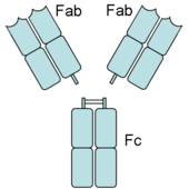
An antibody has Fab (fragment, antigen-binding) and Fc (fragment, crystalizable) regions. Fc receptors bind to the Fc region. Image source: Wikipedia, public domain.
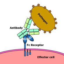
Fc receptor interaction with an antibody-coated microbial pathogen. Image source: Wikipedia, public domain.
The uptake of bacteria by phagocytes is an active process, which requires the triggering of specific receptors on the phagocyte. Fc receptors, which bind antibody-coated bacteria, are one of the receptors capable of triggering phagocytosis. This video is from: Janeway's Immunobiology, 7th Edition Murphy, Travers, & Walport. Source: Garland Science.
There are several different types of Fc receptors, which are classified based on the type of antibody that they recognize. The Latin letter used to identify a type of antibody is converted into the corresponding Greek letter, which is placed after the 'Fc' part of the name:
- Fc that bind the most common class of antibody, IgG, are called Fc-gamma receptors (FcγR)
- Fc that bind IgA are called Fc-alpha receptors (FcαR)
- Fc that bind IgE are called Fc-epsilon receptors (FcεR)
Fc gamma receptors for IgG are CD 16, 32, 64 (double). IgG is transported across the placenta by an IgG-specific Fc receptor called neonatal Fc receptor (FcRn), which resembles a MHC class I molecule. CD16 (FcR-gamma-III) is the most common IgG Fc receptor. CD32 down-regulates IgG. Binding of FcγRIIb is the proposed mechanism of action of IVIG.
Fc gamma receptors for IgG (CD 16, 32, 64) are labeled in "reverse" order:
CD 16 - FcR III - low-affinity
CD 32 - FcR II - intermediate-affinity
CD 64 - FcR I - high-affinity
CD16 and CD56 are present on NK cells. CD16 and CD14 are present on macrophages.
Fc II receptors:
CD 32 - Fc gamma II
CD 23 - Fc epsilon II, Fc receptors for IgE
CD23 is the low affinity IgE receptor (FceRII). CD21 (CR2) binds EBV, HHV8, C3d, and CD23 (low affinity IgE receptor (FceRII).
CD32 down-regulates IgG. Binding of FcγRIIb is the proposed mechanism of action of IVIG.
References
Fc receptor. Wikipedia, the free encyclopedia.
Reviewer: S. Randhawa, M.D., Allergist/Immunologist and Assistant Professor at LSU (Shreveport) Department of Allergy and Immunology
Fc receptor is a protein found on the surface of cells that binds to a part of an antibody known as the Fc (Fragment, crystallizable) region. Each Fc receptor functions as a receptor specific for the CH region of the Ig molecule.
An antibody has Fab (fragment, antigen-binding) and Fc (fragment, crystalizable) regions. Fc receptors bind to the Fc region. Image source: Wikipedia, public domain.

Fc receptor interaction with an antibody-coated microbial pathogen. Image source: Wikipedia, public domain.

The uptake of bacteria by phagocytes is an active process, which requires the triggering of specific receptors on the phagocyte. Fc receptors, which bind antibody-coated bacteria, are one of the receptors capable of triggering phagocytosis. This video is from: Janeway's Immunobiology, 7th Edition Murphy, Travers, & Walport. Source: Garland Science.
There are several different types of Fc receptors, which are classified based on the type of antibody that they recognize. The Latin letter used to identify a type of antibody is converted into the corresponding Greek letter, which is placed after the 'Fc' part of the name:
- Fc that bind the most common class of antibody, IgG, are called Fc-gamma receptors (FcγR)
- Fc that bind IgA are called Fc-alpha receptors (FcαR)
- Fc that bind IgE are called Fc-epsilon receptors (FcεR)
Fc gamma receptors for IgG are CD 16, 32, 64 (double). IgG is transported across the placenta by an IgG-specific Fc receptor called neonatal Fc receptor (FcRn), which resembles a MHC class I molecule. CD16 (FcR-gamma-III) is the most common IgG Fc receptor. CD32 down-regulates IgG. Binding of FcγRIIb is the proposed mechanism of action of IVIG.
Fc gamma receptors for IgG (CD 16, 32, 64) are labeled in "reverse" order:
CD 16 - FcR III - low-affinity
CD 32 - FcR II - intermediate-affinity
CD 64 - FcR I - high-affinity
CD16 and CD56 are present on NK cells. CD16 and CD14 are present on macrophages.
Fc II receptors:
CD 32 - Fc gamma II
CD 23 - Fc epsilon II, Fc receptors for IgE
CD23 is the low affinity IgE receptor (FceRII). CD21 (CR2) binds EBV, HHV8, C3d, and CD23 (low affinity IgE receptor (FceRII).
CD32 down-regulates IgG. Binding of FcγRIIb is the proposed mechanism of action of IVIG.
Immunomodulating effects of IVIG mediated through Fc receptor blockade
- ADCC inhibition by blockade of FcγRIII (CD16) on NK cells
- Inhibition of antibody production by blockade of FcγRIIb (CD32) on B cells
- IVIG blocks membrane Fc receptors on phagocytic cells in spleen and liver in patients with ITP
Place in order the CD designation and the Fc receptor starting with FcγRI, FcγRII, FcγRIII, FceRII:
A. CD23, CD16, CD32, CD64
B. CD64, CD32, CD16, CD23
C. CD64, CD16, CD32, CD23
D. CD16, CD32, CD64, CD23
Answer: B.
Fc receptor. Wikipedia, the free encyclopedia.
Headache After Treatment with Intravenous Immunoglobulin (IVIG)
Author: V. Dimov, M.D., Allergist/Immunologist and Assistant Professor at University of Chicago
Reviewer: S. Randhawa, M.D., Allergist/Immunologist and Assistant Professor at LSU (Shreveport) Department of Allergy and Immunology
A 34-year-old Caucasian female is followed by the allergy clinic for common variable immunodeficiency (CVID) and a treatment with intravenous immune globulin (IVIG) was started 4 months ago. She started to have headaches after the second infusion which are progressively getting worse. Her last dose of IVIG was given 2 days ago and since then she had a persistent pounding headache not relieved by Percocet. She denies any symptoms of aseptic meningitis including photophobia, neck stiffness, nausea, or fever. She rates the pain as 8/10.
Past medical history (PMH)
Common variable immunodeficiency (CVID).
Medications
Intravenous immune globulin (IVIG) 400 mg/kg every 4 weeks, Percocet (oxycodone and acetaminophen)
Social history and family history
Not contributory.
Physical examination
Vital signs stable. Normal nose and throat exam. Respiratory system: Clear to auscultation bilaterally. Cardiovascular system: Clear S1, S2. Abdomen: Soft, non-tender, non-distended. Extremities: no edema. Skin: no rashes.Neurological examination: non focal.
What is the most likely diagnosis?
- Headache associated with intravenous immunoglobulin.
- Migraine.
- Aseptic meningitis is in the differential diagnosis but she did not have features of meningitis.
What tests would you suggest?
No additional tests are needed at this time.
What treatment would you suggest?
After verifying that the patient was not pregnant, she was prescribed zolmitriptan (Zomig) 5 mg po x 1, the dose may be repeated 2 hours later. The headache resolved after the second dose.
How to prevent IVIG-related headache?
Post-infusion headaches may be prevented by the following treatments for as long as 72 hours after the infusion is completed
- NSAIDs such as naproxen or ibuprofen
- Prednisone 30 to 60 mg
- cyproheptadine (an antihistamine and serotonin receptor antagonist)
- migraine medications
In this patient, we recommended prednisone 30 mg po x 1 and zolmitriptan (Zomig) 5 mg po x 1 before the infusion.
Other options that may be considered include "splitting" the dose to every 2 weeks and using subcutaneous immunoglobulin rather than IVIG.
Final diagnosis
Headache associated with intravenous immunoglobulin (IVIG) treatment.
Summary
Headache is a common side effect of intravenous immunoglobulin (IVIG) treatment; it usually resolves shortly after completing or slowing the infusion. Aseptic meningitis occurs in up to 17% of patients afterIVIG treatment, with more than 30 cases described in the literature. Aseptic meningitis usually presents with crescendo headache that also causes changes in sensorium or behavioral changes.
Headaches associated with intravenous immunoglobulin may have features of migraine and may be successfully prevented and/or treated with 5-HT1D receptor agonists.
Many patients develop mild headaches during IVIG infusions, which can be prevented or treated with acetaminophen, aspirin, or NSAIDs; and/or by administering the IGIV at a slower rate.
In some cases, the onset may be delayed until 24 to 48 hours after the infusion is completed. The duration of symptoms is generally less than 48 hours, but occasionally, severe headaches lasting as long as 72 hours have been reported.
Mnemonic: Dose of IVIG in PIDD
400-600 mg/kg/month
4 letter words:
IVIG
CVID
SCID
References
Management of the Acute Migraine Headache. AFP, 2002.
Successful Treatment of Headache Related to Intravenous Immunoglobulin With Antimigraine Medications. Alan G. Finkel, MD ; James F. Howard Jr., MD ; J. Douglas Mann, MD. Headache: The Journal of Head and Face Pain, Volume 38 Issue 4, Pages 317 - 321, 2003.
What Can Be Used to Treat Persistent Headache Caused by Aseptic Meningitis? Randolph Warren Evans, MD. eMedicine, 2000.
Published: 04/19/2009
Updated: 08/18/2010
Reviewer: S. Randhawa, M.D., Allergist/Immunologist and Assistant Professor at LSU (Shreveport) Department of Allergy and Immunology
A 34-year-old Caucasian female is followed by the allergy clinic for common variable immunodeficiency (CVID) and a treatment with intravenous immune globulin (IVIG) was started 4 months ago. She started to have headaches after the second infusion which are progressively getting worse. Her last dose of IVIG was given 2 days ago and since then she had a persistent pounding headache not relieved by Percocet. She denies any symptoms of aseptic meningitis including photophobia, neck stiffness, nausea, or fever. She rates the pain as 8/10.
Past medical history (PMH)
Common variable immunodeficiency (CVID).
Medications
Intravenous immune globulin (IVIG) 400 mg/kg every 4 weeks, Percocet (oxycodone and acetaminophen)
Social history and family history
Not contributory.
Physical examination
Vital signs stable. Normal nose and throat exam. Respiratory system: Clear to auscultation bilaterally. Cardiovascular system: Clear S1, S2. Abdomen: Soft, non-tender, non-distended. Extremities: no edema. Skin: no rashes.Neurological examination: non focal.
What is the most likely diagnosis?
- Headache associated with intravenous immunoglobulin.
- Migraine.
- Aseptic meningitis is in the differential diagnosis but she did not have features of meningitis.
What tests would you suggest?
No additional tests are needed at this time.
What treatment would you suggest?
After verifying that the patient was not pregnant, she was prescribed zolmitriptan (Zomig) 5 mg po x 1, the dose may be repeated 2 hours later. The headache resolved after the second dose.
How to prevent IVIG-related headache?
Post-infusion headaches may be prevented by the following treatments for as long as 72 hours after the infusion is completed
- NSAIDs such as naproxen or ibuprofen
- Prednisone 30 to 60 mg
- cyproheptadine (an antihistamine and serotonin receptor antagonist)
- migraine medications
In this patient, we recommended prednisone 30 mg po x 1 and zolmitriptan (Zomig) 5 mg po x 1 before the infusion.
Other options that may be considered include "splitting" the dose to every 2 weeks and using subcutaneous immunoglobulin rather than IVIG.
Final diagnosis
Headache associated with intravenous immunoglobulin (IVIG) treatment.
Summary
Headache is a common side effect of intravenous immunoglobulin (IVIG) treatment; it usually resolves shortly after completing or slowing the infusion. Aseptic meningitis occurs in up to 17% of patients afterIVIG treatment, with more than 30 cases described in the literature. Aseptic meningitis usually presents with crescendo headache that also causes changes in sensorium or behavioral changes.
Headaches associated with intravenous immunoglobulin may have features of migraine and may be successfully prevented and/or treated with 5-HT1D receptor agonists.
Many patients develop mild headaches during IVIG infusions, which can be prevented or treated with acetaminophen, aspirin, or NSAIDs; and/or by administering the IGIV at a slower rate.
In some cases, the onset may be delayed until 24 to 48 hours after the infusion is completed. The duration of symptoms is generally less than 48 hours, but occasionally, severe headaches lasting as long as 72 hours have been reported.
Anaphylaxis may occur in patients treated with IVIG who have IgE antibodies against IgA, for example patients with selective IgA deficiency (incidence 1 in 500). Preventive measures include using products with the lowest IgA content and pre-medication with antihistamines or corticosteroids.
Mnemonic: Dose of IVIG in PIDD
400-600 mg/kg/month
4 letter words:
IVIG
CVID
SCID
References
Management of the Acute Migraine Headache. AFP, 2002.
Successful Treatment of Headache Related to Intravenous Immunoglobulin With Antimigraine Medications. Alan G. Finkel, MD ; James F. Howard Jr., MD ; J. Douglas Mann, MD. Headache: The Journal of Head and Face Pain, Volume 38 Issue 4, Pages 317 - 321, 2003.
What Can Be Used to Treat Persistent Headache Caused by Aseptic Meningitis? Randolph Warren Evans, MD. eMedicine, 2000.
Published: 04/19/2009
Updated: 08/18/2010
Selective Immunoglobulin M (IgM) Deficiency
Author: V. Dimov, M.D., Allergist/Immunologist and Assistant Professor at University of Chicago
Reviewer: S. Randhawa, M.D., Allergist/Immunologist and Assistant Professor at NSU
A 70-yo Caucasian male (CM) is referred by his primary care physician (PCP) for evaluation for allergic rhinitis/conjunctivitis and chronic sinusitis. He complains of nasal congestion for 25 years which is worse over last 2 years. He has rhinorrhea in spring and fall. He has had 3-4 courses of antibiotics for sinusitis each year for the last 5-7 years.
Past medical history
Diabetes type 2 (DM2), hypertension (HTN).
Medications
Glyburide, amlodipine, Allegra D (fexofenadine and pseudoephedrine) (stopped 5 days ago for skin testing).
Physical examination
Vital signs stable (VSS).
Ears: dull tympanic mebranes (TMs), cerumen present.
Nose: leftward septal deviation, pale boggy turbinates.
Throat: mild cobblestoning.
Lymph: No cervical lymphadenopathy.
Respiratory: CTA (B).
CVS: Clear S1S2.
What tests would you suggest?
Skin prick testing.
CT scan of sinuses.
Would you consider immunodeficiency work-up in this patient?
Immunodeficiency, for example CVID, is in the differential diagnosis of the recurrent episodes of sinusitis in this patient.
Quantitative immunoglobulins were added to the work-up.
What happened?
The CT scan of the sinuses showed pansinusitis, most pronounced in the left maxillary sinus, and moderate leftward deviation of the nasal septum.
Skin prick test was positive for grasses, ragweed, trees, weeds.
We prescribed flunisolide nasal spray qd, saline nasal rinses bid and loratidine qd. He was referred to ENT and a follow-up was arranged in 8 weeks.
What happened next?
The patient came to the follow-up appointment 8 weeks later and reported that he felt 60% better.
His Ig levels were:
The repeated quantitative immunoglobulins showed similar values.

Structure of the pentameric IgM: 1. Base unit, 2. Heavy chains, 3. Light chains, 4. J chain, 5. Intermolecular disulfide bonds. Image source: Wikipedia.
What is the most likely diagnosis?
Selective immunoglobulin M deficiency (SIgMD)
What is the next step in the daignostic work-up of this patient?
Check Anti-ABO antibodies titers. IgM is the antibody against blood group antigens.
Assess biological significance of isolated low IgM level by immunizing the patient with:
- protein vaccines, e.g., tetanus, MMR
- polysaccharide vaccines, e.g. unconjugated Streptococcus pneumoniae
Assess if antigen-specific IgG responses are normal.
How to assess humoral immune response?
Inject patient with Pneumovax 23 (not Prevnar, which is a 7-valent conjugate vaccine). Check anti-polysaccharide IgG antibody to pneumococcus serotypes in 3 weeks. There should be a 3-5 fold increase in the anitbody titer to at least 50% of isotypes.
Which serotypes should be included in the order for Ig?
The same serotypes that were included in the given vaccine. Check the enclosed leaflet for this information. For example, the serotypes included in Pneumovax 23 can be found from the Merck website (PDF):
1 2 3 4 5 6B 7F 8 9N 9V 10A 11A 12F 14 15B 17F 18C 19F 19A 20 22F 23F 33F
How to collect the serum for anti-polysaccharide IgG antibodies?
Collect the serum prior to immunization with Pneumovax 23. Store the pre-immunization serum in the office refrigerator. Give the vaccine. Check the post-immunization serum 3 weeks later. Send both pre- and post-immunization serums to the laboratory at the same time.
The pneumococcal vaccine comprises purified capsular polysaccharide of 23 stereotypes that account for more than 90% of the invasive pneumococcal infections in the USA. It induces anti-polysaccharide IgG antibody levels to most or all of the component polysaccharide antigens in immunocompetent adults. Elderly adults respond equally well to vaccination as do younger adults. The current 23-valent vaccine comprises 25 μg of each of 23 pneumococcal stereotypes (namely, serotypes 1, 2, 3, 4, 5, 6B, 7F, 8, 9N, 9V, 10A, 11A, 12F, 14, 15B, 17F, 18C, 19A, 19F, 20, 22F, 23F and 33F).
The test should be ordered as follows: Pre- and post-IgM and IgG antibody titers to pneumococcal serotypes 1, 2, 3, 4, 5, 6B, 7F, 8, 9N, 9V, 10A, 11A, 12F, 14, 15B, 17F, 18C, 19A, 19F, 20, 22F, 23F and 33F
Final diagnosis
Selective immunoglobulin M deficiency (SIgMD).
What did we learn from this case?
Selective immunoglobulin M deficiency (SIgMD) is a rare form of dysgammaglobulinemia, with an incidence of less than 0.03% in the general population and 1% in hospitalized patients.
Patients with SIgMD are susceptible to infections with encapsulated bacteria, e.g., Streptococcus pneumoniae, Neisseria meningitidis, Haemophilus influenzae.
Quantitative levels of IgM, IgG, IgA are measured to exclude more common immunodeficiency disorders, such as common variable immunodeficiency and IgA deficiency.
Replacement of IgM is not used since IgM is not a significant component of the commercially available intravenous immunoglobulins.
If defective antigen-specific IgG responses are found after immunization with polysaccharide vaccines, IVIG replacement may be an option.
Five immunoglobulin classes (mind map)
In order of their serum concentrations:
IgG 1000 mg/dL
IgA 200 mg/dL
IgM 150 mg/dL
IgD 4 mg/dL
IgE 0.005 mg/dL (extremely low serum concentration compared to other Ig in (GAMED)

Ig structures. Image source: Wikipedia.
Mnemonics: Adaptive Humoral Immunity: B-cells and Immunoglobulins
There are 5 immunoglobulin classes remembered by the mnemonic GAMED: Ig G, A, M, E, D.
Ig
G
Greatest serum concentration, half-life and number of sub-classes
Ig
M
Macro -- largest Ig
Ig
A
Adhesion prevention
Aggregates -- 2 units linked together
Alternative pathway of complement
Activation
References
Immunoglobulin M Deficiency. Iftikhar Hussain. eMedicine, 2006.
How to Diagnose Common Variable Immunodeficiency (CVID)?
Adaptive Humoral Immunity: B-cells and Immunoglobulins
Mnemonics: Adaptive Humoral Immunity: B-cells and Immunoglobulins
Reviewer: S. Randhawa, M.D., Allergist/Immunologist and Assistant Professor at NSU
A 70-yo Caucasian male (CM) is referred by his primary care physician (PCP) for evaluation for allergic rhinitis/conjunctivitis and chronic sinusitis. He complains of nasal congestion for 25 years which is worse over last 2 years. He has rhinorrhea in spring and fall. He has had 3-4 courses of antibiotics for sinusitis each year for the last 5-7 years.
Past medical history
Diabetes type 2 (DM2), hypertension (HTN).
Medications
Glyburide, amlodipine, Allegra D (fexofenadine and pseudoephedrine) (stopped 5 days ago for skin testing).
Physical examination
Vital signs stable (VSS).
Ears: dull tympanic mebranes (TMs), cerumen present.
Nose: leftward septal deviation, pale boggy turbinates.
Throat: mild cobblestoning.
Lymph: No cervical lymphadenopathy.
Respiratory: CTA (B).
CVS: Clear S1S2.
What tests would you suggest?
Skin prick testing.
CT scan of sinuses.
Would you consider immunodeficiency work-up in this patient?
Immunodeficiency, for example CVID, is in the differential diagnosis of the recurrent episodes of sinusitis in this patient.
Quantitative immunoglobulins were added to the work-up.
What happened?
The CT scan of the sinuses showed pansinusitis, most pronounced in the left maxillary sinus, and moderate leftward deviation of the nasal septum.
Skin prick test was positive for grasses, ragweed, trees, weeds.
We prescribed flunisolide nasal spray qd, saline nasal rinses bid and loratidine qd. He was referred to ENT and a follow-up was arranged in 8 weeks.
What happened next?
The patient came to the follow-up appointment 8 weeks later and reported that he felt 60% better.
His Ig levels were:
| Result mg/dL | Reference range | |
| IgA | 283 | 70 to 400 |
| IgM | 25 | 40 to 230 |
| IgG | 1045 | 700 to 1600 |
The repeated quantitative immunoglobulins showed similar values.

Structure of the pentameric IgM: 1. Base unit, 2. Heavy chains, 3. Light chains, 4. J chain, 5. Intermolecular disulfide bonds. Image source: Wikipedia.
What is the most likely diagnosis?
Selective immunoglobulin M deficiency (SIgMD)
What is the next step in the daignostic work-up of this patient?
Check Anti-ABO antibodies titers. IgM is the antibody against blood group antigens.
Assess biological significance of isolated low IgM level by immunizing the patient with:
- protein vaccines, e.g., tetanus, MMR
- polysaccharide vaccines, e.g. unconjugated Streptococcus pneumoniae
Assess if antigen-specific IgG responses are normal.
How to assess humoral immune response?
Inject patient with Pneumovax 23 (not Prevnar, which is a 7-valent conjugate vaccine). Check anti-polysaccharide IgG antibody to pneumococcus serotypes in 3 weeks. There should be a 3-5 fold increase in the anitbody titer to at least 50% of isotypes.
Which serotypes should be included in the order for Ig?
The same serotypes that were included in the given vaccine. Check the enclosed leaflet for this information. For example, the serotypes included in Pneumovax 23 can be found from the Merck website (PDF):
1 2 3 4 5 6B 7F 8 9N 9V 10A 11A 12F 14 15B 17F 18C 19F 19A 20 22F 23F 33F
How to collect the serum for anti-polysaccharide IgG antibodies?
Collect the serum prior to immunization with Pneumovax 23. Store the pre-immunization serum in the office refrigerator. Give the vaccine. Check the post-immunization serum 3 weeks later. Send both pre- and post-immunization serums to the laboratory at the same time.
The pneumococcal vaccine comprises purified capsular polysaccharide of 23 stereotypes that account for more than 90% of the invasive pneumococcal infections in the USA. It induces anti-polysaccharide IgG antibody levels to most or all of the component polysaccharide antigens in immunocompetent adults. Elderly adults respond equally well to vaccination as do younger adults. The current 23-valent vaccine comprises 25 μg of each of 23 pneumococcal stereotypes (namely, serotypes 1, 2, 3, 4, 5, 6B, 7F, 8, 9N, 9V, 10A, 11A, 12F, 14, 15B, 17F, 18C, 19A, 19F, 20, 22F, 23F and 33F).
The test should be ordered as follows: Pre- and post-IgM and IgG antibody titers to pneumococcal serotypes 1, 2, 3, 4, 5, 6B, 7F, 8, 9N, 9V, 10A, 11A, 12F, 14, 15B, 17F, 18C, 19A, 19F, 20, 22F, 23F and 33F
Final diagnosis
Selective immunoglobulin M deficiency (SIgMD).
What did we learn from this case?
Selective immunoglobulin M deficiency (SIgMD) is a rare form of dysgammaglobulinemia, with an incidence of less than 0.03% in the general population and 1% in hospitalized patients.
Patients with SIgMD are susceptible to infections with encapsulated bacteria, e.g., Streptococcus pneumoniae, Neisseria meningitidis, Haemophilus influenzae.
Quantitative levels of IgM, IgG, IgA are measured to exclude more common immunodeficiency disorders, such as common variable immunodeficiency and IgA deficiency.
Replacement of IgM is not used since IgM is not a significant component of the commercially available intravenous immunoglobulins.
If defective antigen-specific IgG responses are found after immunization with polysaccharide vaccines, IVIG replacement may be an option.
Five immunoglobulin classes (mind map)
In order of their serum concentrations:
IgG 1000 mg/dL
IgA 200 mg/dL
IgM 150 mg/dL
IgD 4 mg/dL
IgE 0.005 mg/dL (extremely low serum concentration compared to other Ig in (GAMED)

Ig structures. Image source: Wikipedia.
Mnemonics: Adaptive Humoral Immunity: B-cells and Immunoglobulins
There are 5 immunoglobulin classes remembered by the mnemonic GAMED: Ig G, A, M, E, D.
Ig
G
Greatest serum concentration, half-life and number of sub-classes
Ig
M
Macro -- largest Ig
Ig
A
Adhesion prevention
Aggregates -- 2 units linked together
Alternative pathway of complement
Activation
References
Immunoglobulin M Deficiency. Iftikhar Hussain. eMedicine, 2006.
How to Diagnose Common Variable Immunodeficiency (CVID)?
Adaptive Humoral Immunity: B-cells and Immunoglobulins
Mnemonics: Adaptive Humoral Immunity: B-cells and Immunoglobulins
Interpretation of pneumococcal antibody titers - AAAAI Ask the Expert, 2011.
Cost-effectiveness of Pneumococcal Conjugate Vaccine vs. Polysaccharide Vaccine in Adults: PCV13 was better than PPSV23. JAMA, 2012.
Published: 08/14/2008
Updated: 02/06/2012
Cost-effectiveness of Pneumococcal Conjugate Vaccine vs. Polysaccharide Vaccine in Adults: PCV13 was better than PPSV23. JAMA, 2012.
Published: 08/14/2008
Updated: 02/06/2012
How to Diagnose Common Variable Immunodeficiency (CVID)?
Author: V. Dimov, M.D., Allergist/Immunologist and Assistant Professor at University of Chicago
Reviewer: S. Randhawa, M.D., Allergist/Immunologist and Assistant Professor at NSU
A 65-year-old Caucasian female (CF) is referred by her hematologist and primary care physician (PCP) for work-up of suspected immunodeficiency. She was referred to hematology by her PCP for work-up of frequent urinary tract infections (UTIs) for the last year. Complete blood count with differential (CBCD), bone marrow and chromosomal analysis were negative. Serum immunoglobulins showed low levels of IgG 515 mg/dL (reference range 700 to 1600), IgM 30 (reference range 40 to 230) , IgA 50 (reference range 70 to 400). UA: bacteriuria, no protein. ANA was negative.
Physical examination
Normal.
What is the reason for the low levels of immunoglobulins?
Differential diagnosis:
1. Immunoglobulin loss from GI tract, for example, protein-losing enteropathy. These patients usually have diarrhea but our patient complains of constipation.
2. Immunoglobulin loss in the urine, for example, nephrotic syndrome. IgM (pentamer) and IgA (dimer) are larger molecules than IgG. It is very unlikely to see low levels for IgM in patients with nephrotic syndrome or protein-losing enteropathy. IgG gets filtered in the urine but the large IgM pentamer does not. A 24-hour urine collection is the best method to determine the amount of protein in the urine.
3. Low production of Ig due to bone marrow process or CLL. Both were excluded by BM biopsy and normal CBCD.
4. Common variable immunodeficiency (CVID)
Common variable immunodeficiency (CVID) is the most likely diagnosis in this patient.
How do you diagnose CVID?
A patient with suspected CVID can be tested for the ability to mount humoral and cellular immune response as follows:
1. Humoral immune response.
Inject patient with Pneumovax 23 (not Prevnar, which is a 7-valent conjugate vaccine). Check anti-polysaccharide IgG antibody to pneumococcus serotypes in 3 weeks. There should be a 3-5 fold increase in the anitbody titer to at least 50% of isotypes.
Which serotypes should be included in the order for Ig?
The same serotypes that were included in the given vaccine. Check the enclosed leaflet for this information. For example, the serotypes included in Pneumovax 23 can be found from the Merck website (PDF):
1 2 3 4 5 6B 7F 8 9N 9V 10A 11A 12F 14 15B 17F 18C 19F 19A 20 22F 23F 33F
How to collect the serum for anti-polysaccharide IgG antibodies?
Collect the serum prior to immunization with Pneumovax 23. Store the pre-immunization serum in the office refrigerator. Give the vaccine. Check the post-immunization serum 3 weeks later. Send both pre- and post-immunization serums to the laboratory at the same time.
The pneumococcal vaccine comprises purified capsular polysaccharide of 23 stereotypes that account for more than 90% of the invasive pneumococcal infections in the USA. It induces anti-polysaccharide IgG antibody levels to most or all of the component polysaccharide antigens in immunocompetent adults. Elderly adults respond equally well to vaccination as do younger adults. The current 23-valent vaccine comprises 25 μg of each of 23 pneumococcal stereotypes (namely, serotypes 1, 2, 3, 4, 5, 6B, 7F, 8, 9N, 9V, 10A, 11A, 12F, 14, 15B, 17F, 18C, 19A, 19F, 20, 22F, 23F and 33F).
The test should be ordered as follows: Pre- and post-IgM and IgG antibody titers to pneumococcal serotypes 1, 2, 3, 4, 5, 6B, 7F, 8, 9N, 9V, 10A, 11A, 12F, 14, 15B, 17F, 18C, 19A, 19F, 20, 22F, 23F and 33F
2. Cellular immune response.
Some patients with CVID have decreased cellular immunity as well. Cellular immunity is tested by anergy panel with common antigens to which the patient was likely exposed in the past: Tetanus toxoid, Trichophyton, Candida. An intradermal injection of 0.1 mL of each antigen is necessary to perform the test. The test is read in 48-72 hours. A positive test result indicates intact delayed-type hypersensitivity. A negative test result to all antigens suggests impaired type IV immunity.
Flow cytometry should also be ordered. Lymphocyte proliferation is a more sensitive test to evaluate cellular immune response.
Delayed-type hypersensitivity (DTH) response
The standardized DTH test includes Candida, tetanus, mumps, and TB. Trichophyton is also commonly used. However, the only FDA-approved reagents for DTH are PPD, Candida and mumps.
What are the normal serum immunoglobulin levels (IgG, IgA, IgM)?
Serum levels of IgM, IgG and IgA vary with age, gender and race.
The IgG and IgA concentrations in children show a gradual rise with increasing age. The IgA level is generally about the same in both sexes. Girls typically have higher IgM and IgG levels than boys.
The confidence interval bounded by two standard deviations about the mean excludes 5% of apparently healthy controls.
Elevated IgM, low IgA, low IgG, low IgM, and elevated IgA are the commonest changes observed in apparently healthy humans.
Humoral immunodeficiency is commonly defined as IgG, IgM or IgA level that is two standard deviations (2 SD) below the mean level for IgG, IgM or IgA, respectively, for the particular age group and gender.
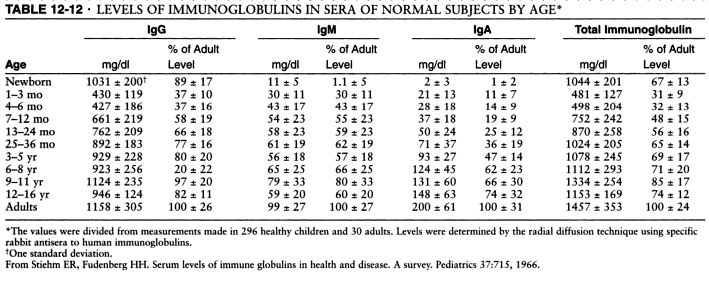
Serum levels of IgM, IgG and IgA. Source: Pediatrics, 1966 and Immunologic disorders in infants and children, by E. Richard Stiehm, Hans D. Ochs, Jerry A. Winkelstein.
Typical pattern of immunoglobulin levels (IgG, IgA, IgM) in humoral immunodeficiency. Click here to enlarge the table.
References
Diagnostic Criteria for Common Variable Immunodeficiency (CVID): Probable and Possible Diagnosis
The diagnostic criteria are divided into three categories: definitive, probable, and possible. There are no criteria for definitive diagnosis of Common Variable Immunodeficiency (CVID) at this time.
To guard against the inclusion of patients who have polymorphic variants in the genes associated with immunodeficiency and to specify the clinical or laboratory finding that is most consistently abnormal in a particular disorder, the patient must fulfill an inclusion criterion that is characteristic of the disorder.
Definitive diagnosis
Patients with a definitive diagnosis are assumed to have a greater than 98% probability that in 20 years they will still be given the same diagnosis. Mutation detection is the most reliable method of making a diagnosis but a single mutation is rarely found in CVID.
Probable diagnosis
Patients with a probable diagnosis are those with all of the clinical and laboratory characteristics of a particular disorder but who do not have a documented abnormality in the gene, the mRNA, or the protein that is known to be abnormal in the disorder. They are assumed to have a greater than 85% probability that in 20 years they will be given the same diagnosis.
Probable diagnosis of CVID:
Male or female patient who has a marked decrease (at least 2 SD below the mean for age) in serum IgG AND IgA and fulfills all of the following criteria:
1. Onset of immunodeficiency at greater than 2 years of age.
2. Absent isohemagglutinins and/or poor response to vaccines.
3. Defined causes of hypogammaglobulinemia have been excluded
Possible diagnosis
Patients with a possible diagnosis are those that have some but not all of the characteristic clinical or laboratory findings of a particular disorder.
Possible diagnosis if CVID:
Male or female patient who has a marked decrease (at least 2 SD below the mean for age) in one of the major isotypes (IgM, IgG, and IgA) and fulfills all of the following criteria:
1. Onset of immunodeficiency at greater than 2 years of age.
2. Absent isohemagglutinins and/or poor response to vaccines.
3. Defined causes of hypogammaglobulinemia have been excluded
Clinical features of CVID
Most patients with CVID are diagnosed with immunodeficiency in the second, third, or fourth decade of life, after they have had several pneumonias; however, children and older adults may be affected.
Viral, fungal, and parasitic infections as well as bacterial infections may be found.
The serum concentration of IgM is normal in about half of the patients.
Abnormalities in T cell numbers or function are common. The majority of patients have normal numbers of B cells; however, some have low or absent B cells.
Approximately 50% of patients have autoimmune manifestations. There is an increased risk of malignancy.
Differential diagnosis of hypogammaglobulinemia includes drug-induced, for example secondary to glucocorticoids (steroids).
Diagnostic Criteria for Primary Immunodeficiencies. Mary Ellen Conley, Luigi D. Notarangelo, and Amos Etzioni Representing PAGID (Pan-American Group for Immunodeficiency) and ESID (European Society for Immunodeficiencies). Clinical Immunology, Vol. 93, No. 3, December, pp. 190–197, 1999.
What is the treatment for CVID?
Intravenous immunoglobulin (IVIG) administered at outpatient clinic, monthly, this is the most common treatment.
Subcutaneous immunoglobulin G (SCIG), administered at home, given weekly.
Summary
CVID is characterized by:
1. Low levels of most or all of the immunoglobulin (Ig) classes
2. Lack of B lymphocytes or plasma cells that are capable of producing antibodies
3. Frequent bacterial infections
CVID is the most common primary immunodeficiency with an incidence of 1 case per 10,000-50,000 population. More than two thirds of patients are aged 21 years or older when CVID is diagnosed. CVID represents a group of heterogeneous conditions. Approximately 50% of patients with the deficiency also have diminished serum immunoglobulin M (IgM) levels and T-lymphocyte dysfunction. About 20% of those with CVID have an autoimmune disease.
The prognosis for patients with CVID is good if they do not have bronchiectasis, autoimmune disease or malignancy.
Five immunoglobulin classes (mind map)
In order of their serum concentrations:
IgG 1000 mg/dL
IgA 200 mg/dL
IgM 150 mg/dL
IgD 4 mg/dL
IgE 0.005 mg/dL (extremely low serum concentration compared to other Ig in (GAMED)
IgG and A are divided in subclasses: 4 for IgG -- IgG1, IgG2, IgG3, IgG4, and 2 for IgA -- IgA1 and IgA2.
Ig structures. Image source: Wikipedia.
Can patients with CVID receive live vaccines?
Patients with CVID should never receive live vaccines and should be advised to avoid contact with children or adults who have recently been given a live vaccine, for example, FluMist® nasal flu vaccine spray.
Can patients with CVID travel to endemic area for hepatitis A or B?
Yes but they should be given hyperimmune Ig prior to travel to endemic areas. The hyperimmune Ig provides protection for 3 weeks.
What is the half-life of IVIG?
3 weeks.
Mnemonic: Dose of IVIG in PIDD
400-600 mg/kg/month
4 letter words:
IVIG
CVID
SCID
What are the long-term risks for patients with CVID?
Malignancies, autoimmune diseases, infections.
FDA guidelines for IVIG state the product should be prepared out of at least 1,000 different human donors. Most commercial IVIG preparations contain products from over 10,000 human donors which increases the risk of acquiring diseases such as HIV, HCV or Creutzfeldt-Jakob disease. The risk is small but nevertheless, it should be discussed with the patient.
What HIV screening test would you do in a patient with CVID?
Patients with CVID can not produce an adequate antibody response to the HIV virus if infected and ELISA or Western blot are not useful. PCR RNA is the test of choise for HIV diagnosis in patients with CVID.
Can patients with CVID still have allergies?
Yes, the production of IgE may not be affected in patients with CVID can have symtpoms of allergic rhinitis, urticaria and other IgE-mediated diseases.
Differences between commercial IVIG preparations: Gammagard and Octagam
Gammagard Liquid is the first 10% IVIG solution with no added carbohydrates or sodium. Baxter is replacing Gammagard S/D with Gammagard Liquid.
Sodium concentration
Gammagard: No added sodium. When reconstituted with the total volume of diluent (Sterile Water for Injection, USP) supplied to 5%, Gammagard contains a physiological concentration of sodium chloride (approximately 8.5 mg/mL) and has a pH of 6.8 ± 0.4.
Octagam: sodium content of the final solution is ≤ 30 mmol/l and the pH is between 5.1 and 6.0.
Osmolality
Gammagard osmolality is 240-300 mOsmol/kg, similar to physiological osmolality (285 to 295 mOsmol/kg).
Octagam osmolality is 310 - 380 mosmol/kg.
Cost
Gammagard Liquid, Baxter, $129.60/gm
Octagam, Octapharma, $118.34/gm
References
Antibody response of pneumococcal vaccine: need for booster dosing? International Journal of Antimicrobial Agents, Volume 14, Issue 2, March 2000, Pages 107-112.
How to identify a possible specific antibody deficiency to pneumococcus. AAAAI, 2007.
Common Variable Immunodeficiency. eMedicine.
Common Variable Immunodeficiency. eMedicine.
Common Variable Immunodeficiency: A Multifaceted And Puzzling Disorder. Expert Review of Clinical Immunology, Medscape, 2009.
Published: 07/12/2008
Updated: 02/06/2012
Reviewer: S. Randhawa, M.D., Allergist/Immunologist and Assistant Professor at NSU
A 65-year-old Caucasian female (CF) is referred by her hematologist and primary care physician (PCP) for work-up of suspected immunodeficiency. She was referred to hematology by her PCP for work-up of frequent urinary tract infections (UTIs) for the last year. Complete blood count with differential (CBCD), bone marrow and chromosomal analysis were negative. Serum immunoglobulins showed low levels of IgG 515 mg/dL (reference range 700 to 1600), IgM 30 (reference range 40 to 230) , IgA 50 (reference range 70 to 400). UA: bacteriuria, no protein. ANA was negative.
Physical examination
Normal.
What is the reason for the low levels of immunoglobulins?
Differential diagnosis:
1. Immunoglobulin loss from GI tract, for example, protein-losing enteropathy. These patients usually have diarrhea but our patient complains of constipation.
2. Immunoglobulin loss in the urine, for example, nephrotic syndrome. IgM (pentamer) and IgA (dimer) are larger molecules than IgG. It is very unlikely to see low levels for IgM in patients with nephrotic syndrome or protein-losing enteropathy. IgG gets filtered in the urine but the large IgM pentamer does not. A 24-hour urine collection is the best method to determine the amount of protein in the urine.
3. Low production of Ig due to bone marrow process or CLL. Both were excluded by BM biopsy and normal CBCD.
4. Common variable immunodeficiency (CVID)
Common variable immunodeficiency (CVID) is the most likely diagnosis in this patient.
How do you diagnose CVID?
A patient with suspected CVID can be tested for the ability to mount humoral and cellular immune response as follows:
1. Humoral immune response.
Inject patient with Pneumovax 23 (not Prevnar, which is a 7-valent conjugate vaccine). Check anti-polysaccharide IgG antibody to pneumococcus serotypes in 3 weeks. There should be a 3-5 fold increase in the anitbody titer to at least 50% of isotypes.
Which serotypes should be included in the order for Ig?
The same serotypes that were included in the given vaccine. Check the enclosed leaflet for this information. For example, the serotypes included in Pneumovax 23 can be found from the Merck website (PDF):
1 2 3 4 5 6B 7F 8 9N 9V 10A 11A 12F 14 15B 17F 18C 19F 19A 20 22F 23F 33F
How to collect the serum for anti-polysaccharide IgG antibodies?
Collect the serum prior to immunization with Pneumovax 23. Store the pre-immunization serum in the office refrigerator. Give the vaccine. Check the post-immunization serum 3 weeks later. Send both pre- and post-immunization serums to the laboratory at the same time.
The pneumococcal vaccine comprises purified capsular polysaccharide of 23 stereotypes that account for more than 90% of the invasive pneumococcal infections in the USA. It induces anti-polysaccharide IgG antibody levels to most or all of the component polysaccharide antigens in immunocompetent adults. Elderly adults respond equally well to vaccination as do younger adults. The current 23-valent vaccine comprises 25 μg of each of 23 pneumococcal stereotypes (namely, serotypes 1, 2, 3, 4, 5, 6B, 7F, 8, 9N, 9V, 10A, 11A, 12F, 14, 15B, 17F, 18C, 19A, 19F, 20, 22F, 23F and 33F).
The test should be ordered as follows: Pre- and post-IgM and IgG antibody titers to pneumococcal serotypes 1, 2, 3, 4, 5, 6B, 7F, 8, 9N, 9V, 10A, 11A, 12F, 14, 15B, 17F, 18C, 19A, 19F, 20, 22F, 23F and 33F
2. Cellular immune response.
Some patients with CVID have decreased cellular immunity as well. Cellular immunity is tested by anergy panel with common antigens to which the patient was likely exposed in the past: Tetanus toxoid, Trichophyton, Candida. An intradermal injection of 0.1 mL of each antigen is necessary to perform the test. The test is read in 48-72 hours. A positive test result indicates intact delayed-type hypersensitivity. A negative test result to all antigens suggests impaired type IV immunity.
Flow cytometry should also be ordered. Lymphocyte proliferation is a more sensitive test to evaluate cellular immune response.
Delayed-type hypersensitivity (DTH) response
The standardized DTH test includes Candida, tetanus, mumps, and TB. Trichophyton is also commonly used. However, the only FDA-approved reagents for DTH are PPD, Candida and mumps.
What are the normal serum immunoglobulin levels (IgG, IgA, IgM)?
Serum levels of IgM, IgG and IgA vary with age, gender and race.
The IgG and IgA concentrations in children show a gradual rise with increasing age. The IgA level is generally about the same in both sexes. Girls typically have higher IgM and IgG levels than boys.
The confidence interval bounded by two standard deviations about the mean excludes 5% of apparently healthy controls.
Elevated IgM, low IgA, low IgG, low IgM, and elevated IgA are the commonest changes observed in apparently healthy humans.
Humoral immunodeficiency is commonly defined as IgG, IgM or IgA level that is two standard deviations (2 SD) below the mean level for IgG, IgM or IgA, respectively, for the particular age group and gender.
Serum levels of IgM, IgG and IgA. Source: Pediatrics, 1966 and Immunologic disorders in infants and children, by E. Richard Stiehm, Hans D. Ochs, Jerry A. Winkelstein.
Typical pattern of immunoglobulin levels (IgG, IgA, IgM) in humoral immunodeficiency. Click here to enlarge the table.
References
Serum immunoglobulin levels in healthy children and adults. J. W. Stoop, B. J. M. Zegers, P. C. Sander, and R. E. Ballieux. Clin Exp Immunol. 1969 January; 4(1): 101–112.
The relationship of race, sex, and age to concentrations of serum immunoglobulins expressed in international units in healthy adults in the USA. S. E. Maddison, C. C. Stewart, C. E. Farshy, and C. B. Reimer. Bull World Health Organ. 1975; 52(2): 179–185.
Serum immunoglobulin concentrations in preschool children measured by laser nephelometry: reference ranges for IgG, IgA, IgM. D Isaacs, D G Altman, C E Tidmarsh, H B Valman, and A D Webster. J Clin Pathol. 1983 October; 36(10): 1193–1196.
D Isaacs, A D Webster, and H B Valman. Clin Exp Immunol. 1984 November; 58(2): 335–340.
Serum Immunoglobulin Levels Throughout the Life-Span of Healthy Man. Ann of Int Med, November 1, 1971, Vol. 75 no. 5 673-682.
Diagnostic Criteria for Common Variable Immunodeficiency (CVID): Probable and Possible Diagnosis
The diagnostic criteria are divided into three categories: definitive, probable, and possible. There are no criteria for definitive diagnosis of Common Variable Immunodeficiency (CVID) at this time.
To guard against the inclusion of patients who have polymorphic variants in the genes associated with immunodeficiency and to specify the clinical or laboratory finding that is most consistently abnormal in a particular disorder, the patient must fulfill an inclusion criterion that is characteristic of the disorder.
Definitive diagnosis
Patients with a definitive diagnosis are assumed to have a greater than 98% probability that in 20 years they will still be given the same diagnosis. Mutation detection is the most reliable method of making a diagnosis but a single mutation is rarely found in CVID.
Probable diagnosis
Patients with a probable diagnosis are those with all of the clinical and laboratory characteristics of a particular disorder but who do not have a documented abnormality in the gene, the mRNA, or the protein that is known to be abnormal in the disorder. They are assumed to have a greater than 85% probability that in 20 years they will be given the same diagnosis.
Probable diagnosis of CVID:
Male or female patient who has a marked decrease (at least 2 SD below the mean for age) in serum IgG AND IgA and fulfills all of the following criteria:
1. Onset of immunodeficiency at greater than 2 years of age.
2. Absent isohemagglutinins and/or poor response to vaccines.
3. Defined causes of hypogammaglobulinemia have been excluded
Possible diagnosis
Patients with a possible diagnosis are those that have some but not all of the characteristic clinical or laboratory findings of a particular disorder.
Possible diagnosis if CVID:
Male or female patient who has a marked decrease (at least 2 SD below the mean for age) in one of the major isotypes (IgM, IgG, and IgA) and fulfills all of the following criteria:
1. Onset of immunodeficiency at greater than 2 years of age.
2. Absent isohemagglutinins and/or poor response to vaccines.
3. Defined causes of hypogammaglobulinemia have been excluded
Clinical features of CVID
Most patients with CVID are diagnosed with immunodeficiency in the second, third, or fourth decade of life, after they have had several pneumonias; however, children and older adults may be affected.
Viral, fungal, and parasitic infections as well as bacterial infections may be found.
The serum concentration of IgM is normal in about half of the patients.
Abnormalities in T cell numbers or function are common. The majority of patients have normal numbers of B cells; however, some have low or absent B cells.
Approximately 50% of patients have autoimmune manifestations. There is an increased risk of malignancy.
Differential diagnosis of hypogammaglobulinemia includes drug-induced, for example secondary to glucocorticoids (steroids).
References
Diagnostic Criteria for Primary Immunodeficiencies. Mary Ellen Conley, Luigi D. Notarangelo, and Amos Etzioni Representing PAGID (Pan-American Group for Immunodeficiency) and ESID (European Society for Immunodeficiencies). Clinical Immunology, Vol. 93, No. 3, December, pp. 190–197, 1999.
Recognizing Primary Immune Deficiency in Clinical Practice. Clinical and Vaccine Immunology, March 2006, p. 329-332, Vol. 13, No. 3.
What is the treatment for CVID?
Intravenous immunoglobulin (IVIG) administered at outpatient clinic, monthly, this is the most common treatment.
Subcutaneous immunoglobulin G (SCIG), administered at home, given weekly.
Summary
CVID is characterized by:
1. Low levels of most or all of the immunoglobulin (Ig) classes
2. Lack of B lymphocytes or plasma cells that are capable of producing antibodies
3. Frequent bacterial infections
CVID is the most common primary immunodeficiency with an incidence of 1 case per 10,000-50,000 population. More than two thirds of patients are aged 21 years or older when CVID is diagnosed. CVID represents a group of heterogeneous conditions. Approximately 50% of patients with the deficiency also have diminished serum immunoglobulin M (IgM) levels and T-lymphocyte dysfunction. About 20% of those with CVID have an autoimmune disease.
The prognosis for patients with CVID is good if they do not have bronchiectasis, autoimmune disease or malignancy.
Five immunoglobulin classes (mind map)
In order of their serum concentrations:
IgG 1000 mg/dL
IgA 200 mg/dL
IgM 150 mg/dL
IgD 4 mg/dL
IgE 0.005 mg/dL (extremely low serum concentration compared to other Ig in (GAMED)
IgG and A are divided in subclasses: 4 for IgG -- IgG1, IgG2, IgG3, IgG4, and 2 for IgA -- IgA1 and IgA2.
Ig structures. Image source: Wikipedia.
Can patients with CVID receive live vaccines?
Patients with CVID should never receive live vaccines and should be advised to avoid contact with children or adults who have recently been given a live vaccine, for example, FluMist® nasal flu vaccine spray.
Can patients with CVID travel to endemic area for hepatitis A or B?
Yes but they should be given hyperimmune Ig prior to travel to endemic areas. The hyperimmune Ig provides protection for 3 weeks.
What is the half-life of IVIG?
3 weeks.
Mnemonic: Dose of IVIG in PIDD
400-600 mg/kg/month
4 letter words:
IVIG
CVID
SCID
What are the long-term risks for patients with CVID?
Malignancies, autoimmune diseases, infections.
FDA guidelines for IVIG state the product should be prepared out of at least 1,000 different human donors. Most commercial IVIG preparations contain products from over 10,000 human donors which increases the risk of acquiring diseases such as HIV, HCV or Creutzfeldt-Jakob disease. The risk is small but nevertheless, it should be discussed with the patient.
What HIV screening test would you do in a patient with CVID?
Patients with CVID can not produce an adequate antibody response to the HIV virus if infected and ELISA or Western blot are not useful. PCR RNA is the test of choise for HIV diagnosis in patients with CVID.
Can patients with CVID still have allergies?
Yes, the production of IgE may not be affected in patients with CVID can have symtpoms of allergic rhinitis, urticaria and other IgE-mediated diseases.
Differences between commercial IVIG preparations: Gammagard and Octagam
Gammagard Liquid is the first 10% IVIG solution with no added carbohydrates or sodium. Baxter is replacing Gammagard S/D with Gammagard Liquid.
Sodium concentration
Gammagard: No added sodium. When reconstituted with the total volume of diluent (Sterile Water for Injection, USP) supplied to 5%, Gammagard contains a physiological concentration of sodium chloride (approximately 8.5 mg/mL) and has a pH of 6.8 ± 0.4.
Octagam: sodium content of the final solution is ≤ 30 mmol/l and the pH is between 5.1 and 6.0.
Osmolality
Gammagard osmolality is 240-300 mOsmol/kg, similar to physiological osmolality (285 to 295 mOsmol/kg).
Octagam osmolality is 310 - 380 mosmol/kg.
Cost
Gammagard Liquid, Baxter, $129.60/gm
Octagam, Octapharma, $118.34/gm
References
Antibody response of pneumococcal vaccine: need for booster dosing? International Journal of Antimicrobial Agents, Volume 14, Issue 2, March 2000, Pages 107-112.
How to identify a possible specific antibody deficiency to pneumococcus. AAAAI, 2007.
Common Variable Immunodeficiency. eMedicine.
Common Variable Immunodeficiency. eMedicine.
Common Variable Immunodeficiency: A Multifaceted And Puzzling Disorder. Expert Review of Clinical Immunology, Medscape, 2009.
Primary Immunodeficiency Diseases: Definition, Diagnosis, and Management. Nima Rezaei, Luigi D. Notarangelo, Asghar Aghamohammadi, 2008, Google eBook.
Common variable immunodeficiency (CVID). Allergologia et Immunopathologia. November 2006. Volume 34 - Number 06 p. 263 - 275.
Adaptive Humoral Immunity: B-cells and Immunoglobulins
Mnemonics: Adaptive Humoral Immunity: B-cells and Immunoglobulins
Intravenous immune globulin. Hopkins HIV Guide.
Common variable immunodeficiency (CVID). Allergologia et Immunopathologia. November 2006. Volume 34 - Number 06 p. 263 - 275.
Adaptive Humoral Immunity: B-cells and Immunoglobulins
Mnemonics: Adaptive Humoral Immunity: B-cells and Immunoglobulins
Intravenous immune globulin. Hopkins HIV Guide.
Evaluation and diagnosis of common variable immunodeficiency. UpToDate.
Octagam, Octapharma.
Gammagard Liquid, Baxter.
Octagam, Octapharma.
Gammagard Liquid, Baxter.
Interpretation of pneumococcal antibody titers - AAAAI Ask the Expert, 2011.
Assessment of antibody response to pneumococcus immunization. AAAAI, Ask the Expert, 2011.
Cost-effectiveness of Pneumococcal Conjugate Vaccine vs. Polysaccharide Vaccine in Adults: PCV13 was better than PPSV23. JAMA, 2012.
Cost-effectiveness of Pneumococcal Conjugate Vaccine vs. Polysaccharide Vaccine in Adults: PCV13 was better than PPSV23. JAMA, 2012.
Published: 07/12/2008
Updated: 02/06/2012
Subscribe to:
Posts (Atom)