Author: V. Dimov, M.D., Allergist/Immunologist at Cleveland Clinic
Reviewer: S. Randhawa, M.D., Allergist/Immunologist and Assistant Professor at NSU
A 72-year-old male was treated for multiple meyeloma 2 years ago. Subsequently, his IgG was found to be 180 mg/dL, IgA 7, IgM and there was insufficient response on the pneumococcus serotypes. At an outside facility, SCIG was started due to poor IV access. There was no history of infections. The diagnosis was secondary immunodeficiency. He has been receiving SCIG (Hizentra) 25 gm every week.
A year later, his IgG is 1200 mg/dL. The reported last dose of SCIG was 3-4 weeks ago (the prescription ran out).
What dose adjustment would you recommend now?
SCIG dose adjustement:
Based on the typical IVIG starting dose of 400 mg/kg/month, the total monthly dose is 400 mg x 65 kg = 26,000 mg (26 g).
According to the Hizentra dosage calculator (Initial recommended dose of Hizentra = 1.53 x Previous IVIg dose(grams)
Number of weeks between scheduled IVIg doses). Based on an IVIg dose of 26grams and 4weeks between IVIg doses, Hizentra weekly dose is: 9.95 g (49.73).
Patient's states the last SCIG dose was 3-4 weeks ago, total IgG is 1200 mg/dL. It is possible that some of the production of IgG by the B cells is recovering.
The recommended new weekly dose of SCIG is 15 g every week. IgG, IgM, IgA levels should be checked in 3 months, just before the SCIG infusion.
SCIG prescription was changed. The infusion nurse was informed.
Mnemonic: Dose of IVIG in PIDD: 4
400-600 mg/kg/month
IgG trough level should be over 400 mg/dL (over 600 mg/dL if bronchiectasis)
4 letter words:
IVIG
CVID
SCID
Starting doses for IVIG: 400 to 600 mg/kg/month for a target trough level of at least 500 mg/dL.
How monitor CVID patients on IVIG?
Immunoglobulin levels at 3-6 month intervals
CBC and CMP yearly
Spirometry yearly
CT chest every 3-4 years - only if lung disease suspected or lung functions not normal
References
Guidelines on Dosing and Treatment Administration for Hizentra Therapy http://buff.ly/1uCZt4Q
Related reading
AInotes - Common Variable Immunodeficiency http://buff.ly/1uCZFky
AInotes - IVIG and Subcutaneous Ig http://buff.ly/1uCZTYP
Published: 07/12/2010
Updated: 03/15/2014
Showing posts with label Immunodeficiency. Show all posts
Showing posts with label Immunodeficiency. Show all posts

Evaluation for isolated low IgA level
Author: V. Dimov, M.D., Allergist/Immunologist and Assistant Professor at University of Chicago
Reviewer: S. Randhawa, M.D., Allergist/Immunologist and Assistant Professor at NSU
A 27-year-old male is in the allergy clinic for evaluation of suspected IgA deficiency. He has no history of asthma, allergic rhinitis, food allergy, or atopic dermatitis.
He has a history of nonspecific abdominal pain, the screening test for celiac disease (TTGA IgA) showed IgA level below 15 mg/dL, and he is here for evaluation. Biopsies from EGD/colonoscopy were negative for abnormalities. He follows a gluten free diet. IBS was considered but the symptoms worsened with IBS therapy.
He has no history of recurrent or severe infections, but he does feel that when the get a cold it lasts longer. No pneumonia or skin infections.
Physical examination is normal apart from pale boggy turbinates and postnasal drip.
Current Medications: None.
Labs reviewed: ImmunoCAP for common food allergens all negative, below 0.35.
What is the most likely diagnosis?
- allergic rhinitis
- isolated, asymptomatic selective partial IgA deficiency
What diagnostic test would you suggest?
He had a percutaneous skin testing with indoor and outdoor allergens that was positive for Dust mite, Trees, and Ragweed.
What happened next?
This patient may have partial IgA deficiency, asymptomatic.
The majority of patients with IgA deficiency are asymptomatic. These patients require only education about the condition.s seems to fall in this category.
The majority of patients with IgA deficiency are asymptomatic. These patients require only education about the condition.s seems to fall in this category.
Only a minority of IgA deficient individuals are symptomatic. These patients may develop recurrent sinopulmonary infections, autoimmune antibodies and/or disorders, gastrointestinal disorders, and rare anaphylactic reactions to blood products. Evaluation begins with measurement of serum levels of IgA, IgG, and IgM (ordered today). Serum levels of IgG and IgM levels must be normal to consider the diagnosis of selective IgA deficiency. Two severities of IgA deficiency are distinguished: A serum IgA level lower than 7 mg/dL is considered severe deficiency. Partial deficiency refers to a level above 7 mg/dL but below the lower limit of age-adjusted normal.
Patients with recurrent sinopulmonary symptoms should be thoroughly evaluated and treated for other conditions predisposing to upper respiratory tract infections (eg, allergic rhinitis/asthma, chronic rhinosinusitis). In patients who continue to have sinopulmonary infections despite aggressive management of predisposing conditions (eg, allergic rhinitis/asthma, chronic rhinosinusitis), a trial of prophylactic antibiotics can be considered.
He also has allergic rhinitis and conjunctivitis with sensitization to Dust mite, Trees, Ragweed.
What management would you suggest?
Regarding suspected IgA deficiency, screening for immunodeficiency with IgG, A, M, titers for diphtheria, tetanus, 23 pneumococcus serotypes was recommended.
Regarding his allergic rhinitis and conjunctivitis, avoidance of relevant allergens was recommended and detailed instructions provided. He can take Zyrtec (cetirizine) 10 mg po qpm as needed. If not better, intranasal steroid could be added. Immunotherapy can be discussed in the future.
A follow up in 1-3 months was suggested to monitor the effect of therapy and to discuss the results of the laboratory tests.
References
UpToDate, 2013
Published: 02/12/2013
Updated: 05/22/2013
Reviewer: S. Randhawa, M.D., Allergist/Immunologist and Assistant Professor at NSU
A 27-year-old male is in the allergy clinic for evaluation of suspected IgA deficiency. He has no history of asthma, allergic rhinitis, food allergy, or atopic dermatitis.
He has a history of nonspecific abdominal pain, the screening test for celiac disease (TTGA IgA) showed IgA level below 15 mg/dL, and he is here for evaluation. Biopsies from EGD/colonoscopy were negative for abnormalities. He follows a gluten free diet. IBS was considered but the symptoms worsened with IBS therapy.
He has no history of recurrent or severe infections, but he does feel that when the get a cold it lasts longer. No pneumonia or skin infections.
Physical examination is normal apart from pale boggy turbinates and postnasal drip.
Current Medications: None.
Labs reviewed: ImmunoCAP for common food allergens all negative, below 0.35.
What is the most likely diagnosis?
- allergic rhinitis
- isolated, asymptomatic selective partial IgA deficiency
What diagnostic test would you suggest?
He had a percutaneous skin testing with indoor and outdoor allergens that was positive for Dust mite, Trees, and Ragweed.
What happened next?
This patient may have partial IgA deficiency, asymptomatic.
The majority of patients with IgA deficiency are asymptomatic. These patients require only education about the condition.s seems to fall in this category.
The majority of patients with IgA deficiency are asymptomatic. These patients require only education about the condition.s seems to fall in this category.
Only a minority of IgA deficient individuals are symptomatic. These patients may develop recurrent sinopulmonary infections, autoimmune antibodies and/or disorders, gastrointestinal disorders, and rare anaphylactic reactions to blood products. Evaluation begins with measurement of serum levels of IgA, IgG, and IgM (ordered today). Serum levels of IgG and IgM levels must be normal to consider the diagnosis of selective IgA deficiency. Two severities of IgA deficiency are distinguished: A serum IgA level lower than 7 mg/dL is considered severe deficiency. Partial deficiency refers to a level above 7 mg/dL but below the lower limit of age-adjusted normal.
Patients with recurrent sinopulmonary symptoms should be thoroughly evaluated and treated for other conditions predisposing to upper respiratory tract infections (eg, allergic rhinitis/asthma, chronic rhinosinusitis). In patients who continue to have sinopulmonary infections despite aggressive management of predisposing conditions (eg, allergic rhinitis/asthma, chronic rhinosinusitis), a trial of prophylactic antibiotics can be considered.
He also has allergic rhinitis and conjunctivitis with sensitization to Dust mite, Trees, Ragweed.
What management would you suggest?
Regarding suspected IgA deficiency, screening for immunodeficiency with IgG, A, M, titers for diphtheria, tetanus, 23 pneumococcus serotypes was recommended.
Regarding his allergic rhinitis and conjunctivitis, avoidance of relevant allergens was recommended and detailed instructions provided. He can take Zyrtec (cetirizine) 10 mg po qpm as needed. If not better, intranasal steroid could be added. Immunotherapy can be discussed in the future.
A follow up in 1-3 months was suggested to monitor the effect of therapy and to discuss the results of the laboratory tests.
References
UpToDate, 2013
Published: 02/12/2013
Updated: 05/22/2013
Selective IgA deficiency
Author: V. Dimov, M.D., Allergist/Immunologist and Assistant Professor at University of Chicago
Reviewer: S. Randhawa, M.D., Allergist/Immunologist and Assistant Professor at NSU
A 16-year-old boy is here for evaluation of suspected IgA deficiency. He had extensive blood work testing for suspected celiac disease 6 months ago. His IgA level was 17 mg/dL. IgM, IgG were normal. He does not have a history of recurrent infections. The scan for celiac disease was negative. A month ago, his IgA level was found to be 7 mg/dL and he is here for evaluation. He has no history of frequent ear, sinus, or lung infections or any other recurrent infections. He has no diagnosis of asthma, allergic rhinitis, food allergy, or atopic dermatitis.
Past medical history and surgical history is negative. Current medications: none.
Physical examination is unremarkable for acute allergic disease.
What is the most likely diagnosis?
This is a patient with suspected IgA immunodeficiency which occurs in approximately 1 in 500 individuals. The majority of these patients are asymptomatic. The definitive diagnosis of IgA immunodeficiency is made by an IgA level of less than 7 (he had one borderline value), and also by the evidence of normal IgM and IgG values. If the patient is asymptomatic, no further treatment is indicated.
What is the next step in the management?
Regarding his possible diagnosis of IgA immunodeficiency, I suggested repeated IgA, IgM, and IgG levels during the next 3 to 12 months.
If his IgA level is less than 7 mg/dL, he has a definitive diagnosis of selective IgA immunodeficiency.
If the IgA level is higher than 7 mg/dL (but lower than 2 standard deviations below the mean for his age), he has a possible IgA immunodeficiency.
The management consists of avoidance of infections as much as possible, and early treatment of any febrile illnesses and infections with antibiotic. IVIG or subcutaneous IgG replacement is not indicated in patients with IgA immunodeficiency.
I suggested recheck of his IgA, IgM, and IgG levels in 1 year from now. If celiac disease evaluation is still indicated, then endoscopy and biopsy may be a better option rather than tissue transglutaminase IgA, considering that his total IgA level is low.
Summary
Humoral immunodeficiency is commonly defined as IgG, IgM or IgA level that is two standard deviations (2 SD) below the mean level for IgG, IgM or IgA, respectively, for the particular age group and gender.
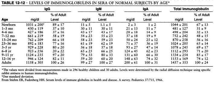
Serum levels of IgM, IgG and IgA. Source: Pediatrics, 1966 and Immunologic disorders in infants and children, by E. Richard Stiehm, Hans D. Ochs, Jerry A. Winkelstein.
Typical pattern of immunoglobulin levels (IgG, IgA, IgM) in humoral immunodeficiency. Click here to enlarge the table.
References
Serum immunoglobulin levels in healthy children and adults. J. W. Stoop, B. J. M. Zegers, P. C. Sander, and R. E. Ballieux. Clin Exp Immunol. 1969 January; 4(1): 101–112.
The relationship of race, sex, and age to concentrations of serum immunoglobulins expressed in international units in healthy adults in the USA. S. E. Maddison, C. C. Stewart, C. E. Farshy, and C. B. Reimer. Bull World Health Organ. 1975; 52(2): 179–185.
Serum immunoglobulin concentrations in preschool children measured by laser nephelometry: reference ranges for IgG, IgA, IgM. D Isaacs, D G Altman, C E Tidmarsh, H B Valman, and A D Webster. J Clin Pathol. 1983 October; 36(10): 1193–1196.
Immunoglobulin levels and function in pre-school children with recurrent respiratory infections.
D Isaacs, A D Webster, and H B Valman. Clin Exp Immunol. 1984 November; 58(2): 335–340.
Serum Immunoglobulin Levels Throughout the Life-Span of Healthy Man. Ann of Int Med, November 1, 1971, Vol. 75 no. 5 673-682.
Diagnostic Criteria for Primary Immunodeficiencies. Mary Ellen Conley, Luigi D. Notarangelo, and Amos Etzioni Representing PAGID (Pan-American Group for Immunodeficiency) and ESID (European Society for Immunodeficiencies). Clinical Immunology, Vol. 93, No. 3, December, pp. 190–197, 1999.
Recognizing Primary Immune Deficiency in Clinical Practice. Clinical and Vaccine Immunology, March 2006, p. 329-332, Vol. 13, No. 3.

Five immunoglobulin classes (mind map)
In order of their serum concentrations:
IgG 1000 mg/dL
IgA 200 mg/dL
IgM 150 mg/dL
IgD 4 mg/dL
IgE 0.005 mg/dL (extremely low serum concentration compared to other Ig in (GAMED)
IgG and A are divided in subclasses: 4 for IgG -- IgG1, IgG2, IgG3, IgG4, and 2 for IgA -- IgA1 and IgA2.

Ig structures. Image source: Wikipedia.
Published: 07/12/2010
Updated: 05/06/2012
Reviewer: S. Randhawa, M.D., Allergist/Immunologist and Assistant Professor at NSU
A 16-year-old boy is here for evaluation of suspected IgA deficiency. He had extensive blood work testing for suspected celiac disease 6 months ago. His IgA level was 17 mg/dL. IgM, IgG were normal. He does not have a history of recurrent infections. The scan for celiac disease was negative. A month ago, his IgA level was found to be 7 mg/dL and he is here for evaluation. He has no history of frequent ear, sinus, or lung infections or any other recurrent infections. He has no diagnosis of asthma, allergic rhinitis, food allergy, or atopic dermatitis.
Past medical history and surgical history is negative. Current medications: none.
Physical examination is unremarkable for acute allergic disease.
What is the most likely diagnosis?
This is a patient with suspected IgA immunodeficiency which occurs in approximately 1 in 500 individuals. The majority of these patients are asymptomatic. The definitive diagnosis of IgA immunodeficiency is made by an IgA level of less than 7 (he had one borderline value), and also by the evidence of normal IgM and IgG values. If the patient is asymptomatic, no further treatment is indicated.
What is the next step in the management?
Regarding his possible diagnosis of IgA immunodeficiency, I suggested repeated IgA, IgM, and IgG levels during the next 3 to 12 months.
If his IgA level is less than 7 mg/dL, he has a definitive diagnosis of selective IgA immunodeficiency.
If the IgA level is higher than 7 mg/dL (but lower than 2 standard deviations below the mean for his age), he has a possible IgA immunodeficiency.
The management consists of avoidance of infections as much as possible, and early treatment of any febrile illnesses and infections with antibiotic. IVIG or subcutaneous IgG replacement is not indicated in patients with IgA immunodeficiency.
I suggested recheck of his IgA, IgM, and IgG levels in 1 year from now. If celiac disease evaluation is still indicated, then endoscopy and biopsy may be a better option rather than tissue transglutaminase IgA, considering that his total IgA level is low.
Summary
Humoral immunodeficiency is commonly defined as IgG, IgM or IgA level that is two standard deviations (2 SD) below the mean level for IgG, IgM or IgA, respectively, for the particular age group and gender.
Serum levels of IgM, IgG and IgA. Source: Pediatrics, 1966 and Immunologic disorders in infants and children, by E. Richard Stiehm, Hans D. Ochs, Jerry A. Winkelstein.
Typical pattern of immunoglobulin levels (IgG, IgA, IgM) in humoral immunodeficiency. Click here to enlarge the table.
References
Serum immunoglobulin levels in healthy children and adults. J. W. Stoop, B. J. M. Zegers, P. C. Sander, and R. E. Ballieux. Clin Exp Immunol. 1969 January; 4(1): 101–112.
The relationship of race, sex, and age to concentrations of serum immunoglobulins expressed in international units in healthy adults in the USA. S. E. Maddison, C. C. Stewart, C. E. Farshy, and C. B. Reimer. Bull World Health Organ. 1975; 52(2): 179–185.
Serum immunoglobulin concentrations in preschool children measured by laser nephelometry: reference ranges for IgG, IgA, IgM. D Isaacs, D G Altman, C E Tidmarsh, H B Valman, and A D Webster. J Clin Pathol. 1983 October; 36(10): 1193–1196.
Immunoglobulin levels and function in pre-school children with recurrent respiratory infections.
D Isaacs, A D Webster, and H B Valman. Clin Exp Immunol. 1984 November; 58(2): 335–340.
Serum Immunoglobulin Levels Throughout the Life-Span of Healthy Man. Ann of Int Med, November 1, 1971, Vol. 75 no. 5 673-682.
Diagnostic Criteria for Primary Immunodeficiencies. Mary Ellen Conley, Luigi D. Notarangelo, and Amos Etzioni Representing PAGID (Pan-American Group for Immunodeficiency) and ESID (European Society for Immunodeficiencies). Clinical Immunology, Vol. 93, No. 3, December, pp. 190–197, 1999.
Recognizing Primary Immune Deficiency in Clinical Practice. Clinical and Vaccine Immunology, March 2006, p. 329-332, Vol. 13, No. 3.

Five immunoglobulin classes (mind map)
In order of their serum concentrations:
IgG 1000 mg/dL
IgA 200 mg/dL
IgM 150 mg/dL
IgD 4 mg/dL
IgE 0.005 mg/dL (extremely low serum concentration compared to other Ig in (GAMED)
IgG and A are divided in subclasses: 4 for IgG -- IgG1, IgG2, IgG3, IgG4, and 2 for IgA -- IgA1 and IgA2.

Ig structures. Image source: Wikipedia.

Published: 07/12/2010
Updated: 05/06/2012
Idiopathic CD4 lymphocytopenia (ICL)
Author: V. Dimov, M.D., Allergist/Immunologist and Assistant Professor at University of Chicago
Reviewer: S. Randhawa, M.D., Allergist/Immunologist, Fort Lauderdale, FL
A 38-year-old male is at the allergy clinic here for evaluation of low CD4 count. The reason for checking his CD4 was that he volunteered to be involved in a research project 5 years ago. His absolute CD4 was 456 at the time. At that time, he was negative for both HIV-1 and HIV-2 type viruses, and the test was repeated in 3 years later. Since then, his CD4 count continues to be low. It was 432 three years ago. Despite the low CD4 count, however, he has no history of any infections or abnormalities related to it. He has history of difficulty gaining immunity against hepatitis B after vaccination, and after 3 separate vaccinations, he was not able to develop detectable IgG antibody against it. His flow cytometry from 3 years ago showed absolute CD4 count of 432, CD8 count of 379, CD19 count of 261, and CD56 count of 278. He had positive immunoglobulin G against measles and rubella 10 years ago. He does not recall any family history of immunodeficiency. He has no family history of allergic rhinitis, asthma or eczema or food allergy.
Past Medical History: As above. Past Surgical History: Negative. Current Medications: He is not on any medications on a regular basis. Family History: Unremarkable. Physical Examination: unremarkable.
What is the most likely diagnosis?
Idiopathic CD4 lymphocytopenia of unclear etiology at this point.
Any clues about the etiology of his idiopathic CD4 lymphocytopenia?
A variety of past or latent viral diseases can induce CD4 lymphocytopenia. Autoimmune conditions can also pay a role. In patients with asymptomatic CD4 lymphocytopenia in the absence of HIV infection and opportunistic infections, a genetic defect could be the cause.
What laboratory workup would you suggest?
Evaluate the function of the other parts of his immune system including B cells and T cells and humoral immunity.
Regarding his idiopathic CD4 lymphocytopenia, the suggested workup would include flow cytometry with CD4, CD3, CD8, CD19, CD26, CD56, and CD16, also immunoglobulin G subclasses, immunoglobulin G, A, M, and E, CBC with differential, immunoglobulin G titers for 23 pneumococcal serotypes, tetanus, diphtheria, hepatitis, and mumps, and also mitogen stimulation test for lymphocyte proliferation for B and T cells.
ANA, CMP, ESR and UA can also be addded to his laboratory work.
An evaluation by an infectious disease specialist for his lymphocytopenia is also suggested in order to rule out any latent viral infection that may play a role in his etiology.
Published: 06/12/2011
Updated: 08/23/2011
Reviewer: S. Randhawa, M.D., Allergist/Immunologist, Fort Lauderdale, FL
A 38-year-old male is at the allergy clinic here for evaluation of low CD4 count. The reason for checking his CD4 was that he volunteered to be involved in a research project 5 years ago. His absolute CD4 was 456 at the time. At that time, he was negative for both HIV-1 and HIV-2 type viruses, and the test was repeated in 3 years later. Since then, his CD4 count continues to be low. It was 432 three years ago. Despite the low CD4 count, however, he has no history of any infections or abnormalities related to it. He has history of difficulty gaining immunity against hepatitis B after vaccination, and after 3 separate vaccinations, he was not able to develop detectable IgG antibody against it. His flow cytometry from 3 years ago showed absolute CD4 count of 432, CD8 count of 379, CD19 count of 261, and CD56 count of 278. He had positive immunoglobulin G against measles and rubella 10 years ago. He does not recall any family history of immunodeficiency. He has no family history of allergic rhinitis, asthma or eczema or food allergy.
Past Medical History: As above. Past Surgical History: Negative. Current Medications: He is not on any medications on a regular basis. Family History: Unremarkable. Physical Examination: unremarkable.
What is the most likely diagnosis?
Idiopathic CD4 lymphocytopenia of unclear etiology at this point.
Any clues about the etiology of his idiopathic CD4 lymphocytopenia?
A variety of past or latent viral diseases can induce CD4 lymphocytopenia. Autoimmune conditions can also pay a role. In patients with asymptomatic CD4 lymphocytopenia in the absence of HIV infection and opportunistic infections, a genetic defect could be the cause.
What laboratory workup would you suggest?
Evaluate the function of the other parts of his immune system including B cells and T cells and humoral immunity.
Regarding his idiopathic CD4 lymphocytopenia, the suggested workup would include flow cytometry with CD4, CD3, CD8, CD19, CD26, CD56, and CD16, also immunoglobulin G subclasses, immunoglobulin G, A, M, and E, CBC with differential, immunoglobulin G titers for 23 pneumococcal serotypes, tetanus, diphtheria, hepatitis, and mumps, and also mitogen stimulation test for lymphocyte proliferation for B and T cells.
ANA, CMP, ESR and UA can also be addded to his laboratory work.
An evaluation by an infectious disease specialist for his lymphocytopenia is also suggested in order to rule out any latent viral infection that may play a role in his etiology.
Published: 06/12/2011
Updated: 08/23/2011
Defects in T cells - part of severe combined immunodeficiencies (SCID)
Author: V. Dimov, M.D., Allergist/Immunologist and Assistant Professor at University of Chicago
Reviewer: S. Randhawa, M.D., Allergist/Immunologist and Assistant Professor at LSU (Shreveport) Department of Allergy and Immunology
Severe combined immunodeficiencies (SCIDs) represent a heterogeneous group of disorders. The most common SCID is X-linked and, therefore, most cases are diagnosed in male infants.
Defects in T cells
Defects in T cells
- CD8 lymphopenia can be caused by BLS1 (MHC1 deficiency) and ZAP70 deficiency
- CD4 lymphopenia can be caused by BLS2 (MHCII deficiency), p56lck deficiency,
and HIV infection
- see more examples in the figure below
- see more examples in the figure below
When diagnosing SCID, focus on B and NK phenotype because T cells are aslmost always absent/deficient with few exceptions.
Severe combined immunodeficiency (SCID) - 4 groups according to T/B/NK cells (click to enlarge the image).
Receptors for each of the following cytokines share the common gamma chain except which one?
A. IL-2
B. IL-7
C. IL-9
D. IL-15
E. IL-5
Answer: E.
Lack of a signal through which cytokine receptor accounts for the lack of T cell maturation in X-linked SCID?
A. IL-2
B. IL-7
C. IL-9
D. IL-15
E. IL-5
Answer: B.
Lack of a signal through which cytokine receptor accounts for the lack of NK cell maturation in X-linked SCID?
A. IL-2
B. IL-7
C. IL-9
D. IL-15
E. IL-5
Answer: D.
References
Practice parameter for the diagnosis and management of primary immunodeficiency. Ann Allergy Asthma Immunol 2005 May;94(5 Suppl 1):S1-63.
Updated: 09/12/2010
Practice parameter for the diagnosis and management of primary immunodeficiency. Ann Allergy Asthma Immunol 2005 May;94(5 Suppl 1):S1-63.
Algorithm 1: General Approach for the Diagnosis of Primary Immunodeficiency
Algorithm 2: Diagnosis of Humoral Immunodeficiency
Algorithm 3: Diagnosis of Cellular and Combined Immunodeficiencies
Algorithm 4: Diagnosis of Phagocyte Defects
Algorithm 5: Diagnosis of Complement Deficiency
Algorithm 6: General Considerations for Therapy of Primary Immunodeficiency
Updated: 09/12/2010
Defects in T cells - part of severe combined immunodeficiencies (SCID)
Author: V. Dimov, M.D., Allergist/Immunologist and Assistant Professor at University of Chicago
Reviewer: S. Randhawa, M.D., Allergist/Immunologist and Assistant Professor at LSU (Shreveport) Department of Allergy and Immunology
Severe combined immunodeficiencies (SCIDs) represent a heterogeneous group of disorders. The most common SCID is X-linked and, therefore, most cases are diagnosed in male infants.
Defects in T cells
Defects in T cells
- CD8 lymphopenia can be caused by BLS1 (MHC1 deficiency) and ZAP70 deficiency
- CD4 lymphopenia can be caused by BLS2 (MHCII deficiency), p56lck deficiency,
and HIV infection
- see more examples in the figure below
- see more examples in the figure below
When diagnosing SCID, focus on B and NK phenotype because T cells are aslmost always absent/deficient with few exceptions.
Severe combined immunodeficiency (SCID) - 4 groups according to T/B/NK cells (click to enlarge the image).
References
Practice parameter for the diagnosis and management of primary immunodeficiency. Ann Allergy Asthma Immunol 2005 May;94(5 Suppl 1):S1-63.
Updated: 09/12/2010
Practice parameter for the diagnosis and management of primary immunodeficiency. Ann Allergy Asthma Immunol 2005 May;94(5 Suppl 1):S1-63.
Algorithm 1: General Approach for the Diagnosis of Primary Immunodeficiency
Algorithm 2: Diagnosis of Humoral Immunodeficiency
Algorithm 3: Diagnosis of Cellular and Combined Immunodeficiencies
Algorithm 4: Diagnosis of Phagocyte Defects
Algorithm 5: Diagnosis of Complement Deficiency
Algorithm 6: General Considerations for Therapy of Primary Immunodeficiency
Updated: 09/12/2010
Follow-up of a Patient with Common Variable Immune Deficiency (CVID)
Author: V. Dimov, M.D., Allergist/Immunologist and Assistant Professor at University of Chicago
Reviewer: S. Randhawa, M.D., Allergist/Immunologist and Assistant Professor at LSU (Shreveport) Department of Allergy and Immunology
A 34-year-old female is in the Allergy/Immunology clinic today for a follow-up of Common Variable Immune Deficiency (CVID). She was last seen in the clinic 3 months ago. Since then, her condition has remained stable and the level of immunoglobulin G has been higher than 700. She is receiving IVIG 90 gm IV every month. She is pre-medicated with Tylenol and oral antihistamines and is able to tolerate the infusions well and has not had any infections of the sinuses or lung infections recently. She however reports painful area in her left armpit for the past two weeks, and it looks like she is developing hidradenitis.
Past medical history (PMH)
Common Variable Immune Deficiency (CVID).
Physical examination
The physical examination is positive for induration and erythema in the right axia which was diagnosed as hidradenitis. The rest of her examination is normal.
What is the most likely diagnosis?
This is a patient with CVID which has been stable on the IVIG replacement at the dose of 90 gm of Gammaguard every month. She also has hidradenitis in the left axilla.
What would you do?
Continue Gammaguard at a dose of 90 grams IV every month. Repeat the level in three months. For her hidradenitis, we recommended Keflex 500 mg PO B.I.D. x 7days.
The patient is planning a trip to the Bahamas for a week in the coming month. She wants to know if she should take any antibiotics with her.
What antibiotics would you prescribe, if she needs any?
Regarding her trip to the Bahamas: in case she develops respiratory or gastrointestinal infection we provided her with a prescription for Levaquin 500 PO QD x 7 days. She is to return to the clinic in four months.
What are the normal serum immunoglobulin levels (IgG, IgA, IgM)?
Serum levels of IgM, IgG and IgA vary with age, gender and race.
The IgG and IgA concentrations in children show a gradual rise with increasing age. The IgA level is generally about the same in both sexes. Girls typically have higher IgM and IgG levels than boys.
The confidence interval bounded by two standard deviations about the mean excludes 5% of apparently healthy controls.
Elevated IgM, low IgA, low IgG, low IgM, and elevated IgA are the commonest changes observed in apparently healthy humans.
Humoral immunodeficiency is commonly defined as IgG, IgM or IgA level that is two standard deviations (2 SD) below the mean level for IgG, IgM or IgA, respectively, for the particular age group and gender.
Serum levels of IgM, IgG and IgA. Source: Pediatrics, 1966 and Immunologic disorders in infants and children, by E. Richard Stiehm, Hans D. Ochs, Jerry A. Winkelstein.
Typical pattern of immunoglobulin levels (IgG, IgA, IgM) in humoral immunodeficiency. Click here to enlarge the table.
References
Diagnostic Criteria for Common Variable Immunodeficiency (CVID): Probable and Possible Diagnosis
The diagnostic criteria are divided into three categories: definitive, probable, and possible. There are no criteria for definitive diagnosis of Common Variable Immunodeficiency (CVID) at this time.
To guard against the inclusion of patients who have polymorphic variants in the genes associated with immunodeficiency and to specify the clinical or laboratory finding that is most consistently abnormal in a particular disorder, the patient must fulfill an inclusion criterion that is characteristic of the disorder.
Definitive diagnosis
Patients with a definitive diagnosis are assumed to have a greater than 98% probability that in 20 years they will still be given the same diagnosis. Mutation detection is the most reliable method of making a diagnosis but a single mutation is rarely found in CVID.
Probable diagnosis
Patients with a probable diagnosis are those with all of the clinical and laboratory characteristics of a particular disorder but who do not have a documented abnormality in the gene, the mRNA, or the protein that is known to be abnormal in the disorder. They are assumed to have a greater than 85% probability that in 20 years they will be given the same diagnosis.
Probable diagnosis of CVID:
Male or female patient who has a marked decrease (at least 2 SD below the mean for age) in serum IgG AND IgA and fulfills all of the following criteria:
1. Onset of immunodeficiency at greater than 2 years of age.
2. Absent isohemagglutinins and/or poor response to vaccines.
3. Defined causes of hypogammaglobulinemia have been excluded
Possible diagnosis
Patients with a possible diagnosis are those that have some but not all of the characteristic clinical or laboratory findings of a particular disorder.
Possible diagnosis if CVID:
Male or female patient who has a marked decrease (at least 2 SD below the mean for age) in one of the major isotypes (IgM, IgG, and IgA) and fulfills all of the following criteria:
1. Onset of immunodeficiency at greater than 2 years of age.
2. Absent isohemagglutinins and/or poor response to vaccines.
3. Defined causes of hypogammaglobulinemia have been excluded
Clinical features of CVID
Most patients with CVID are diagnosed with immunodeficiency in the second, third, or fourth decade of life, after they have had several pneumonias; however, children and older adults may be affected.
Viral, fungal, and parasitic infections as well as bacterial infections may be found.
The serum concentration of IgM is normal in about half of the patients.
Abnormalities in T cell numbers or function are common. The majority of patients have normal numbers of B cells; however, some have low or absent B cells.
Approximately 50% of patients have autoimmune manifestations. There is an increased risk of malignancy.
Differential diagnosis of hypogammaglobulinemia includes drug-induced, for example secondary to glucocorticoids (steroids).
Diagnostic Criteria for Primary Immunodeficiencies. Mary Ellen Conley, Luigi D. Notarangelo, and Amos Etzioni Representing PAGID (Pan-American Group for Immunodeficiency) and ESID (European Society for Immunodeficiencies). Clinical Immunology, Vol. 93, No. 3, December, pp. 190–197, 1999.
Published: 05/12/2010
Updated: 11/23/2011
Reviewer: S. Randhawa, M.D., Allergist/Immunologist and Assistant Professor at LSU (Shreveport) Department of Allergy and Immunology
A 34-year-old female is in the Allergy/Immunology clinic today for a follow-up of Common Variable Immune Deficiency (CVID). She was last seen in the clinic 3 months ago. Since then, her condition has remained stable and the level of immunoglobulin G has been higher than 700. She is receiving IVIG 90 gm IV every month. She is pre-medicated with Tylenol and oral antihistamines and is able to tolerate the infusions well and has not had any infections of the sinuses or lung infections recently. She however reports painful area in her left armpit for the past two weeks, and it looks like she is developing hidradenitis.
Past medical history (PMH)
Common Variable Immune Deficiency (CVID).
Physical examination
The physical examination is positive for induration and erythema in the right axia which was diagnosed as hidradenitis. The rest of her examination is normal.
What is the most likely diagnosis?
This is a patient with CVID which has been stable on the IVIG replacement at the dose of 90 gm of Gammaguard every month. She also has hidradenitis in the left axilla.
What would you do?
Continue Gammaguard at a dose of 90 grams IV every month. Repeat the level in three months. For her hidradenitis, we recommended Keflex 500 mg PO B.I.D. x 7days.
The patient is planning a trip to the Bahamas for a week in the coming month. She wants to know if she should take any antibiotics with her.
What antibiotics would you prescribe, if she needs any?
Regarding her trip to the Bahamas: in case she develops respiratory or gastrointestinal infection we provided her with a prescription for Levaquin 500 PO QD x 7 days. She is to return to the clinic in four months.
What are the normal serum immunoglobulin levels (IgG, IgA, IgM)?
Serum levels of IgM, IgG and IgA vary with age, gender and race.
The IgG and IgA concentrations in children show a gradual rise with increasing age. The IgA level is generally about the same in both sexes. Girls typically have higher IgM and IgG levels than boys.
The confidence interval bounded by two standard deviations about the mean excludes 5% of apparently healthy controls.
Elevated IgM, low IgA, low IgG, low IgM, and elevated IgA are the commonest changes observed in apparently healthy humans.
Humoral immunodeficiency is commonly defined as IgG, IgM or IgA level that is two standard deviations (2 SD) below the mean level for IgG, IgM or IgA, respectively, for the particular age group and gender.
Serum levels of IgM, IgG and IgA. Source: Pediatrics, 1966 and Immunologic disorders in infants and children, by E. Richard Stiehm, Hans D. Ochs, Jerry A. Winkelstein.
Typical pattern of immunoglobulin levels (IgG, IgA, IgM) in humoral immunodeficiency. Click here to enlarge the table.
References
Serum immunoglobulin levels in healthy children and adults. J. W. Stoop, B. J. M. Zegers, P. C. Sander, and R. E. Ballieux. Clin Exp Immunol. 1969 January; 4(1): 101–112.
The relationship of race, sex, and age to concentrations of serum immunoglobulins expressed in international units in healthy adults in the USA. S. E. Maddison, C. C. Stewart, C. E. Farshy, and C. B. Reimer. Bull World Health Organ. 1975; 52(2): 179–185.
Serum immunoglobulin concentrations in preschool children measured by laser nephelometry: reference ranges for IgG, IgA, IgM. D Isaacs, D G Altman, C E Tidmarsh, H B Valman, and A D Webster. J Clin Pathol. 1983 October; 36(10): 1193–1196.
D Isaacs, A D Webster, and H B Valman. Clin Exp Immunol. 1984 November; 58(2): 335–340.
Serum Immunoglobulin Levels Throughout the Life-Span of Healthy Man. Ann of Int Med, November 1, 1971, Vol. 75 no. 5 673-682.
Diagnostic Criteria for Common Variable Immunodeficiency (CVID): Probable and Possible Diagnosis
The diagnostic criteria are divided into three categories: definitive, probable, and possible. There are no criteria for definitive diagnosis of Common Variable Immunodeficiency (CVID) at this time.
To guard against the inclusion of patients who have polymorphic variants in the genes associated with immunodeficiency and to specify the clinical or laboratory finding that is most consistently abnormal in a particular disorder, the patient must fulfill an inclusion criterion that is characteristic of the disorder.
Definitive diagnosis
Patients with a definitive diagnosis are assumed to have a greater than 98% probability that in 20 years they will still be given the same diagnosis. Mutation detection is the most reliable method of making a diagnosis but a single mutation is rarely found in CVID.
Probable diagnosis
Patients with a probable diagnosis are those with all of the clinical and laboratory characteristics of a particular disorder but who do not have a documented abnormality in the gene, the mRNA, or the protein that is known to be abnormal in the disorder. They are assumed to have a greater than 85% probability that in 20 years they will be given the same diagnosis.
Probable diagnosis of CVID:
Male or female patient who has a marked decrease (at least 2 SD below the mean for age) in serum IgG AND IgA and fulfills all of the following criteria:
1. Onset of immunodeficiency at greater than 2 years of age.
2. Absent isohemagglutinins and/or poor response to vaccines.
3. Defined causes of hypogammaglobulinemia have been excluded
Possible diagnosis
Patients with a possible diagnosis are those that have some but not all of the characteristic clinical or laboratory findings of a particular disorder.
Possible diagnosis if CVID:
Male or female patient who has a marked decrease (at least 2 SD below the mean for age) in one of the major isotypes (IgM, IgG, and IgA) and fulfills all of the following criteria:
1. Onset of immunodeficiency at greater than 2 years of age.
2. Absent isohemagglutinins and/or poor response to vaccines.
3. Defined causes of hypogammaglobulinemia have been excluded
Clinical features of CVID
Most patients with CVID are diagnosed with immunodeficiency in the second, third, or fourth decade of life, after they have had several pneumonias; however, children and older adults may be affected.
Viral, fungal, and parasitic infections as well as bacterial infections may be found.
The serum concentration of IgM is normal in about half of the patients.
Abnormalities in T cell numbers or function are common. The majority of patients have normal numbers of B cells; however, some have low or absent B cells.
Approximately 50% of patients have autoimmune manifestations. There is an increased risk of malignancy.
Differential diagnosis of hypogammaglobulinemia includes drug-induced, for example secondary to glucocorticoids (steroids).
References
Diagnostic Criteria for Primary Immunodeficiencies. Mary Ellen Conley, Luigi D. Notarangelo, and Amos Etzioni Representing PAGID (Pan-American Group for Immunodeficiency) and ESID (European Society for Immunodeficiencies). Clinical Immunology, Vol. 93, No. 3, December, pp. 190–197, 1999.
Recognizing Primary Immune Deficiency in Clinical Practice. Clinical and Vaccine Immunology, March 2006, p. 329-332, Vol. 13, No. 3.
Frequency of follow-up visits in a patient receiving intravenous immunoglobulin (IVIG) infusions - every 6 months? AAAAI Ask the Expert, 2011.
Outcome of allogeneic stem cell transplantation (ASCT) in adults with common variable immunodeficiency (CVID) (JACI, 2011).
Frequency of follow-up visits in a patient receiving intravenous immunoglobulin (IVIG) infusions - every 6 months? AAAAI Ask the Expert, 2011.
Outcome of allogeneic stem cell transplantation (ASCT) in adults with common variable immunodeficiency (CVID) (JACI, 2011).
Published: 05/12/2010
Updated: 11/23/2011
STAT-1 Deficiency
Author: V. Dimov, M.D., Allergist/Immunologist and Assistant Professor at University of Chicago
Reviewer: S. Randhawa, M.D., Allergist/Immunologist and Assistant Professor at LSU (Shreveport) Department of Allergy and Immunology
The autosomal recessive form of human complete Stat-1 deficiency is a rare disorder. Stat-1 deficiency is associated with impaired cellular responses to both IFN-gamma and IFN-alpha/beta via Stat-1-containing complexes.
Two unrelated patients developed disseminated bacillus Calmette-Guérin (BCG) and subsequently died of viral illnesses. A third patient with complete Stat-1 deficiency and disseminated BCG infection, died 3 mo after bone marrow transplantation.
Stat-1 deficiency is a severe form of innate immunodeficiency. Stat-1 deficiency should be suspected in children with severe infections, notably but not exclusively patients with mycobacterial or viral diseases.
A deficiency in STAT 1 transcription factor can lead to increased susceptibility to which of the following infections?
(A) Herpes encephalitis
(B) Meningococcal meningitis
(C) Pneumococcal pneumonia
(D) Staphylococcal osteomyelitis
(E) Disseminated infection after BCG immunization
Correct answers: A and E
References
Human Complete Stat-1 Deficiency Is Associated with Defective Type I and II IFN Responses In Vitro but Immunity to Some Low Virulence Viruses In Vivo. Ariane Chapgier et al. The Journal of Immunology, 2006, 176: 5078-5083.
http://www.jimmunol.org/cgi/content/abstract/176/8/5078
Published: 05/09/2010
Updated: 05/09/2010
Reviewer: S. Randhawa, M.D., Allergist/Immunologist and Assistant Professor at LSU (Shreveport) Department of Allergy and Immunology
The autosomal recessive form of human complete Stat-1 deficiency is a rare disorder. Stat-1 deficiency is associated with impaired cellular responses to both IFN-gamma and IFN-alpha/beta via Stat-1-containing complexes.
Two unrelated patients developed disseminated bacillus Calmette-Guérin (BCG) and subsequently died of viral illnesses. A third patient with complete Stat-1 deficiency and disseminated BCG infection, died 3 mo after bone marrow transplantation.
Stat-1 deficiency is a severe form of innate immunodeficiency. Stat-1 deficiency should be suspected in children with severe infections, notably but not exclusively patients with mycobacterial or viral diseases.
A deficiency in STAT 1 transcription factor can lead to increased susceptibility to which of the following infections?
(A) Herpes encephalitis
(B) Meningococcal meningitis
(C) Pneumococcal pneumonia
(D) Staphylococcal osteomyelitis
(E) Disseminated infection after BCG immunization
Correct answers: A and E
References
Human Complete Stat-1 Deficiency Is Associated with Defective Type I and II IFN Responses In Vitro but Immunity to Some Low Virulence Viruses In Vivo. Ariane Chapgier et al. The Journal of Immunology, 2006, 176: 5078-5083.
http://www.jimmunol.org/cgi/content/abstract/176/8/5078
Published: 05/09/2010
Updated: 05/09/2010
Neutrophil actin dysfunction
Author: V. Dimov, M.D., Allergist/Immunologist and Assistant Professor at University of Chicago
Reviewer: S. Randhawa, M.D., Allergist/Immunologist and Assistant Professor at LSU (Shreveport) Department of Allergy and Immunology
Neutrophils and other phagocytes migrate to the site of infection, ingest pathogens, and destroy them after releasing granule contents and active oxygen. These activities are closely associated with a rapid reorganization of the cytoskeleton, in which actin polymerizes, cross-links, anchors to the membrane and depolymerizes under the control of various actin-associated proteins.
Defect in actin results in neutrophil cytoskeletal disease where abnormality primarily appears as motility or chemotactic defect of the cells.
The first case of neutrophil actin dysfunction (NAD) was reported in 1974, a male infant with a severe neutrophil motility disorder and poorly polymerizable actin in PMN extracts. NAD is associated with a true defect in PMN actin assembly and is a genetic disorder that is recessively inherited.
Which of the the following conditions is associated with impaired phagocytic uptake:
(A) neutrophil actin dysfunction
(B) cyclic neutropenia
(C) hyper-IgM syndrome
(D) CVID
(E) IgA deficiency
(F) Kostmann' syndrome, severe congenital neutropenia (SCN)
Correct answer: A
References
Neutrophil actin dysfunction is a genetic disorder associated with partial impairment of neutrophil actin assembly in three family members. F S Southwick, G A Dabiri, and T P Stossel. J Clin Invest. 1988 November; 82(5): 1525–1531.
Neutrophil Cytoskeletal Disease. International Journal of Hematology, 2001.
Published: 05/09/2010
Updated: 05/09/2010
Reviewer: S. Randhawa, M.D., Allergist/Immunologist and Assistant Professor at LSU (Shreveport) Department of Allergy and Immunology
Neutrophils and other phagocytes migrate to the site of infection, ingest pathogens, and destroy them after releasing granule contents and active oxygen. These activities are closely associated with a rapid reorganization of the cytoskeleton, in which actin polymerizes, cross-links, anchors to the membrane and depolymerizes under the control of various actin-associated proteins.
Defect in actin results in neutrophil cytoskeletal disease where abnormality primarily appears as motility or chemotactic defect of the cells.
The first case of neutrophil actin dysfunction (NAD) was reported in 1974, a male infant with a severe neutrophil motility disorder and poorly polymerizable actin in PMN extracts. NAD is associated with a true defect in PMN actin assembly and is a genetic disorder that is recessively inherited.
Which of the the following conditions is associated with impaired phagocytic uptake:
(A) neutrophil actin dysfunction
(B) cyclic neutropenia
(C) hyper-IgM syndrome
(D) CVID
(E) IgA deficiency
(F) Kostmann' syndrome, severe congenital neutropenia (SCN)
Correct answer: A
References
Neutrophil actin dysfunction is a genetic disorder associated with partial impairment of neutrophil actin assembly in three family members. F S Southwick, G A Dabiri, and T P Stossel. J Clin Invest. 1988 November; 82(5): 1525–1531.
Neutrophil Cytoskeletal Disease. International Journal of Hematology, 2001.
Published: 05/09/2010
Updated: 05/09/2010
Hyperimmunoglobulin E syndrome (Job's syndrome) with acute coccidioidal meningoencephalitis
Authors: Sean Stanga, M.D.; Maria Victoria Dajud, M.D., Blank Children's Hospital
Reviewer: V. Dimov, M.D., Allergist/Immunologist and Assistant Professor at University of Chicago
A 4-year-old female was admitted on transfer from a regional medical center for symptoms of nausea, vomiting, dehydration, and complaints of headache and visual changes for approximately four days. Her symptoms had progressively worsened over time and the patient was admitted for suspected meningitis.
Past medical history (PMH)
Past medical history was significant for recurrent upper respiratory infections as well as sinus and skin infections, most notably eczema herpeticum. Previous metabolic and immunologic workup for her symptoms was significant only for an elevated immunoglobulin E, 805 (nl. 0-230 U/mL), and no definitive diagnosis was established at that time.
Medications
No medications.
Allergies
No allergies.
Physical Examination
Fever, nuchal rigidity and photophobia. Dry mucosal membranes and mild tachycardia. The rest of the physical examination was unremarkable.
What is the most likely diagnosis?
Likely bacterial vs. viral meningitis.
What tests would you suggest?
CBC with differential
Comprehensive Metabolic Panel
Urinalysis
Blood Culture
Lumbar Pucture with cell count, differential, culture for bacteria and HSV PCR
Chest X-ray
What happened?
On admission, her WBC count was 12,970/mm3 with 78.5% neutrophils, 12.6% lymphocytes, 6.8% monocytes, 1.9% eosinophils. Hemoglobin, hematocrit, platelets, comprehensive metabolic panel, and urinalysis were all within normal limits. Lumbar puncture was performed which yielded cloudy spinal fluid with 623 white blood cells, and 2 red blood cells. Differential showed 14% neutrophils, 55% lymphocytes, 9% monocytes, and 18% eosinophils. CSF glucose 53, protein 43.2, and Gram stain was negative for microorganisms. Blood culture was negative. Chest x-ray and KUB were normal. CT performed at the outlying regional medical center was negative for any acute intracranial process. HSV PCR along with spinal fluid cultures were obtained and sent.
The patient was initially started on intravenous ceftriaxone for bacterial coverage and acyclovir for possible herpetic meningoencephalitis. On the second day of admission, the patient experienced several simultaneous tonic/clonic seizures that resolved with IV phenytoin. MRI of the brain revealed a small area of infarction within the left basal ganglia and the right superior cerebellar hemisphere. The patient was started on oxycarbazepine for seizure prophylaxis. Immunoglobulin and complement titers were sent. On day three of admission, the patient awoke with severe lethargy, bilateral lower extremity spasticity, right eye ptosis, horizontal nystagmus, and gross left-sided hemiplegia. Repeat MRI demonstrated very large infarcts in the left and right basal ganglia and the right temporal lobe not previously visualized. Blood cultures revealed no growth and HSV PCR was found to be negative on the third day. Immunoglobulin titers revealed an immunoglobulin E of 9147 (nl. 0-230 U/mL). Immunoglobulins A, G, M and complement C3, C4 were within normal limits.
What happened next?
Out of concern for a fungal infectious etiology, the patient was immediately transferred to a tertiary intensive care unit for infectious disease consult. The patient was started on intravenous voriconazole. Further workup and repeat lumbar puncture demonstrated CSF that grew Coccidioides immitus and antifungal therapy was switched to Amphotericin B. The patient recovered well after a prolonged hospital stay and suffered minimal deficits from the acute episodes of stroke. She was discharged on long term fluconazole therapy with extensive rehabilitation therapy in progress.
Final diagnosis
Hyperimmunoglobulin E syndrome (Job's syndrome) with acute coccidioidal meningoencephalitis.
What did we learn from this case?
While the most common source of CNS infection is dissemination from a primary pulmonary infection, our patient in question never manifested any respiratory symptoms or significant radiographic evidence of lung involvement. Her CNS infection likely was the result of a disseminated skin infection given her longstanding history of dermatitis. This patient had an underlying hyperimmunoglobulin E disease pathology which predisposed her to multiple superficial and systemic fungal infections. The fact that her underlying susceptibility to fungemia was not elicited until after severe CNS infection with an invasive fungal pathogen demonstrates the importance of obtaining immunoglobulin titers early in the course of an illness if an immunodeficiency is suspected.
References
1. Gottfredsson M, Perfect JR. Fungal meningitis. Semin Neurol 2000;20:307-322
2. Bronnimann DA, Adam RD, Galgiani JN, et al. Coccidioidomycosis in the acquired immunodeficiency syndrome. Ann Intern Med 1987;106:372-379
3. Chiller TM, Galgiani JN, Stevens DA. Coccidioidomycosis. Infect Dis Clin North Am 2003;17:41-57, viii
4. Johnson RH, Einstein HE. Coccidioidal meningitis. Clin Infect Dis 2006;42:103-107
5. Ragland AS, Arsura E, Ismail Y, Johnson R. Eosinophilic pleocytosis in coccidioidal meningitis: frequency and significance. Am J Med 1993;95:254-257
6. Beard JS, Benson PM, Skillman L. Rapid diagnosis of coccidioidomycosis with a DNA probe to ribosomal RNA. Arch Dermatol 1993;129:1589-1593
7. Kleinschmidt-DeMasters BK, Mazowiecki M, Bonds LA, Cohn DL, Wilson ML. Coccidioidomycosis meningitis with massive dural and cerebral venous thrombosis and tissue arthroconidia. Arch Pathol Lab Med 2000;124:310-314
8. de Carvalho CA, Allen JN, Zafranis A, Yates AJ. Coccidioidal meningitis complicated by cerebral arteritis and infarction. Hum Pathol 1980;11:293-296
9. Williams PL, Johnson R, Pappagianis D, et al. Vasculitic and encephalitic complications associated with Coccidioides immitis infection of the central nervous system in humans: report of 10 cases and review. Clin Infect Dis 1992;14:673-682
10. Antony SJ, Jurczyk P, Brumble L. Successful use of combination antifungal therapy in the treatment of coccidioides meningitis. J Natl Med Assoc 2006;98:940-942
11. Capilla J, Clemons KV, Sobel RA, Stevens DA. Efficacy of amphotericin B lipid complex in a rabbit model of coccidioidal meningitis. J Antimicrob Chemother 2007;60:673-676
12. Clemons KV, Sobel RA, Williams PL, Pappagianis D, Stevens DA. Efficacy of intravenous liposomal amphotericin B (AmBisome) against coccidioidal meningitis in rabbits. Antimicrob Agents Chemother 2002;46:2420-2426
13. Li RK, Ciblak MA, Nordoff N, Pasarell L, Warnock DW, McGinnis MR. In vitro activities of voriconazole, itraconazole, and amphotericin B against Blastomyces dermatitidis, Coccidioides immitis, and Histoplasma capsulatum. Antimicrob Agents Chemother 2000;44:1734-1736
14. Proia LA, Tenorio AR. Successful use of voriconazole for treatment of Coccidioides meningitis. Antimicrob Agents Chemother 2004;48:2341
15. Sorensen KN, Sobel RA, Clemons KV, et al. Comparative efficacies of terbinafine and fluconazole in treatment of experimental coccidioidal meningitis in a rabbit model. Antimicrob Agents Chemother 2000;44:3087-3091
16. Tucker RM, Galgiani JN, Denning DW, et al. Treatment of coccidioidal meningitis with fluconazole. Rev Infect Dis 1990;12 Suppl 3:S380-389
17. Tucker RM, Williams PL, Arathoon EG, et al. Pharmacokinetics of fluconazole in cerebrospinal fluid and serum in human coccidioidal meningitis. Antimicrob Agents Chemother 1988;32:369-373
18. Kamberi P, Sobel RA, Clemons KV, et al. Comparison of itraconazole and fluconazole treatments in a murine model of coccidioidal meningitis. Antimicrob Agents Chemother 2007;51:998-1003
19. Saitoh A, Homans J, Kovacs A. Fluconazole treatment of coccidioidal meningitis in children: two case reports and a review of the literature. Pediatr Infect Dis J 2000;19:1204-1208
Published: 12/07/2009
Updated: 12/07/2009
Reviewer: V. Dimov, M.D., Allergist/Immunologist and Assistant Professor at University of Chicago
A 4-year-old female was admitted on transfer from a regional medical center for symptoms of nausea, vomiting, dehydration, and complaints of headache and visual changes for approximately four days. Her symptoms had progressively worsened over time and the patient was admitted for suspected meningitis.
Past medical history (PMH)
Past medical history was significant for recurrent upper respiratory infections as well as sinus and skin infections, most notably eczema herpeticum. Previous metabolic and immunologic workup for her symptoms was significant only for an elevated immunoglobulin E, 805 (nl. 0-230 U/mL), and no definitive diagnosis was established at that time.
Medications
No medications.
Allergies
No allergies.
Physical Examination
Fever, nuchal rigidity and photophobia. Dry mucosal membranes and mild tachycardia. The rest of the physical examination was unremarkable.
What is the most likely diagnosis?
Likely bacterial vs. viral meningitis.
What tests would you suggest?
CBC with differential
Comprehensive Metabolic Panel
Urinalysis
Blood Culture
Lumbar Pucture with cell count, differential, culture for bacteria and HSV PCR
Chest X-ray
What happened?
On admission, her WBC count was 12,970/mm3 with 78.5% neutrophils, 12.6% lymphocytes, 6.8% monocytes, 1.9% eosinophils. Hemoglobin, hematocrit, platelets, comprehensive metabolic panel, and urinalysis were all within normal limits. Lumbar puncture was performed which yielded cloudy spinal fluid with 623 white blood cells, and 2 red blood cells. Differential showed 14% neutrophils, 55% lymphocytes, 9% monocytes, and 18% eosinophils. CSF glucose 53, protein 43.2, and Gram stain was negative for microorganisms. Blood culture was negative. Chest x-ray and KUB were normal. CT performed at the outlying regional medical center was negative for any acute intracranial process. HSV PCR along with spinal fluid cultures were obtained and sent.
The patient was initially started on intravenous ceftriaxone for bacterial coverage and acyclovir for possible herpetic meningoencephalitis. On the second day of admission, the patient experienced several simultaneous tonic/clonic seizures that resolved with IV phenytoin. MRI of the brain revealed a small area of infarction within the left basal ganglia and the right superior cerebellar hemisphere. The patient was started on oxycarbazepine for seizure prophylaxis. Immunoglobulin and complement titers were sent. On day three of admission, the patient awoke with severe lethargy, bilateral lower extremity spasticity, right eye ptosis, horizontal nystagmus, and gross left-sided hemiplegia. Repeat MRI demonstrated very large infarcts in the left and right basal ganglia and the right temporal lobe not previously visualized. Blood cultures revealed no growth and HSV PCR was found to be negative on the third day. Immunoglobulin titers revealed an immunoglobulin E of 9147 (nl. 0-230 U/mL). Immunoglobulins A, G, M and complement C3, C4 were within normal limits.
What happened next?
Out of concern for a fungal infectious etiology, the patient was immediately transferred to a tertiary intensive care unit for infectious disease consult. The patient was started on intravenous voriconazole. Further workup and repeat lumbar puncture demonstrated CSF that grew Coccidioides immitus and antifungal therapy was switched to Amphotericin B. The patient recovered well after a prolonged hospital stay and suffered minimal deficits from the acute episodes of stroke. She was discharged on long term fluconazole therapy with extensive rehabilitation therapy in progress.
Final diagnosis
Hyperimmunoglobulin E syndrome (Job's syndrome) with acute coccidioidal meningoencephalitis.
What did we learn from this case?
While the most common source of CNS infection is dissemination from a primary pulmonary infection, our patient in question never manifested any respiratory symptoms or significant radiographic evidence of lung involvement. Her CNS infection likely was the result of a disseminated skin infection given her longstanding history of dermatitis. This patient had an underlying hyperimmunoglobulin E disease pathology which predisposed her to multiple superficial and systemic fungal infections. The fact that her underlying susceptibility to fungemia was not elicited until after severe CNS infection with an invasive fungal pathogen demonstrates the importance of obtaining immunoglobulin titers early in the course of an illness if an immunodeficiency is suspected.
References
1. Gottfredsson M, Perfect JR. Fungal meningitis. Semin Neurol 2000;20:307-322
2. Bronnimann DA, Adam RD, Galgiani JN, et al. Coccidioidomycosis in the acquired immunodeficiency syndrome. Ann Intern Med 1987;106:372-379
3. Chiller TM, Galgiani JN, Stevens DA. Coccidioidomycosis. Infect Dis Clin North Am 2003;17:41-57, viii
4. Johnson RH, Einstein HE. Coccidioidal meningitis. Clin Infect Dis 2006;42:103-107
5. Ragland AS, Arsura E, Ismail Y, Johnson R. Eosinophilic pleocytosis in coccidioidal meningitis: frequency and significance. Am J Med 1993;95:254-257
6. Beard JS, Benson PM, Skillman L. Rapid diagnosis of coccidioidomycosis with a DNA probe to ribosomal RNA. Arch Dermatol 1993;129:1589-1593
7. Kleinschmidt-DeMasters BK, Mazowiecki M, Bonds LA, Cohn DL, Wilson ML. Coccidioidomycosis meningitis with massive dural and cerebral venous thrombosis and tissue arthroconidia. Arch Pathol Lab Med 2000;124:310-314
8. de Carvalho CA, Allen JN, Zafranis A, Yates AJ. Coccidioidal meningitis complicated by cerebral arteritis and infarction. Hum Pathol 1980;11:293-296
9. Williams PL, Johnson R, Pappagianis D, et al. Vasculitic and encephalitic complications associated with Coccidioides immitis infection of the central nervous system in humans: report of 10 cases and review. Clin Infect Dis 1992;14:673-682
10. Antony SJ, Jurczyk P, Brumble L. Successful use of combination antifungal therapy in the treatment of coccidioides meningitis. J Natl Med Assoc 2006;98:940-942
11. Capilla J, Clemons KV, Sobel RA, Stevens DA. Efficacy of amphotericin B lipid complex in a rabbit model of coccidioidal meningitis. J Antimicrob Chemother 2007;60:673-676
12. Clemons KV, Sobel RA, Williams PL, Pappagianis D, Stevens DA. Efficacy of intravenous liposomal amphotericin B (AmBisome) against coccidioidal meningitis in rabbits. Antimicrob Agents Chemother 2002;46:2420-2426
13. Li RK, Ciblak MA, Nordoff N, Pasarell L, Warnock DW, McGinnis MR. In vitro activities of voriconazole, itraconazole, and amphotericin B against Blastomyces dermatitidis, Coccidioides immitis, and Histoplasma capsulatum. Antimicrob Agents Chemother 2000;44:1734-1736
14. Proia LA, Tenorio AR. Successful use of voriconazole for treatment of Coccidioides meningitis. Antimicrob Agents Chemother 2004;48:2341
15. Sorensen KN, Sobel RA, Clemons KV, et al. Comparative efficacies of terbinafine and fluconazole in treatment of experimental coccidioidal meningitis in a rabbit model. Antimicrob Agents Chemother 2000;44:3087-3091
16. Tucker RM, Galgiani JN, Denning DW, et al. Treatment of coccidioidal meningitis with fluconazole. Rev Infect Dis 1990;12 Suppl 3:S380-389
17. Tucker RM, Williams PL, Arathoon EG, et al. Pharmacokinetics of fluconazole in cerebrospinal fluid and serum in human coccidioidal meningitis. Antimicrob Agents Chemother 1988;32:369-373
18. Kamberi P, Sobel RA, Clemons KV, et al. Comparison of itraconazole and fluconazole treatments in a murine model of coccidioidal meningitis. Antimicrob Agents Chemother 2007;51:998-1003
19. Saitoh A, Homans J, Kovacs A. Fluconazole treatment of coccidioidal meningitis in children: two case reports and a review of the literature. Pediatr Infect Dis J 2000;19:1204-1208
Published: 12/07/2009
Updated: 12/07/2009
Immunodeficiency and Frequent or Recurrent Infections
 Editor: V. Dimov, M.D., Allergist/Immunologist and Assistant Professor at University of Chicago
Information For Patients
Children PIDD (Immunodeficiency) (PDF)
Children PIDD (Immunodeficiency) (illustrated PDF)
Adult PIDD (Immunodeficiency) (PDF)
4 Stages of Immunologic Testing (PDF). Source: JMFworld.com
Patient & Family Handbook from Immune Deficiency Foundation
Publications (PDF books) from Immune Deficiency Foundation
Sinusitis
Saline sinus rinse
Rhinosinusitis: Saline sinus rinse recipe
Allergy Testing
Kids Connection, Books and Toys from Immune Deficiency Foundation (IDF)
Primary immunodeficiency disorders (PIDD) (click to enlarge the image).
Information For Doctors
Immunodeficiency: Teaching Cases
How to Diagnose Common Variable Immunodeficiency (CVID)?
Antibody titer responses to pneumococcal vaccination in common variable immunodeficiency (CVID)
Follow-up of a Patient with Common Variable Immune Deficiency (CVID)
Evaluation for isolated low IgA level
Selective IgA deficiency
Selective immunoglobulin M deficiency
Headache After Treatment with Intravenous Immunoglobulin (IVIG)
Hyperimmunoglobulin E syndrome (Job's syndrome) with acute coccidioidal meningoencephalitis
Recurrent Pneumonia due to CVID (IgG and IgM Deficiency)
Idiopathic CD4 lymphocytopenia (ICL)
Alpha-1 antitrypsin (AAT) deficiency and panniculitis
Case studies from Essentials of Clinical Immunology, 5th edition
Immunology Cases
MMR Immunization and Egg Allergy
Related Reading
Primary Immunodeficiency (PID) in Adults - free video lectures from ACAAI 2012 http://bit.ly/ZeuONf
Use and interpretation of diagnostic vaccination in primary immunodeficiency: AAAAI working group report. JACI, 2012.
Primary immunodeficiency disorders (PIDD)
Phagocyte Deficiencies
Chronic Granulomatous Disease (CGD)
Chediak-Higashi Syndrome (CHS)
Leukocyte adhesion deficiency (LAD)
Leukocyte adhesion deficiency type I (LAD I)
Leukocyte adhesion deficiency type II (LAD II)
Leukocyte adhesion deficiency type III (LAD III)
Hyper IgE Syndrome (HIES)
IPEX (immunodysregulation, polyendocrinopathy, enteropathy, X linked) syndrome
Autoimmune lymphoproliferative syndrome (ALPS)
Chronic Mucocutaneous Candidiasis (CMCC)
Diagnosis of T-cell Immunodeficiency
DiGeorge Syndrome (DGS)
Wiskott-Aldrich Syndrome (WAS)
Ataxia-Telangiectasia (A-T)
Complement Deficiencies
Mind maps: Primary Immunodeficiency Disorders (PIDD)
Mnemonics: Primary Immunodeficiency
Innate Immune System
Adaptive Humoral Immunity: B-cells and Immunoglobulins
Mast Cells and Basophils
Eosinophils
T Lymphocytes and Interferons
Immunology Resources: Audio and Video Lectures, PowerPoint Presentations, Q&A
Blog articles from AllergyNotes
Primary Immunodeficiency: Update on Classification from International Union of Immunological Societies http://buff.ly/1mDZ9SL
Image source: Wikipedia.
Published: 06/28/2007
Updated: 02/07/2013
Complement Deficiencies
Author: V. Dimov, M.D., Allergist/Immunologist and Assistant Professor at University of Chicago
Reviewer: S. Randhawa, M.D., Allergist/Immunologist and Assistant Professor at LSU (Shreveport) Department of Allergy and Immunology
Deficiencies of almost all complement components have been reported but most are very uncommon. C2 deficiency is most common, at 1:10,000.

Complement deficiencies (click to enlarge the image).
What is CH50?
All 9 components of classical pathway (C1-C9) are required for a normal CH50 value, which is 150 to 250 units/mL.
CH50 of 200 units/mL means that a serum sample diluted 1:200 lysed 50% of the antibody-coated sheep erythrocytes in the test mixture.
SLE
Terminal Complement Deficiency
Terminal complement deficiency is associated with disseminated Neisseria infections and may be associated with various autoimmune diseases. Lysis, not merely opsonization, is required to clear Neisseria infection. These patients have normal C3 levels.
Related Reading
Chapter 6: The Complement System, Part 1 and Part 2. Allergy and Immunology Review Corner: Chapter 6, Part 1 of Middleton’s Allergy Principles and Practice, 7th Edition, edited by N. Franklin Adkinson, et al. FIT Corner Q&A.
Mnemonics: Complement
Published: 08/29/2009
Updated: 08/29/2010
Reviewer: S. Randhawa, M.D., Allergist/Immunologist and Assistant Professor at LSU (Shreveport) Department of Allergy and Immunology
Deficiencies of almost all complement components have been reported but most are very uncommon. C2 deficiency is most common, at 1:10,000.

Complement deficiencies (click to enlarge the image).
What is CH50?
All 9 components of classical pathway (C1-C9) are required for a normal CH50 value, which is 150 to 250 units/mL.
CH50 of 200 units/mL means that a serum sample diluted 1:200 lysed 50% of the antibody-coated sheep erythrocytes in the test mixture.
SLE
SLE is seen much more frequently with deficiency of C1q than any other complement
deficiency
Terminal Complement Deficiency
Terminal complement deficiency is associated with disseminated Neisseria infections and may be associated with various autoimmune diseases. Lysis, not merely opsonization, is required to clear Neisseria infection. These patients have normal C3 levels.
Related Reading
Chapter 6: The Complement System, Part 1 and Part 2. Allergy and Immunology Review Corner: Chapter 6, Part 1 of Middleton’s Allergy Principles and Practice, 7th Edition, edited by N. Franklin Adkinson, et al. FIT Corner Q&A.
Mnemonics: Complement
Published: 08/29/2009
Updated: 08/29/2010
Hyper IgE Syndrome (HIES)
Author: V. Dimov, M.D., Allergist/Immunologist and Assistant Professor at University of Chicago
Reviewer: S. Randhawa, M.D., Allergist/Immunologist and Assistant Professor at NSU
Phagocyte immunodeficiencies (click to enlarge the image):

Hyper-IgE syndrome (HIES) is a complex primary immunodeficiency characterized by atopic dermatitis associated with extremely high serum IgE levels and susceptibility to infections of the lung and skin with extracellular bacteria.
Genetic basis:
- Dominant-negative mutations in the signal transducer and activator of transcription 3 (STAT3) gene result in the classical multisystem form of HIES
- A null mutation in the tyrosine kinase 2 (TYK2) gene causes an autosomal recessive HIES associated with viral and mycobacterial infections
Signal transduction for multiple cytokines, including IL-6 and IL-23, is was defective, resulting in impaired TH17 function.
Clinical features of HIES
- Recurrent infections of the lower respiratory system and skin
- Chronic eczema
- Extremely elevated IgE levels
- Eosinophilia
The patients with HIES are not neutropenic. Neutrophils engulf and kill bacteria normally but they do have intermittent chemotactic defects in 80% of cases. Inflammatory cytokine production is impaired and inflammation is minimal leading to cold (non inflamed) abscesses.
Nonimmunological abnormalities include:
- distinctive facial appearance
- fracture following minor trauma
- scoliosis
- hyperextensive joints
- retention of deciduous teeth
In HIES, there is hypertelorism, prominent mandible, broad nasal bridge, wide nasal tip, and increased interalar distance (a larger distance between alae nasi) ("boxer's nose").
In HIES, lung abscesses are almost always staphylococcal. A pneumatocele often develops, and becomes a substrate for superinfections with Aspergillus and Pseudomonas.
Patients with HIES characteristically will have a pneumatocele on chest X-ray.
Laboratory findings in HIES
- IgE is usually above 2000 IU/ml
- T cell dysfunction may underlie the excessive production of IgE
- IgG is normal
- Antibody responses to polysaccharide and protein antigens are abnormal
- Eosinophilia is common
Reviewer: S. Randhawa, M.D., Allergist/Immunologist and Assistant Professor at NSU
Phagocyte immunodeficiencies (click to enlarge the image):
Hyper-IgE syndrome (HIES) is a complex primary immunodeficiency characterized by atopic dermatitis associated with extremely high serum IgE levels and susceptibility to infections of the lung and skin with extracellular bacteria.
HIES is associated with heterozygous dominant-negative mutations in the signal transducer and activator of transcription 3 (STAT3) and severe reductions of T(H)17 cells.
Genetic basis:
- Dominant-negative mutations in the signal transducer and activator of transcription 3 (STAT3) gene result in the classical multisystem form of HIES
- A null mutation in the tyrosine kinase 2 (TYK2) gene causes an autosomal recessive HIES associated with viral and mycobacterial infections
Signal transduction for multiple cytokines, including IL-6 and IL-23, is was defective, resulting in impaired TH17 function.
Clinical features of HIES
- Recurrent infections of the lower respiratory system and skin
- Chronic eczema
- Extremely elevated IgE levels
- Eosinophilia
The level of IgE increases during childhood until about 10 years of age. At age 10, the total IgE reaches a value that is typically maintained throughout adult life.
The patients with HIES are not neutropenic. Neutrophils engulf and kill bacteria normally but they do have intermittent chemotactic defects in 80% of cases. Inflammatory cytokine production is impaired and inflammation is minimal leading to cold (non inflamed) abscesses.
Nonimmunological abnormalities include:
- distinctive facial appearance
- fracture following minor trauma
- scoliosis
- hyperextensive joints
- retention of deciduous teeth
In HIES, there is hypertelorism, prominent mandible, broad nasal bridge, wide nasal tip, and increased interalar distance (a larger distance between alae nasi) ("boxer's nose").
In HIES, lung abscesses are almost always staphylococcal. A pneumatocele often develops, and becomes a substrate for superinfections with Aspergillus and Pseudomonas.
Patients with HIES characteristically will have a pneumatocele on chest X-ray.
Laboratory findings in HIES
- IgE is usually above 2000 IU/ml
- T cell dysfunction may underlie the excessive production of IgE
- IgG is normal
- Antibody responses to polysaccharide and protein antigens are abnormal
- Eosinophilia is common
A combination of 5 clinical features predicted STAT3 mutations with 85% accuracy.
Diagnostic guidelines for STAT3-deficient HIES:
- Possible: IgE greater than 1000 IU/mL plus a weighted score of clinical features greater than 30 based on recurrent pneumonia, newborn rash, pathologic bone fractures, characteristic face, and high palate.
- Probable: These characteristics plus lack of T(H)17 cells or a family history for definitive HIES.
- Definitive: These characteristics plus a dominant-negative heterozygous mutation in STAT3.
References
Hyper-IgE syndrome. Yoshiyuki Minegishia. Current Opinion in Immunology, 2009.
doi:10.1016/j.coi.2009.07.013
Diagnostic guidelines for STAT3-deficient HIES:
- Possible: IgE greater than 1000 IU/mL plus a weighted score of clinical features greater than 30 based on recurrent pneumonia, newborn rash, pathologic bone fractures, characteristic face, and high palate.
- Probable: These characteristics plus lack of T(H)17 cells or a family history for definitive HIES.
- Definitive: These characteristics plus a dominant-negative heterozygous mutation in STAT3.
How to make the diagnosis of HIES from a practical point of view?
A clinical diagnosis is made based on the classic manifestations and laboratory tests such as elevated IgE levels and hypereosinophilia. A specific confirmatory diagnosis would require demonstration of one of the following genetic defects:
- dominant mutations in signal transducer and activator of transcription 3 (STAT3)
- mutations in the tyrosine-kinase 2 gene (Tyk2)
- autosomal recessive mutations in DOC8
However, no commercial labs are offering these tests as of year 2010. Research laboratories might be willing to perform the assays.
Treatment of HIES
Anti-staphylococcal antibiotics long-term.
The prognosis is generally good for those who undergo treatment.
Anti-staphylococcal antibiotics long-term.
The prognosis is generally good for those who undergo treatment.
Conditions with elevated IgE
Atopic dermatitis, Asthma, ABPA, and allergic fungal sinusitis
Infections (parasites, HIV, TB, EBV, and CMV)
Malignancy (IgE myeloma and lymphoma)
Churg-Strauss syndrome
Kimura’s disease, painless, unilateral cervical lymphadenopathy or subcutaneous masses in the head or neck region
Immunodeficiency diseases with elevated IgE
Hyper IgE syndrome (HIES)
Wiskott-Aldrich syndrome (WAS)
Omenn syndrome
DiGeorge syndrome (DGS)
Netherton syndrome, form of ichthyosis associated with SPINK5
Nezelof syndrome, congenital hypoplasia of the thymus with retention of normal parathyroid function (in contrast to complete DiGeorge syndrome in which there is absence of the parathyroids)
References
Hyper-IgE syndrome. Yoshiyuki Minegishia. Current Opinion in Immunology, 2009.
doi:10.1016/j.coi.2009.07.013
Mutations in STAT3 and diagnostic guidelines for hyper-IgE syndrome. J Allergy Clin Immunol. 2010 Feb;125(2):424-432.e8.
Successful long-term immunologic reconstitution by allogeneic hematopoietic stem cell transplantation cures patients with autosomal dominant hyper-IgE syndrome. JACI, 2010.
Published: 08/29/2009
Updated: 03/27/2012
Diagnosis of hyper-IgE syndrome. Ask The Expert, 2010.
Cutaneous Manifestations of Hyper IgE Syndrome (full text PDF) and http://goo.gl/uJWMT
Cutaneous Manifestations of Hyper IgE Syndrome (full text PDF) and http://goo.gl/uJWMT
Updated: 03/27/2012
Leukocyte adhesion deficiency type III (LAD III)
Author: V. Dimov, M.D., Allergist/Immunologist and Assistant Professor at University of Chicago
Reviewer: S. Randhawa, M.D., Allergist/Immunologist and Assistant Professor at LSU (Shreveport) Department of Allergy and Immunology
Phagocyte immunodeficiencies (click to enlarge the image):
In leukocyte adhesion deficiency type III, there is a normal expression, but defective activation of β1, β2, and β3 integrins suggesting defect in one or more intracellular signaling molecules.
Published: 08/29/2009
Updated: 08/29/2009
Reviewer: S. Randhawa, M.D., Allergist/Immunologist and Assistant Professor at LSU (Shreveport) Department of Allergy and Immunology
Phagocyte immunodeficiencies (click to enlarge the image):
In leukocyte adhesion deficiency type III, there is a normal expression, but defective activation of β1, β2, and β3 integrins suggesting defect in one or more intracellular signaling molecules.
Published: 08/29/2009
Updated: 08/29/2009
Leukocyte adhesion deficiency type II (LAD II)
Author: V. Dimov, M.D., Allergist/Immunologist and Assistant Professor at University of Chicago
Reviewer: S. Randhawa, M.D., Allergist/Immunologist and Assistant Professor at LSU (Shreveport) Department of Allergy and Immunology
Phagocyte immunodeficiencies (click to enlarge the image):
There is inability to put sialyl-LewisX cap on leukocytes. Sialyl LewisX (SLeX) is a tetrasaccharide carbohydrate attached to O-glycans on the surface of the cells that plays a vital role in cell-cell recognition processes. Sialyl Lewis X is also a blood group antigen.

Sialyl LewisX. Image source: Wikipedia, public domain.
Sialyl Lewis X determinant, E-selectin ligand carbohydrate structure, is expressed on granulocytes and monocytes and mediates extravasation. Defective synthesis of Sialyl Lewis X can be caused by defects in fucose metabolsims.

Fucose is a hexose deoxy sugar. Image source: Wikipedia, GNU Free Documentation License.
CD15
F
Fifteen
Fucose, Sialyl LewisX (SLeX)
FucT-1 gene
LAD 2 is a rare, AR disease due to an absence of fucosylated carbohydrate ligands that leads to a defective rolling of hematopoietic cells.
Glycans that incorporate fucose - SLex (CD15a) and H antigen (Bombay) are not expressed on cells.
Treatment of LAD 2
Fucose supplementation - a trial recommended in all LAD 2 patients. Fucose has been only administered to a handful of patients with one successful case.
Adhesion Molecules

Overview of adhesion molecules, 3 groups remembered by the mnemonic SIS.
LAD type 1 is a problem of PMNs binding to integrins (LAF-1). Integration (tight adhesion) is the second phase of the PMN recruitment (see the "SIP" mnemonic below). LAD type 2 is a problem of PMNs binding to selectins. Selection ("rolling") is the first phase of the PMN recruitment (see the "SIP" mnemonic below).
Neutrophils (PMN) are the most numerous among peripheral leukocytes (70%) and are the first line of defense against pathogens. PMN circulate in the blood for only 6 hours. PMN migration is regulated via adhesion molecules.
Recruitment of Leukocytes to Sites of Infection
SIP of wine:
Selectins
Integrins
Penetration of BM by PMN
Selectins are first in the chain of events. They upregulate TNF and IL-1.
Integrins cause release of VCAM and VLA.
Adhesion molecules, 3 groups = SIS
Selectins
Integrins
Superfamily Ig
LAD type 1 is a problem of PMNs binding to integrins (LAF-1). Integration (tight adhesion) is the second phase of the PMN recruitment (see the "SIP" mnemonic above). LAD type 2 is a problem of PMNs binding to selectins. Selection ("rolling") is the first phase of the PMN recruitment (see the "SIP" mnemonic above).
Selectins
The name selectin comes from the words "selected" and "lectins." Selectins are a type of carbohydrate-recognizing proteins.
There are 3 groups of selectins = LEP
L-selectins, CD62L
Leukocytes
E-selectins, CD62E
Endothelial cells
P-selectins, CD62P
Platelets

Selectin E (endothelial adhesion molecule 1). Image source: Wikipedia.
CD62E - E-selectin is a cell adhesion molecule expressed only on endothelial cells activated by cytokines.
CD62L - L-selectin is a cell adhesion molecule found on leukocytes.
CD62P - P-selectin is a cell adhesion molecule (CAM) found in granules in endothelial cells (cells lining blood vessels) and activated platelets.
Published: 08/29/2009
Updated: 04/29/2010
Reviewer: S. Randhawa, M.D., Allergist/Immunologist and Assistant Professor at LSU (Shreveport) Department of Allergy and Immunology
Phagocyte immunodeficiencies (click to enlarge the image):
There is inability to put sialyl-LewisX cap on leukocytes. Sialyl LewisX (SLeX) is a tetrasaccharide carbohydrate attached to O-glycans on the surface of the cells that plays a vital role in cell-cell recognition processes. Sialyl Lewis X is also a blood group antigen.

Sialyl LewisX. Image source: Wikipedia, public domain.

Sialyl Lewis X determinant, E-selectin ligand carbohydrate structure, is expressed on granulocytes and monocytes and mediates extravasation. Defective synthesis of Sialyl Lewis X can be caused by defects in fucose metabolsims.

Fucose is a hexose deoxy sugar. Image source: Wikipedia, GNU Free Documentation License.

CD15
F
Fifteen
Fucose, Sialyl LewisX (SLeX)
FucT-1 gene
LAD 2 is a rare, AR disease due to an absence of fucosylated carbohydrate ligands that leads to a defective rolling of hematopoietic cells.
Glycans that incorporate fucose - SLex (CD15a) and H antigen (Bombay) are not expressed on cells.
Treatment of LAD 2
Fucose supplementation - a trial recommended in all LAD 2 patients. Fucose has been only administered to a handful of patients with one successful case.
Adhesion Molecules

Overview of adhesion molecules, 3 groups remembered by the mnemonic SIS.
LAD type 1 is a problem of PMNs binding to integrins (LAF-1). Integration (tight adhesion) is the second phase of the PMN recruitment (see the "SIP" mnemonic below). LAD type 2 is a problem of PMNs binding to selectins. Selection ("rolling") is the first phase of the PMN recruitment (see the "SIP" mnemonic below).
Neutrophils (PMN) are the most numerous among peripheral leukocytes (70%) and are the first line of defense against pathogens. PMN circulate in the blood for only 6 hours. PMN migration is regulated via adhesion molecules.
Recruitment of Leukocytes to Sites of Infection
SIP of wine:
Selectins
Integrins
Penetration of BM by PMN
Selectins are first in the chain of events. They upregulate TNF and IL-1.
Integrins cause release of VCAM and VLA.
Adhesion molecules, 3 groups = SIS
Selectins
Integrins
Superfamily Ig
LAD type 1 is a problem of PMNs binding to integrins (LAF-1). Integration (tight adhesion) is the second phase of the PMN recruitment (see the "SIP" mnemonic above). LAD type 2 is a problem of PMNs binding to selectins. Selection ("rolling") is the first phase of the PMN recruitment (see the "SIP" mnemonic above).
Selectins
The name selectin comes from the words "selected" and "lectins." Selectins are a type of carbohydrate-recognizing proteins.
There are 3 groups of selectins = LEP
L-selectins, CD62L
Leukocytes
E-selectins, CD62E
Endothelial cells
P-selectins, CD62P
Platelets

Selectin E (endothelial adhesion molecule 1). Image source: Wikipedia.
CD62E - E-selectin is a cell adhesion molecule expressed only on endothelial cells activated by cytokines.
CD62L - L-selectin is a cell adhesion molecule found on leukocytes.
CD62P - P-selectin is a cell adhesion molecule (CAM) found in granules in endothelial cells (cells lining blood vessels) and activated platelets.
Leukocyte adhesion deficiency II (LAD2 ) is characterized by which of the following?
(A) X-linked inheritance
(B) neutropenia
(C) hypogammaglobulinemia
(D) severe mental retardation
(E) autosomal recessive inheritance
(F) CD18 defect
(G) Sialyl LewisX-related defect
Answers: D, E, G. LAD1 is related to CD18 defect.
Published: 08/29/2009
Updated: 04/29/2010
Leukocyte adhesion deficiency type I (LAD I)
Author: V. Dimov, M.D., Allergist/Immunologist and Assistant Professor at University of Chicago
Reviewer: S. Randhawa, M.D., Allergist/Immunologist and Assistant Professor at LSU (Shreveport) Department of Allergy and Immunology
Phagocyte immunodeficiencies (click to enlarge the image):
First recognized as a distinct clinical entity in the 1970s. The classic descriptions of LAD included recurrent bacterial infections, defects in neutrophil adhesion, and a delay in umbilical cord sloughing. The defects in adhesion result in poor neutrophil chemotaxis and phagocytosis.
LAD 1
CD18 is a beta chain common to all members of beta 2 subclass of integrins.
CD11a, CD11b, and CD11c are alpha chains associated with LFA-1, Mac-1, and p150,95:
- Lymphocyte function associated antigen-1 (LFA-1, CD11a/CD18)
- Mac-1 (CD11b/CD18) and
- gp 150/95 (CD11c/CD18)

Complement receptors (click to enlarge the image).
In LAD 1, there is a leukocytosis due to inability of neutrophils to marginate.
Clinical features of LAD 1:
Lack of pus formation at sites of infection
Recurrent soft tissue infections
Delayed umbilical cord separation
Severe periodontal disease
LAD 1 diagnosis is by flow cytometry for CD18 and heterodimers CD11a/b/c.
Rolling Adhesion. This video describes the way in which leukocytes bind to the blood vessel endothelium. This video is from: Janeway's Immunobiology, 7th Edition Murphy, Travers, & Walport. Source: Garland Science.
Leukocyte Extravasation. This video describes how a leukocyte moves out of the endothelium of a blood vessel. This video is from: Janeway's Immunobiology, 7th Edition Murphy, Travers, & Walport. Source: Garland Science.
CD18
The inherited molecular defect in patients with LAD is a defect in CD18. CD18 is the β2 chain (β-2 integrin subunit) common to LFA-1 and MAC-1.
CD18 is involved in making 3 proteins (LFA-1, Integrin alphaXbeta2, and MAC-1/CR3). The defect results in the lack of important molecules which help neutrophils make their way from the blood stream into the infected areas of the body.
CD18 is integrin, beta 2 (ITGB2) (complement component 3 receptor 3 and 4 subunit). Integrins are surface proteins composed of an alpha chain and a beta chain. A given chain may combine with multiple partners resulting in different integrins. For example, beta 2 combines with the alpha L chain to form the integrin LFA-1, and combines with the alpha M chain to form the integrin Mac-1.
CD18 is the beta subunit of 3 different structures (paired with CD11 a, b, c):
- LFA-1 (paired with CD11a)
- Macrophage-1 antigen, MAC-1 (paired with CD11b)
- Integrin alphaXbeta2 (paired with CD11c)
Lymphocyte function-associated antigen 1 (LFA-1) is found on leukocytes is involved in recruitment to the site of infection. It binds to ICAM-1 on antigen-presenting cells and functions as an adhesion molecule. LFA-1 is part of the family of leukocyte integrins which are recognised by their common β-chains (CD18). LFA-1 also has a distinct α-chain (CD11a).
Macrophage-1 antigen, MAC-1 (integrin alphaMbeta2) is a complement receptor (CR3) consisting of CD11b and CD18. It binds to C3b and C4b.
Integrin alphaXbeta2 (CR4) is a complement receptor composed of CD11c and CD18.
CD11 a, b, c
Integrin, alpha L (antigen CD11A, lymphocyte function-associated antigen 1) is also known as ITGAL or CD11a. CD11a is one of the two components, along with CD18, which form lymphocyte function-associated antigen-1. Efalizumab (Raptiva), used to treat psoriasis, is a recombinant humanized monoclonal antibody that binds to CD11a and acts as an immunosuppressant. Due to risk for progressive multifocal leukoencephalopathy (PML), Efalizumab (Raptiva) was withdrawn from the market in 2009.
Integrin alpha M (ITGAM) is one protein subunit that forms the heterodimeric integrin alpha-M beta-2 (αMβ2) molecule, also known as macrophage-1 antigen (Mac-1) or complement receptor 3 (CR3) ITGAM is also known as CR3A, and cluster of differentiation molecule 11B (CD11B).
CD11c is also known as Integrin, alpha X (complement component 3 receptor 4 subunit) (ITGAX).
The integrin superfamily consists of 30 proteins that promote cell-cell or cell-matrix interactions. The name integrins derives from the idea that they coordinate (i.e., "integrate") signals.
All integrins are cell surface proteins composed of 2 polypeptide chains, α and β.
Integrins are classified into several subfamilies based on the β chains.
The β1-containing integrins are also called VLA molecules. VLA ("very late antigens") received their name because α1β1 and α2β1 were expressed on T cells 2 to 4 weeks after repetitive stimulation in vitro in the early experiments.
The β1 integrins are also called CD49a-fCD29. CD49a-f refers to different α chains (α1 to α6). CD29 refers to the common β1 subunit.
The β2 integrins are also known as the LFA-1 family or CD11a-cCD18. CD11 refers to different α chains and CD18 to the common β2 subunit. LFA-1 is also called CD11aCD18.
Other members of the LFA-1 family include CD11bCD18 (Mac-1 or CR3) and CD11cCD18 (p150,95 or CR4), both of which have the same β subunit as LFA-1.
Diagnosis
A WBC differential count reveals extremely elevated levels of neutrophils (on the order of 6-10x normal) because they are unable to leave the blood vessels. Specific diagnosis is made through monoclonal antibody testing for CR3, one of the three complete proteins which fail to form properly as a result of β-2 integrin subunit deficiency.
Treatment
Once the diagnosis of LAD is made, bone marrow transplantation is the current standard of care.
Adhesion molecules, 3 groups = SIS
Selectins
Integrins
Superfamily Ig
Integrins
LFA (leukocyte function Ag), VLA (very late Ag). For example, LFA 1-3 bind to CD (cluster of differentiation) cell adhesion molecules on the surface of T cells.
Ig Superfamily = cell adhesion molecules (CAM)
VIP:
VCAM (vascular cell adhesion molecule)
ICAM (intercellular adhesion molecule)
PECAM (platelet-endothelial cell adhesion molecule)

Intercellular adhesion molecule 2 (ICAM 2). Image source: Wikipedia.
Published: 08/29/2009
Updated: 08/29/2010
Reviewer: S. Randhawa, M.D., Allergist/Immunologist and Assistant Professor at LSU (Shreveport) Department of Allergy and Immunology
Phagocyte immunodeficiencies (click to enlarge the image):
First recognized as a distinct clinical entity in the 1970s. The classic descriptions of LAD included recurrent bacterial infections, defects in neutrophil adhesion, and a delay in umbilical cord sloughing. The defects in adhesion result in poor neutrophil chemotaxis and phagocytosis.
LAD 1
CD18 is a beta chain common to all members of beta 2 subclass of integrins.
CD11a, CD11b, and CD11c are alpha chains associated with LFA-1, Mac-1, and p150,95:
- Lymphocyte function associated antigen-1 (LFA-1, CD11a/CD18)
- Mac-1 (CD11b/CD18) and
- gp 150/95 (CD11c/CD18)
Complement receptors (click to enlarge the image).
In LAD 1, there is a leukocytosis due to inability of neutrophils to marginate.
Clinical features of LAD 1:
Lack of pus formation at sites of infection
Recurrent soft tissue infections
Delayed umbilical cord separation
Severe periodontal disease
LAD 1 diagnosis is by flow cytometry for CD18 and heterodimers CD11a/b/c.
Rolling Adhesion. This video describes the way in which leukocytes bind to the blood vessel endothelium. This video is from: Janeway's Immunobiology, 7th Edition Murphy, Travers, & Walport. Source: Garland Science.
Leukocyte Extravasation. This video describes how a leukocyte moves out of the endothelium of a blood vessel. This video is from: Janeway's Immunobiology, 7th Edition Murphy, Travers, & Walport. Source: Garland Science.
CD18
The inherited molecular defect in patients with LAD is a defect in CD18. CD18 is the β2 chain (β-2 integrin subunit) common to LFA-1 and MAC-1.
CD18 is involved in making 3 proteins (LFA-1, Integrin alphaXbeta2, and MAC-1/CR3). The defect results in the lack of important molecules which help neutrophils make their way from the blood stream into the infected areas of the body.
CD18 is integrin, beta 2 (ITGB2) (complement component 3 receptor 3 and 4 subunit). Integrins are surface proteins composed of an alpha chain and a beta chain. A given chain may combine with multiple partners resulting in different integrins. For example, beta 2 combines with the alpha L chain to form the integrin LFA-1, and combines with the alpha M chain to form the integrin Mac-1.
CD18 is the beta subunit of 3 different structures (paired with CD11 a, b, c):
- LFA-1 (paired with CD11a)
- Macrophage-1 antigen, MAC-1 (paired with CD11b)
- Integrin alphaXbeta2 (paired with CD11c)
Lymphocyte function-associated antigen 1 (LFA-1) is found on leukocytes is involved in recruitment to the site of infection. It binds to ICAM-1 on antigen-presenting cells and functions as an adhesion molecule. LFA-1 is part of the family of leukocyte integrins which are recognised by their common β-chains (CD18). LFA-1 also has a distinct α-chain (CD11a).
Macrophage-1 antigen, MAC-1 (integrin alphaMbeta2) is a complement receptor (CR3) consisting of CD11b and CD18. It binds to C3b and C4b.
Integrin alphaXbeta2 (CR4) is a complement receptor composed of CD11c and CD18.
CD11 a, b, c
Integrin, alpha L (antigen CD11A, lymphocyte function-associated antigen 1) is also known as ITGAL or CD11a. CD11a is one of the two components, along with CD18, which form lymphocyte function-associated antigen-1. Efalizumab (Raptiva), used to treat psoriasis, is a recombinant humanized monoclonal antibody that binds to CD11a and acts as an immunosuppressant. Due to risk for progressive multifocal leukoencephalopathy (PML), Efalizumab (Raptiva) was withdrawn from the market in 2009.
Integrin alpha M (ITGAM) is one protein subunit that forms the heterodimeric integrin alpha-M beta-2 (αMβ2) molecule, also known as macrophage-1 antigen (Mac-1) or complement receptor 3 (CR3) ITGAM is also known as CR3A, and cluster of differentiation molecule 11B (CD11B).
CD11c is also known as Integrin, alpha X (complement component 3 receptor 4 subunit) (ITGAX).
The integrin superfamily consists of 30 proteins that promote cell-cell or cell-matrix interactions. The name integrins derives from the idea that they coordinate (i.e., "integrate") signals.
All integrins are cell surface proteins composed of 2 polypeptide chains, α and β.
Integrins are classified into several subfamilies based on the β chains.
The β1-containing integrins are also called VLA molecules. VLA ("very late antigens") received their name because α1β1 and α2β1 were expressed on T cells 2 to 4 weeks after repetitive stimulation in vitro in the early experiments.
The β1 integrins are also called CD49a-fCD29. CD49a-f refers to different α chains (α1 to α6). CD29 refers to the common β1 subunit.
The β2 integrins are also known as the LFA-1 family or CD11a-cCD18. CD11 refers to different α chains and CD18 to the common β2 subunit. LFA-1 is also called CD11aCD18.
Other members of the LFA-1 family include CD11bCD18 (Mac-1 or CR3) and CD11cCD18 (p150,95 or CR4), both of which have the same β subunit as LFA-1.
Diagnosis
A WBC differential count reveals extremely elevated levels of neutrophils (on the order of 6-10x normal) because they are unable to leave the blood vessels. Specific diagnosis is made through monoclonal antibody testing for CR3, one of the three complete proteins which fail to form properly as a result of β-2 integrin subunit deficiency.
Treatment
Once the diagnosis of LAD is made, bone marrow transplantation is the current standard of care.
Adhesion molecules, 3 groups = SIS
Selectins
Integrins
Superfamily Ig
Integrins
LFA (leukocyte function Ag), VLA (very late Ag). For example, LFA 1-3 bind to CD (cluster of differentiation) cell adhesion molecules on the surface of T cells.
Ig Superfamily = cell adhesion molecules (CAM)
VIP:
VCAM (vascular cell adhesion molecule)
ICAM (intercellular adhesion molecule)
PECAM (platelet-endothelial cell adhesion molecule)

Intercellular adhesion molecule 2 (ICAM 2). Image source: Wikipedia.
Leukocyte adhesion deficiency II (LAD2 ) is characterized by which of the following?
(A) X-linked inheritance
(B) neutropenia
(C) hypogammaglobulinemia
(D) severe mental retardation
(E) autosomal recessive inheritance
(F) CD18 defect
(G) Sialyl LewisX-related defect
Answers: D, E, G. LAD1 is related to CD18 defect (not LAD2).
Published: 08/29/2009
Updated: 08/29/2010
Chediak-Higashi Syndrome (CHS)
Author: V. Dimov, M.D., Allergist/Immunologist and Assistant Professor at University of Chicago
Reviewer: S. Randhawa, M.D., Allergist/Immunologist and Assistant Professor at LSU (Shreveport) Department of Allergy and Immunology
Phagocyte immunodeficiencies (click to enlarge the image):
In Chediak-Higashi syndrome (CHS), there are giant abnormal granules in all granule-containing cells – including melanocytes. CHS patients are unable to form normal lysosomes and cytoplasmic granules.
Clinical features of Chediak-Higashi Syndrome (CHS)
- neutropenia
Prognosis of CHS
Only 10% of patients survive early childhood. Patients with CHS who do not die from infection, enter the "accelerated phase" of the disease. In the "accelerated phase" of CHS, a massive lymphohistiocytic infiltration affects all organ systems and even more profound immune deficiency takes place. This phase is usually lethal.
Related reading:
Chediak-Higashi syndrome: pathognomonic feature - The Lancet http://bit.ly/1aArcdH
Published: 08/29/2009
Updated: 08/29/2010
Reviewer: S. Randhawa, M.D., Allergist/Immunologist and Assistant Professor at LSU (Shreveport) Department of Allergy and Immunology
Phagocyte immunodeficiencies (click to enlarge the image):
In Chediak-Higashi syndrome (CHS), there are giant abnormal granules in all granule-containing cells – including melanocytes. CHS patients are unable to form normal lysosomes and cytoplasmic granules.
Clinical features of Chediak-Higashi Syndrome (CHS)
- neutropenia
- recurrent pyogenic infections
- partial oculocutaneous albinism
- progressive neurologic abnormalities
- mild coagulation defects
- lymphoma-like accelerated phase which often leads to death
Less than 500 cases of CHS were reported worldwide in the past 20 years. Parental consanguinity is common. CHS is linked to a mutation at 1q42 leading to an absent CHS1/LYST protein, a lysosomal trafficking regulator.
CHS1/LYST is part of the BEACH family of vesicle trafficking regulatory proteins. BEACH is a family of vesicle trafficking regulatory proteins, BEACH domain (named after BEige And Chediak-Higashi), related to WD40 repeats (proteins in histone recognition).
Diagnosis of CHS
- partial oculocutaneous albinism
- progressive neurologic abnormalities
- mild coagulation defects
- lymphoma-like accelerated phase which often leads to death
Less than 500 cases of CHS were reported worldwide in the past 20 years. Parental consanguinity is common. CHS is linked to a mutation at 1q42 leading to an absent CHS1/LYST protein, a lysosomal trafficking regulator.
CHS1/LYST is part of the BEACH family of vesicle trafficking regulatory proteins. BEACH is a family of vesicle trafficking regulatory proteins, BEACH domain (named after BEige And Chediak-Higashi), related to WD40 repeats (proteins in histone recognition).
Diagnosis of CHS
The diagnosis is usually made with the giant granules secondary to chemotactic defect, and mutations in LYST 1q42.
Only 10% of patients survive early childhood. Patients with CHS who do not die from infection, enter the "accelerated phase" of the disease. In the "accelerated phase" of CHS, a massive lymphohistiocytic infiltration affects all organ systems and even more profound immune deficiency takes place. This phase is usually lethal.
Related reading:
Chediak-Higashi syndrome: pathognomonic feature - The Lancet http://bit.ly/1aArcdH
Published: 08/29/2009
Updated: 08/29/2010
Chronic Granulomatous Disease (CGD)
Author: V. Dimov, M.D., Allergist/Immunologist and Assistant Professor at University of Chicago
Reviewer: S. Randhawa, M.D., Allergist/Immunologist, Assistant Professor at NSU
Phagocyte immunodeficiencies (click to enlarge the image):
CGD mnemonic: 3
3 letters - CGD
3 stages of testing: DHR, immunoblot, genotyping
3 prophylaxis modalities: TMP-SMX, antifungal, INF-gamma
There is a defective intracellular killing of bacteria (catalase positive) and fungi. CGD is X-linked (2/3) or autosomal recessive (1/3). The incidence of CGD is 1/100,000. There are defects in phagocyte NADPH oxidase (phox).
NADPH oxidase is a 5-protein complex:
- Membrane-bound cytochrome b-245/b588 = gp91phox and p22phox, embedded in walls of secondary granules
- Proteins in cytosol - p47phox, p67phox, and p40phox
Mutations in 4 of the 5 genes (gp91phox, p22phox, p47phox, p67phox) account for all known cases of CGD:
- gp91phox is encoded by CYBB, located at Xp21.1, 65-70% of cases
- p22phox is encoded by CYBA, located at chromosome 16q24
- cytosolic factor p47phox is encoded by NCF1, located at 7q11.23, 25% of cases.
- cytosolic factor p67phox is encoded by NCF2, located at chromosome 1q25
In CGD, phagocytic cells ingest but do not kill bacteria due to failure to form oxygen radicals.
PMN kills microbes by using 2 enzymes: ROS and NO. This "respiratory burst" is impaired in chronic granulomatous disease (CGD). In CGD, PMN function migration is normal bu there is no respiratory burst.
CGD was first described in 1957 as "a fatal granulomatosus of childhood". CGD is a diverse group of hereditary diseases in which PMN cannot form the reactive oxygen compounds (superoxide radicals) to kill ingested pathogens. This leads to the formation of granulomata in many organs, hence the name of the condition. CGD affects 1 in 200,000 people in the U.S. and 20 new cases are diagnosed each year.

Two neutrophils (PMN) among red blood cells. PMN are the type cells affected by chronic granulomatous disease. Image source: Wikipedia, GNU Free Documentation license.

Superoxide anion. Image source: Wikipedia, GNU Free Documentation license.
Phenotype expression in CGD
Infections caused by catalase-positive bacteria and fungi (Staph aureus, Burkholderia cepacia, Serratia marcescens, Aspergillus, and Nocardia) are common but not streptococcal infections. The granuloma formation is due to a lack of phagocytic activity, it commonly affects GI, GU, and liver. Common infections are pneumonia, lymphadenitis, liver abscess, skin abscess, perianal abscess, and osteomyelitis.
Diagnosis of CGD
There are 2 tests to diagnose CGD. The diagnosis of CGD is based on flow cytometry (dihydro-rhodamine 123 assay) (DHR) or nitroblue tetrazolium dye test (NBT).
NBT test (classic test) in CGD - interpretation:
- Normal PMNs stimulated with phorbol esters or calcium ionophore, phagocytose and reduce NBT. There are large cells with blue cytoplasm (NBT).
- Unstimulated PMNs or stimulated PMNs derived from a CGD patient do not reduce NBT. There are small cells with colorless (or slightly yellow) cytoplasm.
The original test for diagnosis of CGD from a historical perpsective is nitroblue-tetrazolium (NBT). NBT test depends on the direct reduction of NBT by superoxide free radical to form an insoluble formazan. NBT is a simple rapid test but is only a "yes/no" test (qualitative test) which detects whether or not there is a problem with the oxidative enzymes. It cannot provide information on how much the enzymes are affected, i.e. NBT is not a quantitative test. DHR test is preferred because it is a quantitative assay.
In recent years, there has been a shift in the diagnostic paradigm for evaluation of male patients with CGD, based on the rhodamine assay. Rhodamine dyes fluoresce and can be detected easily with fluorometers. Rhodamine takes up SOD and its level is measured by flow cytometry, thus providing information about how much superoxide a patient's phagocytes can produce.

A Rhodamine 6G-based dye laser. The dye solution is the orange fluid in the tubes. Image source: Wikipedia, GNU Free Documentation license.
CGD diagnostic tests
Dihydrorhodamine 123 (DHR) test
Non-fluorescent rhodamine derivative, DHR, is taken up by phagocytes and oxidized to a green fluorescent by products of the NADPH oxidase.
DHR flow cytometry quantifies neutrophil oxidative capacity. This is the most sensitive option and the most commonly used test. Phagocytic cells reduce DHR to rhodamine.
Immunoblot can be used to confirm CGD: Failure to detect p47phox or p67phox.
Diagnostic testing in CGD consists of 3 stages:
1. DHR
2. Western blot (immunoblot) for gp91phox, p22phox, p47phox, p67phox, and p40phox
3. Genotyping.
Stages 2 and 3 confirm the findings in stage 1.
Treatment relies on antimicrobial and antifungal agents, followed by prophylaxis with 3 agents (TMP-SMX, itraconazole, gamma-interferon).
The patients benefit from consultations with a genetics specialist for genotyping and family evaluation, and a hematologist for a potential hematopoetic cell transplantation (HCT) which can be curative.
Treatment of CGD
Treatment of acute infections depends on the type of microorganism involved, and relies on antibacterial and antifungal agents.
Antimicrobial prophylaxis in CGD relies on a triad of therapies:
- Antibacterial: TMP-SMX
- Antifungal: Itraconazole
- Immunomodulatory: Interferon-gamma (IFN-gamma)
Prophylactic TMP-SMX reduces frequency of staphylococcal infections, but does not affect fungal infections. Long-term itraconazole or voriconazole reduces Aspergillus and Candida infections. IFN-gamma 3x weekly also reduces infections. Interferon-gamma is used for prophylaxis but not for acute treatment of infections in CGD.
BMT is with variable success in CGD since patients develop bacterial and fungal infections during chemoablation. Gene therapy is not successful in humans due to vector issues. A 2011 study reported excellent survival after stem cell transplantation for chronic granulomatous disease (CGD) (JACI, 2011).
The median survival is 38 years for X-linked CGD and 50 years for AR CGD.
All patients with CGD should receive lifelong antifungal plus antibacterial prophylaxis with or without immunomodulatory therapy (INF-gamma).

The 3 types of interferons, remembered by the mnemonic ABG: alpha, beta, gamma (click to enlarge the image).
Interferons bind to membrane receptors, which initiate the activation of Janus kinase (JAK)-signal transducer and activator of transcription (STAT)-signaling pathways. This leads to gene transcription.
Various interferons have been approved for clinical use:
IFN-γ 1b (Actimmune) - Used in chronic granulomatous disease (CGD).
Recombinant IFN-α-2a (Roferon-A) - Used in chronic hepatitis C, hairy cell leukemia, chronic myeloid leukemia (CML).
Recombinant IFN-α-2b (Intron-A) - Used in Kaposi’s sarcoma, chronic hepatitis B or C, malignant melanoma, follicular lymphoma, and condylomata acuminate.
IFN-α -n3 (Alferon-N) - Used for treatment of genital warts.
Pegylated IFN-α-2a (Pegasys) - Used in chronic hepatitis B or C.
IFN-β-1a (Avonex/Rebif) - Used in relapsing multiple sclerosis (MS).
IFN-β-1b (Betaseron) - Used in early or relapsing multiple sclerosis (MS).
The most common cause of chronic granulomatous disease (CGD) is:
A. phox47 mutation
B. IL-12 receptor defect
C. phox91 mutation
D. IFN-gamma alpha chain defect
E. JAK mutation
Answer: C.
Related reading
Pediatric Chronic Granulomatous Disease. eMedicine, 2010.
Chronic Granulomatous Disease. eMedicine, 2012.
Published: 08/29/2009
Updated: 01/29/2012
Reviewer: S. Randhawa, M.D., Allergist/Immunologist, Assistant Professor at NSU
Phagocyte immunodeficiencies (click to enlarge the image):
CGD mnemonic: 3
3 letters - CGD
3 stages of testing: DHR, immunoblot, genotyping
3 prophylaxis modalities: TMP-SMX, antifungal, INF-gamma
There is a defective intracellular killing of bacteria (catalase positive) and fungi. CGD is X-linked (2/3) or autosomal recessive (1/3). The incidence of CGD is 1/100,000. There are defects in phagocyte NADPH oxidase (phox).
NADPH oxidase is a 5-protein complex:
- Membrane-bound cytochrome b-245/b588 = gp91phox and p22phox, embedded in walls of secondary granules
- Proteins in cytosol - p47phox, p67phox, and p40phox
Mutations in 4 of the 5 genes (gp91phox, p22phox, p47phox, p67phox) account for all known cases of CGD:
- gp91phox is encoded by CYBB, located at Xp21.1, 65-70% of cases
- p22phox is encoded by CYBA, located at chromosome 16q24
- cytosolic factor p47phox is encoded by NCF1, located at 7q11.23, 25% of cases.
- cytosolic factor p67phox is encoded by NCF2, located at chromosome 1q25
In CGD, phagocytic cells ingest but do not kill bacteria due to failure to form oxygen radicals.
PMN kills microbes by using 2 enzymes: ROS and NO. This "respiratory burst" is impaired in chronic granulomatous disease (CGD). In CGD, PMN function migration is normal bu there is no respiratory burst.
CGD was first described in 1957 as "a fatal granulomatosus of childhood". CGD is a diverse group of hereditary diseases in which PMN cannot form the reactive oxygen compounds (superoxide radicals) to kill ingested pathogens. This leads to the formation of granulomata in many organs, hence the name of the condition. CGD affects 1 in 200,000 people in the U.S. and 20 new cases are diagnosed each year.

Two neutrophils (PMN) among red blood cells. PMN are the type cells affected by chronic granulomatous disease. Image source: Wikipedia, GNU Free Documentation license.


Superoxide anion. Image source: Wikipedia, GNU Free Documentation license.

Phenotype expression in CGD
Infections caused by catalase-positive bacteria and fungi (Staph aureus, Burkholderia cepacia, Serratia marcescens, Aspergillus, and Nocardia) are common but not streptococcal infections. The granuloma formation is due to a lack of phagocytic activity, it commonly affects GI, GU, and liver. Common infections are pneumonia, lymphadenitis, liver abscess, skin abscess, perianal abscess, and osteomyelitis.
Diagnosis of CGD
There are 2 tests to diagnose CGD. The diagnosis of CGD is based on flow cytometry (dihydro-rhodamine 123 assay) (DHR) or nitroblue tetrazolium dye test (NBT).
NBT test (classic test) in CGD - interpretation:
- Normal PMNs stimulated with phorbol esters or calcium ionophore, phagocytose and reduce NBT. There are large cells with blue cytoplasm (NBT).
- Unstimulated PMNs or stimulated PMNs derived from a CGD patient do not reduce NBT. There are small cells with colorless (or slightly yellow) cytoplasm.
The original test for diagnosis of CGD from a historical perpsective is nitroblue-tetrazolium (NBT). NBT test depends on the direct reduction of NBT by superoxide free radical to form an insoluble formazan. NBT is a simple rapid test but is only a "yes/no" test (qualitative test) which detects whether or not there is a problem with the oxidative enzymes. It cannot provide information on how much the enzymes are affected, i.e. NBT is not a quantitative test. DHR test is preferred because it is a quantitative assay.
In recent years, there has been a shift in the diagnostic paradigm for evaluation of male patients with CGD, based on the rhodamine assay. Rhodamine dyes fluoresce and can be detected easily with fluorometers. Rhodamine takes up SOD and its level is measured by flow cytometry, thus providing information about how much superoxide a patient's phagocytes can produce.

A Rhodamine 6G-based dye laser. The dye solution is the orange fluid in the tubes. Image source: Wikipedia, GNU Free Documentation license.

CGD diagnostic tests
Dihydrorhodamine 123 (DHR) test
Non-fluorescent rhodamine derivative, DHR, is taken up by phagocytes and oxidized to a green fluorescent by products of the NADPH oxidase.
DHR flow cytometry quantifies neutrophil oxidative capacity. This is the most sensitive option and the most commonly used test. Phagocytic cells reduce DHR to rhodamine.
Immunoblot can be used to confirm CGD: Failure to detect p47phox or p67phox.
Diagnostic testing in CGD consists of 3 stages:
1. DHR
2. Western blot (immunoblot) for gp91phox, p22phox, p47phox, p67phox, and p40phox
3. Genotyping.
Stages 2 and 3 confirm the findings in stage 1.
Treatment relies on antimicrobial and antifungal agents, followed by prophylaxis with 3 agents (TMP-SMX, itraconazole, gamma-interferon).
The patients benefit from consultations with a genetics specialist for genotyping and family evaluation, and a hematologist for a potential hematopoetic cell transplantation (HCT) which can be curative.
Treatment of CGD
Treatment of acute infections depends on the type of microorganism involved, and relies on antibacterial and antifungal agents.
Antimicrobial prophylaxis in CGD relies on a triad of therapies:
- Antibacterial: TMP-SMX
- Antifungal: Itraconazole
- Immunomodulatory: Interferon-gamma (IFN-gamma)
Prophylactic TMP-SMX reduces frequency of staphylococcal infections, but does not affect fungal infections. Long-term itraconazole or voriconazole reduces Aspergillus and Candida infections. IFN-gamma 3x weekly also reduces infections. Interferon-gamma is used for prophylaxis but not for acute treatment of infections in CGD.
BMT is with variable success in CGD since patients develop bacterial and fungal infections during chemoablation. Gene therapy is not successful in humans due to vector issues. A 2011 study reported excellent survival after stem cell transplantation for chronic granulomatous disease (CGD) (JACI, 2011).
The median survival is 38 years for X-linked CGD and 50 years for AR CGD.
All patients with CGD should receive lifelong antifungal plus antibacterial prophylaxis with or without immunomodulatory therapy (INF-gamma).

The 3 types of interferons, remembered by the mnemonic ABG: alpha, beta, gamma (click to enlarge the image).
Interferons bind to membrane receptors, which initiate the activation of Janus kinase (JAK)-signal transducer and activator of transcription (STAT)-signaling pathways. This leads to gene transcription.
Various interferons have been approved for clinical use:
IFN-γ 1b (Actimmune) - Used in chronic granulomatous disease (CGD).
Recombinant IFN-α-2a (Roferon-A) - Used in chronic hepatitis C, hairy cell leukemia, chronic myeloid leukemia (CML).
Recombinant IFN-α-2b (Intron-A) - Used in Kaposi’s sarcoma, chronic hepatitis B or C, malignant melanoma, follicular lymphoma, and condylomata acuminate.
IFN-α -n3 (Alferon-N) - Used for treatment of genital warts.
Pegylated IFN-α-2a (Pegasys) - Used in chronic hepatitis B or C.
IFN-β-1a (Avonex/Rebif) - Used in relapsing multiple sclerosis (MS).
IFN-β-1b (Betaseron) - Used in early or relapsing multiple sclerosis (MS).
The most common cause of chronic granulomatous disease (CGD) is:
A. phox47 mutation
B. IL-12 receptor defect
C. phox91 mutation
D. IFN-gamma alpha chain defect
E. JAK mutation
Answer: C.
Related reading
Pediatric Chronic Granulomatous Disease. eMedicine, 2010.
Chronic Granulomatous Disease. eMedicine, 2012.
Published: 08/29/2009
Updated: 01/29/2012
Phagocyte Deficiencies
Author: V. Dimov, M.D., Allergist/Immunologist and Assistant Professor at University of Chicago
Reviewer: S. Randhawa, M.D., Allergist/Immunologist and Assistant Professor at NSU
Phagocyte immunodeficiencies (click to enlarge the image):
Chronic Granulomatous Disease (CGD)
Chediak-Higashi Syndrome (CHS)
Leukocyte Adhesion Deficiency (LAD)
Leukocyte adhesion deficiency type I (LAD I)
Leukocyte adhesion deficiency type II (LAD II)
Leukocyte adhesion deficiency type III (LAD III)
Hyper IgE Syndrome (HIES)
Related reading
Interferon regulatory factor 8 (IRF8) Mutations and Human Dendritic-Cell Immunodeficiency. NEJM, 2011.
Published: 08/29/2009
Updated: 08/29/2012
Reviewer: S. Randhawa, M.D., Allergist/Immunologist and Assistant Professor at NSU
Phagocyte immunodeficiencies (click to enlarge the image):
Chronic Granulomatous Disease (CGD)
Chediak-Higashi Syndrome (CHS)
Leukocyte Adhesion Deficiency (LAD)
Leukocyte adhesion deficiency type I (LAD I)
Leukocyte adhesion deficiency type II (LAD II)
Leukocyte adhesion deficiency type III (LAD III)
Hyper IgE Syndrome (HIES)
Related reading
Interferon regulatory factor 8 (IRF8) Mutations and Human Dendritic-Cell Immunodeficiency. NEJM, 2011.
Published: 08/29/2009
Updated: 08/29/2012
Diagnosis of T-cell Immunodeficiency
Author: V. Dimov, M.D., Allergist/Immunologist and Assistant Professor at University of Chicago
Reviewer: S. Randhawa, M.D., Allergist/Immunologist and Assistant Professor at NSU

T-cell Immunodeficiencies (click to enlarge the image).
Reviewer: S. Randhawa, M.D., Allergist/Immunologist and Assistant Professor at NSU

T-cell Immunodeficiencies (click to enlarge the image).
Bare lymphocyte syndromes include MHC class I and MHC class II deficiencies. These are primary immune deficiency disorders (PIDD) due to a lack of expression of either MHC I or MHC II.
Screening Tests for T-cell Immunodeficiency
Absolute lymphocyte count
Chest X-ray for thymus shadow in the newborn period
Delayed skin hypersensitivity to recall antigens
Quantitation of T-cell subsets
Candida (most cost effective), MMR, Histoplasma, Diphtheria
One-test sensitivity ~ 65%
Three tests ~ 96%
In the past: dinitrochlorobenzene (DNCB) 2.5% followed in 3-4 weeks by 0.1% to see if “sensitized”
Delayed-type hypersensitivity (DTH) response
The standardized DTH test includes Candida, tetanus, mumps, and TB. Trichophyton is also commonly used. However, the only FDA-approved reagents for DTH are PPD, Candida and mumps.
Advanced T-cell testing
Lymphocyte proliferative response to mitogens, Ag, and allogenic cells
Lymphocyte mediated cytotoxicity – NK and ADCC activity
Production of cytokines, TH2/TH1 and functional response to cytokines
Signal transduction studies
Northern blot analysis for mRNA
TREC
Formation of TRECs
During their passage through the thymus, T-cell precursors rearrange their TCR genes. There are excisions of segments of DNA, the ends are ligated to form small circles called T-cell receptor excision circles (TRECs).
T-cell receptor excision circles (TRECs) can be used as a routine newborn screening protocol for SCID. DNA was extracted from DBSs NBS cards, and real-time quantitative PCR determined the number of TRECs (dried blood spots (DBSs), newborn screening (NBS) cards). No TRECs were detected in either the SCID or naive T-cell-depleted samples.
Population-based screening for SCID in neonates: The winner is T-cell receptor excision circles. JACI, 2012. See the TRECs figure here: http://goo.gl/dAXHv

Severe combined immunodeficiency (SCID) - 4 groups according to T/B/NK cells (click to enlarge the image).
Treatment
Published: 08/29/2009
Updated: 03/09/2012
Screening Tests for T-cell Immunodeficiency
Absolute lymphocyte count
Chest X-ray for thymus shadow in the newborn period
Delayed skin hypersensitivity to recall antigens
Quantitation of T-cell subsets
Candida (most cost effective), MMR, Histoplasma, Diphtheria
One-test sensitivity ~ 65%
Three tests ~ 96%
In the past: dinitrochlorobenzene (DNCB) 2.5% followed in 3-4 weeks by 0.1% to see if “sensitized”
Delayed-type hypersensitivity (DTH) response
The standardized DTH test includes Candida, tetanus, mumps, and TB. Trichophyton is also commonly used. However, the only FDA-approved reagents for DTH are PPD, Candida and mumps.
Advanced T-cell testing
Lymphocyte proliferative response to mitogens, Ag, and allogenic cells
Lymphocyte mediated cytotoxicity – NK and ADCC activity
Production of cytokines, TH2/TH1 and functional response to cytokines
Signal transduction studies
Northern blot analysis for mRNA
TREC
Formation of TRECs
During their passage through the thymus, T-cell precursors rearrange their TCR genes. There are excisions of segments of DNA, the ends are ligated to form small circles called T-cell receptor excision circles (TRECs).
T-cell receptor excision circles (TRECs) can be used as a routine newborn screening protocol for SCID. DNA was extracted from DBSs NBS cards, and real-time quantitative PCR determined the number of TRECs (dried blood spots (DBSs), newborn screening (NBS) cards). No TRECs were detected in either the SCID or naive T-cell-depleted samples.
Population-based screening for SCID in neonates: The winner is T-cell receptor excision circles. JACI, 2012. See the TRECs figure here: http://goo.gl/dAXHv
Severe combined immunodeficiency (SCID) - 4 groups according to T/B/NK cells (click to enlarge the image).
Treatment
In SCID, the younger the age of the patient at the time of transplantation, the better the prognosis. There is a 95% survival rate in an infant who undergoes a transplant before 3 months of age. After six months, the survival rate decreases dramatically, to 50%.
References
Screening for T-cell lymphopenia and SCID recommended as an addition to the newborn screening programs in all states. Expert Rev Clin Immunol. 2011 Nov;7(6):761-8.
The state of Wisconsin approach to newborn screening for SCID: 5 infants with SCID detected in 3 years. JACI, 2012.
References
Screening for T-cell lymphopenia and SCID recommended as an addition to the newborn screening programs in all states. Expert Rev Clin Immunol. 2011 Nov;7(6):761-8.
The state of Wisconsin approach to newborn screening for SCID: 5 infants with SCID detected in 3 years. JACI, 2012.
Published: 08/29/2009
Updated: 03/09/2012
Subscribe to:
Posts (Atom)