Author: V. Dimov, M.D., Allergist/Immunologist and Assistant Professor at University of Chicago
Reviewer: S. Randhawa, M.D., Allergist/Immunologist, Fort Lauderdale, FL
A 38-year-old male is at the allergy clinic here for evaluation of low CD4 count. The reason for checking his CD4 was that he volunteered to be involved in a research project 5 years ago. His absolute CD4 was 456 at the time. At that time, he was negative for both HIV-1 and HIV-2 type viruses, and the test was repeated in 3 years later. Since then, his CD4 count continues to be low. It was 432 three years ago. Despite the low CD4 count, however, he has no history of any infections or abnormalities related to it. He has history of difficulty gaining immunity against hepatitis B after vaccination, and after 3 separate vaccinations, he was not able to develop detectable IgG antibody against it. His flow cytometry from 3 years ago showed absolute CD4 count of 432, CD8 count of 379, CD19 count of 261, and CD56 count of 278. He had positive immunoglobulin G against measles and rubella 10 years ago. He does not recall any family history of immunodeficiency. He has no family history of allergic rhinitis, asthma or eczema or food allergy.
Past Medical History: As above. Past Surgical History: Negative. Current Medications: He is not on any medications on a regular basis. Family History: Unremarkable. Physical Examination: unremarkable.
What is the most likely diagnosis?
Idiopathic CD4 lymphocytopenia of unclear etiology at this point.
Any clues about the etiology of his idiopathic CD4 lymphocytopenia?
A variety of past or latent viral diseases can induce CD4 lymphocytopenia. Autoimmune conditions can also pay a role. In patients with asymptomatic CD4 lymphocytopenia in the absence of HIV infection and opportunistic infections, a genetic defect could be the cause.
What laboratory workup would you suggest?
Evaluate the function of the other parts of his immune system including B cells and T cells and humoral immunity.
Regarding his idiopathic CD4 lymphocytopenia, the suggested workup would include flow cytometry with CD4, CD3, CD8, CD19, CD26, CD56, and CD16, also immunoglobulin G subclasses, immunoglobulin G, A, M, and E, CBC with differential, immunoglobulin G titers for 23 pneumococcal serotypes, tetanus, diphtheria, hepatitis, and mumps, and also mitogen stimulation test for lymphocyte proliferation for B and T cells.
ANA, CMP, ESR and UA can also be addded to his laboratory work.
An evaluation by an infectious disease specialist for his lymphocytopenia is also suggested in order to rule out any latent viral infection that may play a role in his etiology.
Published: 06/12/2011
Updated: 08/23/2011
Showing posts with label PIDD. Show all posts
Showing posts with label PIDD. Show all posts
Defects in T cells - part of severe combined immunodeficiencies (SCID)
Author: V. Dimov, M.D., Allergist/Immunologist and Assistant Professor at University of Chicago
Reviewer: S. Randhawa, M.D., Allergist/Immunologist and Assistant Professor at LSU (Shreveport) Department of Allergy and Immunology
Severe combined immunodeficiencies (SCIDs) represent a heterogeneous group of disorders. The most common SCID is X-linked and, therefore, most cases are diagnosed in male infants.
Defects in T cells
Defects in T cells
- CD8 lymphopenia can be caused by BLS1 (MHC1 deficiency) and ZAP70 deficiency
- CD4 lymphopenia can be caused by BLS2 (MHCII deficiency), p56lck deficiency,
and HIV infection
- see more examples in the figure below
- see more examples in the figure below
When diagnosing SCID, focus on B and NK phenotype because T cells are aslmost always absent/deficient with few exceptions.
Severe combined immunodeficiency (SCID) - 4 groups according to T/B/NK cells (click to enlarge the image).
References
Practice parameter for the diagnosis and management of primary immunodeficiency. Ann Allergy Asthma Immunol 2005 May;94(5 Suppl 1):S1-63.
Updated: 09/12/2010
Practice parameter for the diagnosis and management of primary immunodeficiency. Ann Allergy Asthma Immunol 2005 May;94(5 Suppl 1):S1-63.
Algorithm 1: General Approach for the Diagnosis of Primary Immunodeficiency
Algorithm 2: Diagnosis of Humoral Immunodeficiency
Algorithm 3: Diagnosis of Cellular and Combined Immunodeficiencies
Algorithm 4: Diagnosis of Phagocyte Defects
Algorithm 5: Diagnosis of Complement Deficiency
Algorithm 6: General Considerations for Therapy of Primary Immunodeficiency
Updated: 09/12/2010
Hyper IgE Syndrome (HIES)
Author: V. Dimov, M.D., Allergist/Immunologist and Assistant Professor at University of Chicago
Reviewer: S. Randhawa, M.D., Allergist/Immunologist and Assistant Professor at NSU
Phagocyte immunodeficiencies (click to enlarge the image):

Hyper-IgE syndrome (HIES) is a complex primary immunodeficiency characterized by atopic dermatitis associated with extremely high serum IgE levels and susceptibility to infections of the lung and skin with extracellular bacteria.
Genetic basis:
- Dominant-negative mutations in the signal transducer and activator of transcription 3 (STAT3) gene result in the classical multisystem form of HIES
- A null mutation in the tyrosine kinase 2 (TYK2) gene causes an autosomal recessive HIES associated with viral and mycobacterial infections
Signal transduction for multiple cytokines, including IL-6 and IL-23, is was defective, resulting in impaired TH17 function.
Clinical features of HIES
- Recurrent infections of the lower respiratory system and skin
- Chronic eczema
- Extremely elevated IgE levels
- Eosinophilia
The patients with HIES are not neutropenic. Neutrophils engulf and kill bacteria normally but they do have intermittent chemotactic defects in 80% of cases. Inflammatory cytokine production is impaired and inflammation is minimal leading to cold (non inflamed) abscesses.
Nonimmunological abnormalities include:
- distinctive facial appearance
- fracture following minor trauma
- scoliosis
- hyperextensive joints
- retention of deciduous teeth
In HIES, there is hypertelorism, prominent mandible, broad nasal bridge, wide nasal tip, and increased interalar distance (a larger distance between alae nasi) ("boxer's nose").
In HIES, lung abscesses are almost always staphylococcal. A pneumatocele often develops, and becomes a substrate for superinfections with Aspergillus and Pseudomonas.
Patients with HIES characteristically will have a pneumatocele on chest X-ray.
Laboratory findings in HIES
- IgE is usually above 2000 IU/ml
- T cell dysfunction may underlie the excessive production of IgE
- IgG is normal
- Antibody responses to polysaccharide and protein antigens are abnormal
- Eosinophilia is common
Reviewer: S. Randhawa, M.D., Allergist/Immunologist and Assistant Professor at NSU
Phagocyte immunodeficiencies (click to enlarge the image):
Hyper-IgE syndrome (HIES) is a complex primary immunodeficiency characterized by atopic dermatitis associated with extremely high serum IgE levels and susceptibility to infections of the lung and skin with extracellular bacteria.
HIES is associated with heterozygous dominant-negative mutations in the signal transducer and activator of transcription 3 (STAT3) and severe reductions of T(H)17 cells.
Genetic basis:
- Dominant-negative mutations in the signal transducer and activator of transcription 3 (STAT3) gene result in the classical multisystem form of HIES
- A null mutation in the tyrosine kinase 2 (TYK2) gene causes an autosomal recessive HIES associated with viral and mycobacterial infections
Signal transduction for multiple cytokines, including IL-6 and IL-23, is was defective, resulting in impaired TH17 function.
Clinical features of HIES
- Recurrent infections of the lower respiratory system and skin
- Chronic eczema
- Extremely elevated IgE levels
- Eosinophilia
The level of IgE increases during childhood until about 10 years of age. At age 10, the total IgE reaches a value that is typically maintained throughout adult life.
The patients with HIES are not neutropenic. Neutrophils engulf and kill bacteria normally but they do have intermittent chemotactic defects in 80% of cases. Inflammatory cytokine production is impaired and inflammation is minimal leading to cold (non inflamed) abscesses.
Nonimmunological abnormalities include:
- distinctive facial appearance
- fracture following minor trauma
- scoliosis
- hyperextensive joints
- retention of deciduous teeth
In HIES, there is hypertelorism, prominent mandible, broad nasal bridge, wide nasal tip, and increased interalar distance (a larger distance between alae nasi) ("boxer's nose").
In HIES, lung abscesses are almost always staphylococcal. A pneumatocele often develops, and becomes a substrate for superinfections with Aspergillus and Pseudomonas.
Patients with HIES characteristically will have a pneumatocele on chest X-ray.
Laboratory findings in HIES
- IgE is usually above 2000 IU/ml
- T cell dysfunction may underlie the excessive production of IgE
- IgG is normal
- Antibody responses to polysaccharide and protein antigens are abnormal
- Eosinophilia is common
A combination of 5 clinical features predicted STAT3 mutations with 85% accuracy.
Diagnostic guidelines for STAT3-deficient HIES:
- Possible: IgE greater than 1000 IU/mL plus a weighted score of clinical features greater than 30 based on recurrent pneumonia, newborn rash, pathologic bone fractures, characteristic face, and high palate.
- Probable: These characteristics plus lack of T(H)17 cells or a family history for definitive HIES.
- Definitive: These characteristics plus a dominant-negative heterozygous mutation in STAT3.
References
Hyper-IgE syndrome. Yoshiyuki Minegishia. Current Opinion in Immunology, 2009.
doi:10.1016/j.coi.2009.07.013
Diagnostic guidelines for STAT3-deficient HIES:
- Possible: IgE greater than 1000 IU/mL plus a weighted score of clinical features greater than 30 based on recurrent pneumonia, newborn rash, pathologic bone fractures, characteristic face, and high palate.
- Probable: These characteristics plus lack of T(H)17 cells or a family history for definitive HIES.
- Definitive: These characteristics plus a dominant-negative heterozygous mutation in STAT3.
How to make the diagnosis of HIES from a practical point of view?
A clinical diagnosis is made based on the classic manifestations and laboratory tests such as elevated IgE levels and hypereosinophilia. A specific confirmatory diagnosis would require demonstration of one of the following genetic defects:
- dominant mutations in signal transducer and activator of transcription 3 (STAT3)
- mutations in the tyrosine-kinase 2 gene (Tyk2)
- autosomal recessive mutations in DOC8
However, no commercial labs are offering these tests as of year 2010. Research laboratories might be willing to perform the assays.
Treatment of HIES
Anti-staphylococcal antibiotics long-term.
The prognosis is generally good for those who undergo treatment.
Anti-staphylococcal antibiotics long-term.
The prognosis is generally good for those who undergo treatment.
Conditions with elevated IgE
Atopic dermatitis, Asthma, ABPA, and allergic fungal sinusitis
Infections (parasites, HIV, TB, EBV, and CMV)
Malignancy (IgE myeloma and lymphoma)
Churg-Strauss syndrome
Kimura’s disease, painless, unilateral cervical lymphadenopathy or subcutaneous masses in the head or neck region
Immunodeficiency diseases with elevated IgE
Hyper IgE syndrome (HIES)
Wiskott-Aldrich syndrome (WAS)
Omenn syndrome
DiGeorge syndrome (DGS)
Netherton syndrome, form of ichthyosis associated with SPINK5
Nezelof syndrome, congenital hypoplasia of the thymus with retention of normal parathyroid function (in contrast to complete DiGeorge syndrome in which there is absence of the parathyroids)
References
Hyper-IgE syndrome. Yoshiyuki Minegishia. Current Opinion in Immunology, 2009.
doi:10.1016/j.coi.2009.07.013
Mutations in STAT3 and diagnostic guidelines for hyper-IgE syndrome. J Allergy Clin Immunol. 2010 Feb;125(2):424-432.e8.
Successful long-term immunologic reconstitution by allogeneic hematopoietic stem cell transplantation cures patients with autosomal dominant hyper-IgE syndrome. JACI, 2010.
Published: 08/29/2009
Updated: 03/27/2012
Diagnosis of hyper-IgE syndrome. Ask The Expert, 2010.
Cutaneous Manifestations of Hyper IgE Syndrome (full text PDF) and http://goo.gl/uJWMT
Cutaneous Manifestations of Hyper IgE Syndrome (full text PDF) and http://goo.gl/uJWMT
Updated: 03/27/2012
DiGeorge Syndrome (DGS)
Author: V. Dimov, M.D., Allergist/Immunologist and Assistant Professor at University of Chicago
Reviewer: S. Randhawa, M.D., Allergist/Immunologist and Assistant Professor at NSU

Combined Immunodeficiencies (click to enlarge the image).
Deletion of 22q11.2 is the cause of DGS in 90% of cases. Hypocalcemia that results from the hypoparathyroidism can result in seizures. There are wide clinical variations of DGS.
DGS is due to dysmorphogenesis of the 3rd and 4th pharyngeal pouches which leads to thymic and parathyroid aplasia or hypoplasia . DGS can be partial or complete, depending on the degree of thymic hypo/aplasia . Microdeletions at 22q11.2 are the most common cause. DGS is rarely familial.
The lack of thymus in DGS leads to a lack of T-cell differentiation. DGS manifests with decreased cellular immunity which leads to an increased susceptibility to certain fungal, viral and bacterial infections. The B-cell function is also affected due to the lack of helper cells.
Features of complete DGS
Micrognathia
Hypertelorism
Low-set malformed ears
Short Philtrum
Fish shaped mouth
Thymic aplasia or hypoplasia, congenital heart disease, hypoparathyroidism
Absent thymus shadow
Treatment of DiGeorge Syndrome
No treatment is needed for children with the partial form. DGS has a generally good prognosis if there are no major cardiac anomalies.
The transplant of thymic epithelial explants from HLA-matched donors has reconstituted immune function in some patients with complete DiGeorge syndrome.
Published: 08/29/2009
Updated: 12/03/2012
Reviewer: S. Randhawa, M.D., Allergist/Immunologist and Assistant Professor at NSU

Combined Immunodeficiencies (click to enlarge the image).
Deletion of 22q11.2 is the cause of DGS in 90% of cases. Hypocalcemia that results from the hypoparathyroidism can result in seizures. There are wide clinical variations of DGS.
DGS is due to dysmorphogenesis of the 3rd and 4th pharyngeal pouches which leads to thymic and parathyroid aplasia or hypoplasia . DGS can be partial or complete, depending on the degree of thymic hypo/aplasia . Microdeletions at 22q11.2 are the most common cause. DGS is rarely familial.
The lack of thymus in DGS leads to a lack of T-cell differentiation. DGS manifests with decreased cellular immunity which leads to an increased susceptibility to certain fungal, viral and bacterial infections. The B-cell function is also affected due to the lack of helper cells.
Features of complete DGS
Micrognathia
Hypertelorism
Low-set malformed ears
Short Philtrum
Fish shaped mouth
Thymic aplasia or hypoplasia, congenital heart disease, hypoparathyroidism
Absent thymus shadow
DiGeorge syndrome: part of CATCH 22
DiGeorge syndrome (DGS) comprises thymic hypoplasia, hypocalcaemia, outflow tract defects of the heart, and dysmorphic facies. It results in almost all cases from a deletion within chromosome 22q11.
DiGeorge syndrome should be seen as the severe end of the clinical spectrum embraced by the acronym CATCH 22 syndrome:
Cardiac defects
Abnormal facies
Thymic hypoplasia
Cleft palate
Hypocalcaemia resulting from 22q11 deletions.
Patients with complete DiGeorge syndrome typically do not have a thymic shadow on chest X-ray.
Cardiac defects
Abnormal facies
Thymic hypoplasia
Cleft palate
Hypocalcaemia resulting from 22q11 deletions.
Patients with complete DiGeorge syndrome typically do not have a thymic shadow on chest X-ray.
Treatment of DiGeorge Syndrome
No treatment is needed for children with the partial form. DGS has a generally good prognosis if there are no major cardiac anomalies.
The transplant of thymic epithelial explants from HLA-matched donors has reconstituted immune function in some patients with complete DiGeorge syndrome.
Conditions with elevated IgE
Atopic dermatitis, Asthma, ABPA, and allergic fungal sinusitis
Infections (parasites, HIV, TB, EBV, and CMV)
Malignancy (IgE myeloma and lymphoma)
Churg-Strauss syndrome
Kimura’s disease, painless, unilateral cervical lymphadenopathy or subcutaneous masses in the head or neck region
Immunodeficiency diseases with elevated IgE
Hyper IgE syndrome (HIES)
Wiskott-Aldrich syndrome (WAS)
Omenn syndrome
DiGeorge syndrome (DGS)
Netherton syndrome, form of ichthyosis associated with SPINK5
Nezelof syndrome, congenital hypoplasia of the thymus with retention of normal parathyroid function (in contrast to complete DiGeorge syndrome in which there is absence of the parathyroids)
References
DiGeorge syndrome: part of CATCH 22. Wilson DI, Burn J, Scambler P, Goodship J. J Med Genet. 1993 Oct;30(10):852-6.
Published: 08/29/2009
Updated: 12/03/2012
Primary immunodeficiency disorders (PIDD)
Author: V. Dimov, M.D., Allergist/Immunologist and Assistant Professor at University of Chicago
Reviewer: S. Randhawa, M.D., Allergist/Immunologist and Assistant Professor at NSU
Since Bruton described X-linked agammaglobulinemia in 1952, more than 180 different primary immunodeficiencies have been described.
10 Warning Signs of Primary Immunodeficiency in Children
1. Four or more new ear infections within one year.
2. Two or more sinus infections within one year, or chronic sinusitis.
3. Two or more months on antibiotics with little effect.
4. Two or more pneumonias within one year, or pneumonia twice over any time.
5. Failure of an infant to gain weight or grow normally.
6. Recurrent, deep skin or organ abscesses.
7. Persistent thrush in mouth or fungal infection on skin.
8. Need for intravenous antibiotics to clear infection.
9. Two or more deep-seated infections including septicemia.
10. A family history of primary immunodeficiency.
10 Warning Signs of Primary Immunodeficiency in Adults
1. Two or more new ear infections within one year.
2. Two or more serious sinus infections within one year, in the absence of an allergy; or chronic sinusitis.
3. One pneumonia per year for more than one year, or pneumonia twice over any time.
4. Chronic diarrhea with weight loss.
5. Recurrent viral infections (colds, herpes, warts, condyloma).
6. Recurrent need for intravenous antibiotics to clear infections.
7. Recurrent, deep abscesses of the skin or internal organs.
8. Persistent thrush or fungal infection on skin or elsewhere.
9. Infection with normally harmless tuberculosis-like bacteria.
10. A family history of primary immunodeficiency.
Source: JMFworld.com. PDF handouts: Children PIDD, Children PIDD (illustrated), Adult PIDD, 4 Stages of Immunologic Testing when PIDD is suspected.

Primary immunodeficiency disorders (PIDD) (click to enlarge the image).
Definition of PIDD: Genetically determined immunodeficiency.
The World Health Organization recognizes more than 100 PIDD. Most of PIDD are rare but selective IgA deficiency is relatively common, prevalence 1:500.
4 Major Host Defense Mechanisms:
- B-cell immunity
- T-cell immunity
- Phagocytic cells
- Complement system
Phagocyte immunodeficiencies (click to enlarge the image):
Read more:
Phagocyte Deficiencies
Chronic Granulomatous Disease (CGD)
Chediak-Higashi Syndrome (CHS)
Leukocyte adhesion deficiency (LAD)
Leukocyte adhesion deficiency type I (LAD I)
Leukocyte adhesion deficiency type II (LAD II)
Leukocyte adhesion deficiency type III (LAD III)
Hyper IgE Syndrome (HIES)

Humoral immunodeficiency (click to enlarge the image).
B and T cells "talk" constantly in T-dependent immune responses where a host of enzymes are involved in class switching - AICD, UNG, CD40, CD40L - if there is a defect in any of these enzymes, B cells cannot class switch - Ig gets "stuck" at IgM level and hyper-IgM immonodeficiency develops.

T-cell Immunodeficiencies (click to enlarge the image).
The autoimmune regulatory gene (AIRE) is expressed in the thymus. AIRE promotes expression of non-thymic tissue antigens in the thymus - a key part of the thymic education of T cells.
Mutation in the AIRE gene produces disorders such as autoimmune polyglandular syndrome or autoimmune polyendocrinopathy-candidiasis-ectodermal dystrophy (APECED). In this syndrome, T cells do not develop tolerance to self-antigens. The endocrine organs are attacked by autoreactive T cells. The AIRE mutation was the first report of a single-gene defect causing a systemic human autoimmune disease.
Mutations in FAS or caspase 10 manifest as autoimmune lymphoproliferative syndrome (ALPS). In ALPS, lymphocytes do not get a signal to "die" and they accumulate in the lymphoid organs.
Read more:
IPEX (immunodysregulation, polyendocrinopathy, enteropathy, X linked) syndrome
Autoimmune lymphoproliferative syndrome (ALPS)
Chronic Mucocutaneous Candidiasis (CMCC)
Diagnosis of T-cell Immunodeficiency
Combined Immunodeficiencies (click to enlarge the image).
Bare lymphocyte syndromes include MHC class I and MHC class II deficiencies. These are primary immune deficiency disorders (PIDD) due to a lack of expression of either MHC I or MHC II. MHC class I deficiency leads to CD8 lymphopenia. MHC class II deficiency leads to CD4 lymphopenia.
Read more:
DiGeorge Syndrome (DGS)
Wiskott-Aldrich Syndrome (WAS)
Ataxia-Telangiectasia (A-T)
.jpg)
Severe Combined Immunodeficiency (SCID) (click to enlarge the image).
In SCID, the younger the age of the patient at the time of transplantation, the better the prognosis. There is a 95% survival rate in an infant who undergoes a transplant before 3 months of age. After six months, the survival rate decreases dramatically, to 50%.
Receptor editing reactivates RAG-1 and RAG-2 when a high affinity self-antigen is recognized by a B cell receptor (BCR). RAG-1 and RAG-2 defects lead to Omenn syndrome, a form of SCID.

Severe combined immunodeficiency (SCID) - 4 groups according to T/B/NK cells (click to enlarge the image).
Receptors for IL-2, IL-4, IL-7, IL-9, IL-15, and IL-21 contain γ chain, which is affected in X-linked SCID.

Cytokine receptors (click to enlarge the image)

Complement deficiencies (click to enlarge the image).
Read more in Complement Deficiencies.
Tests and Workup of Suspected Immunodeficiency - mostly primary immunodeficiency disorders (PIDD)
B-cell PIDD
Flow Cytometry: CD19 and CD20
Ig G, A, M, E
IgG subclasses (IgG1, IgG2, IgG3, IgG4)
IgG titer for Tetanus
IgG titer for Diphtheria
IgG titer for Haemophilus influenzae type B
IgG titer for Mumps
IgG titers for Pneumococcus
IgG titers for Pneumococcal conjugated vaccine (if given PCV7 or PCV13, Prevnar in the U.S.)
IgG titers for Pneumococcal unconjugated vaccine (if given PSPV, Pneumovax in the U.S.)
Blood group Isohemagglutinins (for diagnosis of CVID)
Mitogen stimulation assays for B- and T-cells
T-cell PIDD (T-cell and NK cell)
Absolute lymphocyte count (CBC-Diff)
Flow Cytometry: CD3CD4, CD3CD8, and CD16CD56
Delayed-type hypersensitivity
Enzyme assays (ADA and PNP)
NK cytolysis assay
Mitogen stimulation assays for B- and T-cells
Phagocyte PIDD (neutrophils, macrophages and monocytes)
CBC-Diff with peripheral smear
Absolute neutrophil count
Flow Cytometry: LFA-1 (CD11a or CD18) and CD15
Oxidative function (DHR or NBT or chemiluminescence)
Enzyme assays (MPO and G6PDH)
Phagocyte function assay - chemotaxis and bactericidal function
Complement PIDD
C3 and C4
C1q
C1 esterase inhibitor (C1-INH) - qualitative and quantitative
CH50 (classic pathway)
AH50 (alternative pathway)
References
Practice parameter for the diagnosis and management of primary immunodeficiency. Ann Allergy Asthma Immunol 2005 May;94(5 Suppl 1):S1-63.
Algorithm 1: General Approach for the Diagnosis of Primary Immunodeficiency
Algorithm 2: Diagnosis of Humoral Immunodeficiency
Algorithm 3: Diagnosis of Cellular and Combined Immunodeficiencies
Algorithm 4: Diagnosis of Phagocyte Defects
Algorithm 5: Diagnosis of Complement Deficiency
Algorithm 6: General Considerations for Therapy of Primary Immunodeficiency
Classification of primary immunodeficiencies: Need for a revised approach? http://buff.ly/YBkwI7
Published: 08/29/2009
Updated: 03/02/2014
Reviewer: S. Randhawa, M.D., Allergist/Immunologist and Assistant Professor at NSU
Since Bruton described X-linked agammaglobulinemia in 1952, more than 180 different primary immunodeficiencies have been described.
10 Warning Signs of Primary Immunodeficiency in Children
1. Four or more new ear infections within one year.
2. Two or more sinus infections within one year, or chronic sinusitis.
3. Two or more months on antibiotics with little effect.
4. Two or more pneumonias within one year, or pneumonia twice over any time.
5. Failure of an infant to gain weight or grow normally.
6. Recurrent, deep skin or organ abscesses.
7. Persistent thrush in mouth or fungal infection on skin.
8. Need for intravenous antibiotics to clear infection.
9. Two or more deep-seated infections including septicemia.
10. A family history of primary immunodeficiency.
10 Warning Signs of Primary Immunodeficiency in Adults
1. Two or more new ear infections within one year.
2. Two or more serious sinus infections within one year, in the absence of an allergy; or chronic sinusitis.
3. One pneumonia per year for more than one year, or pneumonia twice over any time.
4. Chronic diarrhea with weight loss.
5. Recurrent viral infections (colds, herpes, warts, condyloma).
6. Recurrent need for intravenous antibiotics to clear infections.
7. Recurrent, deep abscesses of the skin or internal organs.
8. Persistent thrush or fungal infection on skin or elsewhere.
9. Infection with normally harmless tuberculosis-like bacteria.
10. A family history of primary immunodeficiency.
Source: JMFworld.com. PDF handouts: Children PIDD, Children PIDD (illustrated), Adult PIDD, 4 Stages of Immunologic Testing when PIDD is suspected.
Primary immunodeficiency disorders (PIDD) (click to enlarge the image).
Definition of PIDD: Genetically determined immunodeficiency.
The World Health Organization recognizes more than 100 PIDD. Most of PIDD are rare but selective IgA deficiency is relatively common, prevalence 1:500.
4 Major Host Defense Mechanisms:
- B-cell immunity
- T-cell immunity
- Phagocytic cells
- Complement system
Phagocyte immunodeficiencies (click to enlarge the image):
Read more:
Phagocyte Deficiencies
Chronic Granulomatous Disease (CGD)
Chediak-Higashi Syndrome (CHS)
Leukocyte adhesion deficiency (LAD)
Leukocyte adhesion deficiency type I (LAD I)
Leukocyte adhesion deficiency type II (LAD II)
Leukocyte adhesion deficiency type III (LAD III)
Hyper IgE Syndrome (HIES)

Humoral immunodeficiency (click to enlarge the image).
B and T cells "talk" constantly in T-dependent immune responses where a host of enzymes are involved in class switching - AICD, UNG, CD40, CD40L - if there is a defect in any of these enzymes, B cells cannot class switch - Ig gets "stuck" at IgM level and hyper-IgM immonodeficiency develops.

T-cell Immunodeficiencies (click to enlarge the image).
The autoimmune regulatory gene (AIRE) is expressed in the thymus. AIRE promotes expression of non-thymic tissue antigens in the thymus - a key part of the thymic education of T cells.
Mutation in the AIRE gene produces disorders such as autoimmune polyglandular syndrome or autoimmune polyendocrinopathy-candidiasis-ectodermal dystrophy (APECED). In this syndrome, T cells do not develop tolerance to self-antigens. The endocrine organs are attacked by autoreactive T cells. The AIRE mutation was the first report of a single-gene defect causing a systemic human autoimmune disease.
Mutations in FAS or caspase 10 manifest as autoimmune lymphoproliferative syndrome (ALPS). In ALPS, lymphocytes do not get a signal to "die" and they accumulate in the lymphoid organs.
Read more:
IPEX (immunodysregulation, polyendocrinopathy, enteropathy, X linked) syndrome
Autoimmune lymphoproliferative syndrome (ALPS)
Chronic Mucocutaneous Candidiasis (CMCC)
Diagnosis of T-cell Immunodeficiency
Combined Immunodeficiencies (click to enlarge the image).
Bare lymphocyte syndromes include MHC class I and MHC class II deficiencies. These are primary immune deficiency disorders (PIDD) due to a lack of expression of either MHC I or MHC II. MHC class I deficiency leads to CD8 lymphopenia. MHC class II deficiency leads to CD4 lymphopenia.
Read more:
DiGeorge Syndrome (DGS)
Wiskott-Aldrich Syndrome (WAS)
Ataxia-Telangiectasia (A-T)
.jpg)
Severe Combined Immunodeficiency (SCID) (click to enlarge the image).
In SCID, the younger the age of the patient at the time of transplantation, the better the prognosis. There is a 95% survival rate in an infant who undergoes a transplant before 3 months of age. After six months, the survival rate decreases dramatically, to 50%.
Receptor editing reactivates RAG-1 and RAG-2 when a high affinity self-antigen is recognized by a B cell receptor (BCR). RAG-1 and RAG-2 defects lead to Omenn syndrome, a form of SCID.
Severe combined immunodeficiency (SCID) - 4 groups according to T/B/NK cells (click to enlarge the image).
Receptors for IL-2, IL-4, IL-7, IL-9, IL-15, and IL-21 contain γ chain, which is affected in X-linked SCID.
Cytokine receptors (click to enlarge the image)

Complement deficiencies (click to enlarge the image).
Read more in Complement Deficiencies.
Tests and Workup of Suspected Immunodeficiency - mostly primary immunodeficiency disorders (PIDD)
B-cell PIDD
Flow Cytometry: CD19 and CD20
Ig G, A, M, E
IgG subclasses (IgG1, IgG2, IgG3, IgG4)
IgG titer for Tetanus
IgG titer for Diphtheria
IgG titer for Haemophilus influenzae type B
IgG titer for Mumps
IgG titers for Pneumococcus
IgG titers for Pneumococcal conjugated vaccine (if given PCV7 or PCV13, Prevnar in the U.S.)
IgG titers for Pneumococcal unconjugated vaccine (if given PSPV, Pneumovax in the U.S.)
Blood group Isohemagglutinins (for diagnosis of CVID)
Mitogen stimulation assays for B- and T-cells
T-cell PIDD (T-cell and NK cell)
Absolute lymphocyte count (CBC-Diff)
Flow Cytometry: CD3CD4, CD3CD8, and CD16CD56
Delayed-type hypersensitivity
Enzyme assays (ADA and PNP)
NK cytolysis assay
Mitogen stimulation assays for B- and T-cells
Phagocyte PIDD (neutrophils, macrophages and monocytes)
CBC-Diff with peripheral smear
Absolute neutrophil count
Flow Cytometry: LFA-1 (CD11a or CD18) and CD15
Oxidative function (DHR or NBT or chemiluminescence)
Enzyme assays (MPO and G6PDH)
Phagocyte function assay - chemotaxis and bactericidal function
Complement PIDD
C3 and C4
C1q
C1 esterase inhibitor (C1-INH) - qualitative and quantitative
CH50 (classic pathway)
AH50 (alternative pathway)
References
Practice parameter for the diagnosis and management of primary immunodeficiency. Ann Allergy Asthma Immunol 2005 May;94(5 Suppl 1):S1-63.
Algorithm 1: General Approach for the Diagnosis of Primary Immunodeficiency
Algorithm 2: Diagnosis of Humoral Immunodeficiency
Algorithm 3: Diagnosis of Cellular and Combined Immunodeficiencies
Algorithm 4: Diagnosis of Phagocyte Defects
Algorithm 5: Diagnosis of Complement Deficiency
Algorithm 6: General Considerations for Therapy of Primary Immunodeficiency
Classification of primary immunodeficiencies: Need for a revised approach? http://buff.ly/YBkwI7
Published: 08/29/2009
Updated: 03/02/2014
CD15, Sialyl LewisX (SLeX)
Author: V. Dimov, M.D., Fellow, Creighton University Division of Allergy & Immunology
Reviewer: S. Randhawa, M.D., Fellow, LSU (Shreveport) Department of Allergy & Immunology
Sialyl LewisX (SLeX, CD15) is a tetrasaccharide carbohydrate attached to O-glycans on the surface of the cells that plays a vital role in cell-cell recognition processes. Sialyl Lewis X is also a blood group antigen. CD15 is a carbohydrate adhesion molecule (not a protein) that mediates phagocytosis and chemotaxis.
Granulocytes are CD45+ CD15+ cells. Monocytes are CD45+ CD14+ cells.

Sialyl LewisX (SLeX). Image source: Wikipedia, public domain.
Sialyl Lewis X determinant, E-selectin ligand carbohydrate structure, is expressed on granulocytes and monocytes and mediates extravasation. Defective synthesis of Sialyl Lewis X can be caused by defects in fucose metabolsims.

Fucose is a hexose deoxy sugar. Image source: Wikipedia, GNU Free Documentation License.
Leukocyte Extravasation. This video describes how a leukocyte moves out of the endothelium of a blood vessel. This video is from: Janeway's Immunobiology, 7th Edition Murphy, Travers, & Walport. Source: Garland Science.
LAD type 2
There is inability to put sialyl-LewisX (SLeX) cap on leukocytes. CD15 (SLeX) is found on neutrophils; expressed in patients with Hodgkin disease, some B-cell CLL, ALL, and most AML.
LAD type 2
CD15
F
Fifteen
Fucose, Sialyl LewisX (SLeX)
FucT-1 gene
CD18
CD18 helps neutrophils make their way from the blood stream into the infected areas of the body.

Neutrophils extravasate from blood vessels to the site of tissue injury or infection during the innate immune response. Image source: Wikipedia, GNU Free Documentation License.
CD18 is the beta subunit of 3 different structures (paired with CD11 a, b, c):
- LFA-1 (paired with CD11a)
- Macrophage-1 antigen, MAC-1 (paired with CD11b)
- Integrin alphaXbeta2 (paired with CD11c)
CD11a - Subunit of LFA-1, a membrane glycoprotein that provides cell-cell adhesion by interaction with ICAM-1. Lymphocyte function-associated antigen 1 (LFA-1) consists of an alpha-chain (CD11a) and beta-chain (CD18).
CD11b - Subunit of MAC-1, a complement receptor (CR3). MAC-1 consists of an alpha-chain (CD11b) and beta-chain (CD18).
CD11c - Integrin alphaXbeta2 is a subunit of the complement receptor CR4. CR4 consists of an alpha-chain (CD11c) and beta-chain (CD18). CD11c is expressed in hairy cell leukemias, acute nonlymphocytic leukemias, and some B-cell CLL. CD11c is one of the defining markers for dendritic cells.
The integrin superfamily consists of 30 proteins that promote cell-cell or cell-matrix interactions. The name integrins derives from the idea that they coordinate (i.e., "integrate") signals.
All integrins are cell surface proteins composed of 2 polypeptide chains, α and β.
Integrins are classified into several subfamilies based on the β chains.
The β1-containing integrins are also called VLA molecules. VLA ("very late antigens") received their name because α1β1 and α2β1 were expressed on T cells 2 to 4 weeks after repetitive stimulation in vitro in the early experiments.
The β1 integrins are also called CD49a-fCD29. CD49a-f refers to different α chains (α1 to α6). CD29 refers to the common β1 subunit.
The β2 integrins are also known as the LFA-1 family or CD11a-cCD18. CD11 refers to different α chains and CD18 to the common β2 subunit. LFA-1 is also called CD11aCD18.
Other members of the LFA-1 family include CD11bCD18 (Mac-1 or CR3) and CD11cCD18 (p150,95 or CR4), both of which have the same β subunit as LFA-1.
LAD type 1
First recognized as a distinct clinical entity in the 1970s. The inherited molecular defect in patients with LAD type 1 is a defect in CD18. CD18 is the β2 chain (β-2 integrin subunit) common to LFA-1 and MAC-1.
Adhesion Molecules

Overview of adhesion molecules, 3 groups remembered by the mnemonic SIS.
Neutrophils (PMN) are the most numerous among peripheral leukocytes (70%) and are the first line of defense against pathogens. PMN circulate in the blood for only 6 hours. PMN migration is regulated via adhesion molecules.
Recruitment of Leukocytes to Sites of Infection
SIP of wine:
Selectins
Integrins
Penetration of BM by PMN
Selectins are first in the chain of events. They upregulate TNF and IL-1.
Integrins cause release of VCAM and VLA.
Adhesion molecules, 3 groups = SIS
Selectins
Integrins
Superfamily Ig
Selectins
The name selectin comes from the words "selected" and "lectins." Selectins are a type of carbohydrate-recognizing proteins.
There are 3 groups of selectins = LEP
L-selectins, CD62L
Leukocytes
E-selectins, CD62E
Endothelial cells
P-selectins, CD62P
Platelets

Selectin E (endothelial adhesion molecule 1). Image source: Wikipedia.
CD62E - E-selectin is a cell adhesion molecule expressed only on endothelial cells activated by cytokines.
CD62L - L-selectin is a cell adhesion molecule found on leukocytes.
CD62P - P-selectin is a cell adhesion molecule (CAM) found in granules in endothelial cells (cells lining blood vessels) and activated platelets.
LAD type 1 is a problem of PMNs binding to integrins (LAF-1). Integration (tight adhesion) is the second phase of the PMN recruitment (see the "SIP" mnemonic above). LAD type 2 is a problem of PMNs binding to selectins. Selection ("rolling") is the first phase of the PMN recruitment (see the "SIP" mnemonic above).
Integrins
LFA (leukocyte function Ag), VLA (very late Ag). For example, LFA 1-3 bind to CD (cluster of differentiation) cell adhesion molecules on the surface of T cells.
Ig Superfamily = cell adhesion molecules (CAM)
VIP:
VCAM (vascular cell adhesion molecule)
ICAM (intercellular adhesion molecule)
PECAM (platelet-endothelial cell adhesion molecule)

Intercellular adhesion molecule 2 (ICAM 2). Image source: Wikipedia.
(A) X-linked inheritance
References
Leukocyte adhesion deficiency. Wikipedia.
CD18. Wikipedia.
Sialyl Lewis X. Wikipedia.
Published: 05/18/2009
Updated: 08/18/2010
Reviewer: S. Randhawa, M.D., Fellow, LSU (Shreveport) Department of Allergy & Immunology
Sialyl LewisX (SLeX, CD15) is a tetrasaccharide carbohydrate attached to O-glycans on the surface of the cells that plays a vital role in cell-cell recognition processes. Sialyl Lewis X is also a blood group antigen. CD15 is a carbohydrate adhesion molecule (not a protein) that mediates phagocytosis and chemotaxis.
Granulocytes are CD45+ CD15+ cells. Monocytes are CD45+ CD14+ cells.

Sialyl LewisX (SLeX). Image source: Wikipedia, public domain.
Sialyl Lewis X determinant, E-selectin ligand carbohydrate structure, is expressed on granulocytes and monocytes and mediates extravasation. Defective synthesis of Sialyl Lewis X can be caused by defects in fucose metabolsims.

Fucose is a hexose deoxy sugar. Image source: Wikipedia, GNU Free Documentation License.
Leukocyte Extravasation. This video describes how a leukocyte moves out of the endothelium of a blood vessel. This video is from: Janeway's Immunobiology, 7th Edition Murphy, Travers, & Walport. Source: Garland Science.
LAD type 2
There is inability to put sialyl-LewisX (SLeX) cap on leukocytes. CD15 (SLeX) is found on neutrophils; expressed in patients with Hodgkin disease, some B-cell CLL, ALL, and most AML.
LAD type 2
CD15
F
Fifteen
Fucose, Sialyl LewisX (SLeX)
FucT-1 gene
CD18
CD18 helps neutrophils make their way from the blood stream into the infected areas of the body.
Neutrophils extravasate from blood vessels to the site of tissue injury or infection during the innate immune response. Image source: Wikipedia, GNU Free Documentation License.
CD18 is the beta subunit of 3 different structures (paired with CD11 a, b, c):
- LFA-1 (paired with CD11a)
- Macrophage-1 antigen, MAC-1 (paired with CD11b)
- Integrin alphaXbeta2 (paired with CD11c)
CD11a - Subunit of LFA-1, a membrane glycoprotein that provides cell-cell adhesion by interaction with ICAM-1. Lymphocyte function-associated antigen 1 (LFA-1) consists of an alpha-chain (CD11a) and beta-chain (CD18).
CD11b - Subunit of MAC-1, a complement receptor (CR3). MAC-1 consists of an alpha-chain (CD11b) and beta-chain (CD18).
CD11c - Integrin alphaXbeta2 is a subunit of the complement receptor CR4. CR4 consists of an alpha-chain (CD11c) and beta-chain (CD18). CD11c is expressed in hairy cell leukemias, acute nonlymphocytic leukemias, and some B-cell CLL. CD11c is one of the defining markers for dendritic cells.
The integrin superfamily consists of 30 proteins that promote cell-cell or cell-matrix interactions. The name integrins derives from the idea that they coordinate (i.e., "integrate") signals.
All integrins are cell surface proteins composed of 2 polypeptide chains, α and β.
Integrins are classified into several subfamilies based on the β chains.
The β1-containing integrins are also called VLA molecules. VLA ("very late antigens") received their name because α1β1 and α2β1 were expressed on T cells 2 to 4 weeks after repetitive stimulation in vitro in the early experiments.
The β1 integrins are also called CD49a-fCD29. CD49a-f refers to different α chains (α1 to α6). CD29 refers to the common β1 subunit.
The β2 integrins are also known as the LFA-1 family or CD11a-cCD18. CD11 refers to different α chains and CD18 to the common β2 subunit. LFA-1 is also called CD11aCD18.
Other members of the LFA-1 family include CD11bCD18 (Mac-1 or CR3) and CD11cCD18 (p150,95 or CR4), both of which have the same β subunit as LFA-1.
LAD type 1
First recognized as a distinct clinical entity in the 1970s. The inherited molecular defect in patients with LAD type 1 is a defect in CD18. CD18 is the β2 chain (β-2 integrin subunit) common to LFA-1 and MAC-1.
Adhesion Molecules

Overview of adhesion molecules, 3 groups remembered by the mnemonic SIS.
Neutrophils (PMN) are the most numerous among peripheral leukocytes (70%) and are the first line of defense against pathogens. PMN circulate in the blood for only 6 hours. PMN migration is regulated via adhesion molecules.
Recruitment of Leukocytes to Sites of Infection
SIP of wine:
Selectins
Integrins
Penetration of BM by PMN
Selectins are first in the chain of events. They upregulate TNF and IL-1.
Integrins cause release of VCAM and VLA.
Adhesion molecules, 3 groups = SIS
Selectins
Integrins
Superfamily Ig
Selectins
The name selectin comes from the words "selected" and "lectins." Selectins are a type of carbohydrate-recognizing proteins.
There are 3 groups of selectins = LEP
L-selectins, CD62L
Leukocytes
E-selectins, CD62E
Endothelial cells
P-selectins, CD62P
Platelets

Selectin E (endothelial adhesion molecule 1). Image source: Wikipedia.
CD62E - E-selectin is a cell adhesion molecule expressed only on endothelial cells activated by cytokines.
CD62L - L-selectin is a cell adhesion molecule found on leukocytes.
CD62P - P-selectin is a cell adhesion molecule (CAM) found in granules in endothelial cells (cells lining blood vessels) and activated platelets.
CD markers for central vs. effector memory T cells:
Central memory T cells: CD45RA-, CD27+, CCR7+, CD62L+
Effector memory T cells: CD45RA-, CD27-, CCR7-, CD62L-
LAD type 1 is a problem of PMNs binding to integrins (LAF-1). Integration (tight adhesion) is the second phase of the PMN recruitment (see the "SIP" mnemonic above). LAD type 2 is a problem of PMNs binding to selectins. Selection ("rolling") is the first phase of the PMN recruitment (see the "SIP" mnemonic above).
Integrins
LFA (leukocyte function Ag), VLA (very late Ag). For example, LFA 1-3 bind to CD (cluster of differentiation) cell adhesion molecules on the surface of T cells.
Ig Superfamily = cell adhesion molecules (CAM)
VIP:
VCAM (vascular cell adhesion molecule)
ICAM (intercellular adhesion molecule)
PECAM (platelet-endothelial cell adhesion molecule)

Intercellular adhesion molecule 2 (ICAM 2). Image source: Wikipedia.
Leukocyte adhesion deficiency II (LAD2 ) is characterized by which of the following?
(A) X-linked inheritance
(B) neutropenia
(C) hypogammaglobulinemia
(D) severe mental retardation
(E) autosomal recessive inheritance
(F) CD18 defect
(G) Sialyl LewisX-related defect
Answers: D, E, G. LAD1 is related to CD18 defect.
References
Leukocyte adhesion deficiency. Wikipedia.
CD18. Wikipedia.
Sialyl Lewis X. Wikipedia.
Published: 05/18/2009
Updated: 08/18/2010
Mnemonics: Primary Immunodeficiency
Author: V. Dimov, M.D., Allergist/Immunologist and Assistant Professor at University of Chicago
Reviewer: S. Randhawa, M.D., Allergist/Immunologist and Assistant Professor at LSU (Shreveport) Department of Allergy and Immunology
Which PIDD is most common?
Antibody (50%), T cell defects (20%), phagocyte def (20%), Complement (2%). Mnemonic: 50-20-20-2
Primary Immunodeficiency (PIDD): pH CCC
Phagocyte
Humoral
Cellular
Combined
Complement
CGD mnemonic: 3
3 letters - CGD
3 stages of testing: DHR, immunoblot, genotyping
3 prophylaxis modalities: TMP-SMX, antifungal, INF-gamma
JAK3 Deficiency
JAK3 defect causes a form of SCID.
Janus
"Just fine" B cells (T-/B+/NK-)

Key steps of the JAK-STAT pathway. Image source: Wikpedia, public domain.
Cytokine Signaling. This video is from: Janeway's Immunobiology, 7th Edition Murphy, Travers, & Walport. Source: Garland Science.
Type I cytokine receptors are mediated through JAK/STAT and bind: IL-2, 3, 4, 5, 6, 7, 9, 10, 12, 13, 15, GM-CSF.
LAD type 2
CD15
F
Fifteen
Fucose, Sialyl LewisX (SLeX)
FucT-1 gene
Surface Markers
Stem cell CD 34+
Plasma cell CD 38+
Stem cell CD 34
Wiskott-Aldrich CD 43 defect
L
Ligands on
Lymphocytes
CD40L on T helpers (CD4+)
FAS-L on CTL 9CD8+)
CD40 on B cells
FAS ("death signal") on cells to be killed by CTLs
CD40 and CD40L defects are among the causes of HyperIgM syndrome.
Four - CXCR4 (CD4) - also involved in WHIM syndrome (PIDD)
A mutation of CXCR4 causes a phagocytic defect called WHIM syndrome (Warts, Hypogammaglobulinemia, Infection, and Myelokathexis). Myelokathexis refers to retention (kathexis) of neutrophils in the bone marrow (myelo). Myelokathexis causes severe chronic leukopenia and neutropenia.
ZAP70
ZAP 70 binds to the Zeta chain (CD247) of the T-cell receptor.
Z
ZAP-70
Zeta-chain-associated protein kinase 70 (70 is the molecular weight in kDa)
ZAP70 deficiency is a form of SCID (T-/B+/NK+), one of the few SCID forms with palpable LN.

T-cell receptor complex with TCR-α and TCR-β chains. Image source: Wikipedia.
Dose of IVIG in PIDD
400-600 mg/kg/month
4 letter words:
IVIG
CVID
SCID
Published: 03/01/2009
Updated: 01/19/2012
Reviewer: S. Randhawa, M.D., Allergist/Immunologist and Assistant Professor at LSU (Shreveport) Department of Allergy and Immunology
Which PIDD is most common?
Antibody (50%), T cell defects (20%), phagocyte def (20%), Complement (2%). Mnemonic: 50-20-20-2
Primary Immunodeficiency (PIDD): pH CCC
Phagocyte
Humoral
Cellular
Combined
Complement
CGD mnemonic: 3
3 letters - CGD
3 stages of testing: DHR, immunoblot, genotyping
3 prophylaxis modalities: TMP-SMX, antifungal, INF-gamma
JAK3 Deficiency
JAK3 defect causes a form of SCID.
Janus
"Just fine" B cells (T-/B+/NK-)

Key steps of the JAK-STAT pathway. Image source: Wikpedia, public domain.
Cytokine Signaling. This video is from: Janeway's Immunobiology, 7th Edition Murphy, Travers, & Walport. Source: Garland Science.
Type I cytokine receptors are mediated through JAK/STAT and bind: IL-2, 3, 4, 5, 6, 7, 9, 10, 12, 13, 15, GM-CSF.
LAD type 2
CD15
F
Fifteen
Fucose, Sialyl LewisX (SLeX)
FucT-1 gene
Surface Markers
Stem cell CD 34+
Plasma cell CD 38+
Stem cell CD 34
Wiskott-Aldrich CD 43 defect
L
Ligands on
Lymphocytes
CD40L on T helpers (CD4+)
FAS-L on CTL 9CD8+)
CD40 on B cells
FAS ("death signal") on cells to be killed by CTLs
CD40 and CD40L defects are among the causes of HyperIgM syndrome.
Four - CXCR4 (CD4) - also involved in WHIM syndrome (PIDD)
A mutation of CXCR4 causes a phagocytic defect called WHIM syndrome (Warts, Hypogammaglobulinemia, Infection, and Myelokathexis). Myelokathexis refers to retention (kathexis) of neutrophils in the bone marrow (myelo). Myelokathexis causes severe chronic leukopenia and neutropenia.
ZAP70
ZAP 70 binds to the Zeta chain (CD247) of the T-cell receptor.
Z
ZAP-70
Zeta-chain-associated protein kinase 70 (70 is the molecular weight in kDa)
ZAP70 deficiency is a form of SCID (T-/B+/NK+), one of the few SCID forms with palpable LN.

T-cell receptor complex with TCR-α and TCR-β chains. Image source: Wikipedia.
Dose of IVIG in PIDD
400-600 mg/kg/month
4 letter words:
IVIG
CVID
SCID
All of the following are causes of hyper-IgM syndrome except:
A. CD40L deficiency on B cells
B. AID defect (activation-induced cytidine deaminase gene)
C. CD40 deficiency on B cells
D. CD40L deficiency on T cells
E. UNG defect
Answer: A. There is no CD40L on B cells. CD40L is on T cells.
Published: 03/01/2009
Updated: 01/19/2012
How to Diagnose Common Variable Immunodeficiency (CVID)?
Author: V. Dimov, M.D., Allergist/Immunologist and Assistant Professor at University of Chicago
Reviewer: S. Randhawa, M.D., Allergist/Immunologist and Assistant Professor at NSU
A 65-year-old Caucasian female (CF) is referred by her hematologist and primary care physician (PCP) for work-up of suspected immunodeficiency. She was referred to hematology by her PCP for work-up of frequent urinary tract infections (UTIs) for the last year. Complete blood count with differential (CBCD), bone marrow and chromosomal analysis were negative. Serum immunoglobulins showed low levels of IgG 515 mg/dL (reference range 700 to 1600), IgM 30 (reference range 40 to 230) , IgA 50 (reference range 70 to 400). UA: bacteriuria, no protein. ANA was negative.
Physical examination
Normal.
What is the reason for the low levels of immunoglobulins?
Differential diagnosis:
1. Immunoglobulin loss from GI tract, for example, protein-losing enteropathy. These patients usually have diarrhea but our patient complains of constipation.
2. Immunoglobulin loss in the urine, for example, nephrotic syndrome. IgM (pentamer) and IgA (dimer) are larger molecules than IgG. It is very unlikely to see low levels for IgM in patients with nephrotic syndrome or protein-losing enteropathy. IgG gets filtered in the urine but the large IgM pentamer does not. A 24-hour urine collection is the best method to determine the amount of protein in the urine.
3. Low production of Ig due to bone marrow process or CLL. Both were excluded by BM biopsy and normal CBCD.
4. Common variable immunodeficiency (CVID)
Common variable immunodeficiency (CVID) is the most likely diagnosis in this patient.
How do you diagnose CVID?
A patient with suspected CVID can be tested for the ability to mount humoral and cellular immune response as follows:
1. Humoral immune response.
Inject patient with Pneumovax 23 (not Prevnar, which is a 7-valent conjugate vaccine). Check anti-polysaccharide IgG antibody to pneumococcus serotypes in 3 weeks. There should be a 3-5 fold increase in the anitbody titer to at least 50% of isotypes.
Which serotypes should be included in the order for Ig?
The same serotypes that were included in the given vaccine. Check the enclosed leaflet for this information. For example, the serotypes included in Pneumovax 23 can be found from the Merck website (PDF):
1 2 3 4 5 6B 7F 8 9N 9V 10A 11A 12F 14 15B 17F 18C 19F 19A 20 22F 23F 33F
How to collect the serum for anti-polysaccharide IgG antibodies?
Collect the serum prior to immunization with Pneumovax 23. Store the pre-immunization serum in the office refrigerator. Give the vaccine. Check the post-immunization serum 3 weeks later. Send both pre- and post-immunization serums to the laboratory at the same time.
The pneumococcal vaccine comprises purified capsular polysaccharide of 23 stereotypes that account for more than 90% of the invasive pneumococcal infections in the USA. It induces anti-polysaccharide IgG antibody levels to most or all of the component polysaccharide antigens in immunocompetent adults. Elderly adults respond equally well to vaccination as do younger adults. The current 23-valent vaccine comprises 25 μg of each of 23 pneumococcal stereotypes (namely, serotypes 1, 2, 3, 4, 5, 6B, 7F, 8, 9N, 9V, 10A, 11A, 12F, 14, 15B, 17F, 18C, 19A, 19F, 20, 22F, 23F and 33F).
The test should be ordered as follows: Pre- and post-IgM and IgG antibody titers to pneumococcal serotypes 1, 2, 3, 4, 5, 6B, 7F, 8, 9N, 9V, 10A, 11A, 12F, 14, 15B, 17F, 18C, 19A, 19F, 20, 22F, 23F and 33F
2. Cellular immune response.
Some patients with CVID have decreased cellular immunity as well. Cellular immunity is tested by anergy panel with common antigens to which the patient was likely exposed in the past: Tetanus toxoid, Trichophyton, Candida. An intradermal injection of 0.1 mL of each antigen is necessary to perform the test. The test is read in 48-72 hours. A positive test result indicates intact delayed-type hypersensitivity. A negative test result to all antigens suggests impaired type IV immunity.
Flow cytometry should also be ordered. Lymphocyte proliferation is a more sensitive test to evaluate cellular immune response.
Delayed-type hypersensitivity (DTH) response
The standardized DTH test includes Candida, tetanus, mumps, and TB. Trichophyton is also commonly used. However, the only FDA-approved reagents for DTH are PPD, Candida and mumps.
What are the normal serum immunoglobulin levels (IgG, IgA, IgM)?
Serum levels of IgM, IgG and IgA vary with age, gender and race.
The IgG and IgA concentrations in children show a gradual rise with increasing age. The IgA level is generally about the same in both sexes. Girls typically have higher IgM and IgG levels than boys.
The confidence interval bounded by two standard deviations about the mean excludes 5% of apparently healthy controls.
Elevated IgM, low IgA, low IgG, low IgM, and elevated IgA are the commonest changes observed in apparently healthy humans.
Humoral immunodeficiency is commonly defined as IgG, IgM or IgA level that is two standard deviations (2 SD) below the mean level for IgG, IgM or IgA, respectively, for the particular age group and gender.
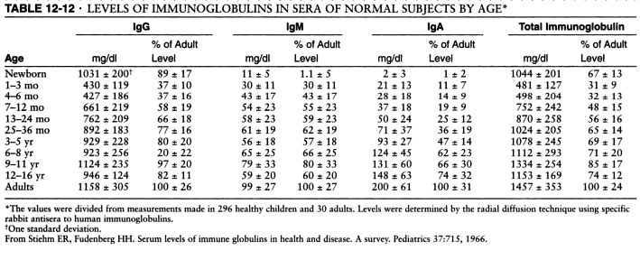
Serum levels of IgM, IgG and IgA. Source: Pediatrics, 1966 and Immunologic disorders in infants and children, by E. Richard Stiehm, Hans D. Ochs, Jerry A. Winkelstein.
Typical pattern of immunoglobulin levels (IgG, IgA, IgM) in humoral immunodeficiency. Click here to enlarge the table.
References
Diagnostic Criteria for Common Variable Immunodeficiency (CVID): Probable and Possible Diagnosis
The diagnostic criteria are divided into three categories: definitive, probable, and possible. There are no criteria for definitive diagnosis of Common Variable Immunodeficiency (CVID) at this time.
To guard against the inclusion of patients who have polymorphic variants in the genes associated with immunodeficiency and to specify the clinical or laboratory finding that is most consistently abnormal in a particular disorder, the patient must fulfill an inclusion criterion that is characteristic of the disorder.
Definitive diagnosis
Patients with a definitive diagnosis are assumed to have a greater than 98% probability that in 20 years they will still be given the same diagnosis. Mutation detection is the most reliable method of making a diagnosis but a single mutation is rarely found in CVID.
Probable diagnosis
Patients with a probable diagnosis are those with all of the clinical and laboratory characteristics of a particular disorder but who do not have a documented abnormality in the gene, the mRNA, or the protein that is known to be abnormal in the disorder. They are assumed to have a greater than 85% probability that in 20 years they will be given the same diagnosis.
Probable diagnosis of CVID:
Male or female patient who has a marked decrease (at least 2 SD below the mean for age) in serum IgG AND IgA and fulfills all of the following criteria:
1. Onset of immunodeficiency at greater than 2 years of age.
2. Absent isohemagglutinins and/or poor response to vaccines.
3. Defined causes of hypogammaglobulinemia have been excluded
Possible diagnosis
Patients with a possible diagnosis are those that have some but not all of the characteristic clinical or laboratory findings of a particular disorder.
Possible diagnosis if CVID:
Male or female patient who has a marked decrease (at least 2 SD below the mean for age) in one of the major isotypes (IgM, IgG, and IgA) and fulfills all of the following criteria:
1. Onset of immunodeficiency at greater than 2 years of age.
2. Absent isohemagglutinins and/or poor response to vaccines.
3. Defined causes of hypogammaglobulinemia have been excluded
Clinical features of CVID
Most patients with CVID are diagnosed with immunodeficiency in the second, third, or fourth decade of life, after they have had several pneumonias; however, children and older adults may be affected.
Viral, fungal, and parasitic infections as well as bacterial infections may be found.
The serum concentration of IgM is normal in about half of the patients.
Abnormalities in T cell numbers or function are common. The majority of patients have normal numbers of B cells; however, some have low or absent B cells.
Approximately 50% of patients have autoimmune manifestations. There is an increased risk of malignancy.
Differential diagnosis of hypogammaglobulinemia includes drug-induced, for example secondary to glucocorticoids (steroids).
Diagnostic Criteria for Primary Immunodeficiencies. Mary Ellen Conley, Luigi D. Notarangelo, and Amos Etzioni Representing PAGID (Pan-American Group for Immunodeficiency) and ESID (European Society for Immunodeficiencies). Clinical Immunology, Vol. 93, No. 3, December, pp. 190–197, 1999.
What is the treatment for CVID?
Intravenous immunoglobulin (IVIG) administered at outpatient clinic, monthly, this is the most common treatment.
Subcutaneous immunoglobulin G (SCIG), administered at home, given weekly.
Summary
CVID is characterized by:
1. Low levels of most or all of the immunoglobulin (Ig) classes
2. Lack of B lymphocytes or plasma cells that are capable of producing antibodies
3. Frequent bacterial infections
CVID is the most common primary immunodeficiency with an incidence of 1 case per 10,000-50,000 population. More than two thirds of patients are aged 21 years or older when CVID is diagnosed. CVID represents a group of heterogeneous conditions. Approximately 50% of patients with the deficiency also have diminished serum immunoglobulin M (IgM) levels and T-lymphocyte dysfunction. About 20% of those with CVID have an autoimmune disease.
The prognosis for patients with CVID is good if they do not have bronchiectasis, autoimmune disease or malignancy.

Five immunoglobulin classes (mind map)
In order of their serum concentrations:
IgG 1000 mg/dL
IgA 200 mg/dL
IgM 150 mg/dL
IgD 4 mg/dL
IgE 0.005 mg/dL (extremely low serum concentration compared to other Ig in (GAMED)
IgG and A are divided in subclasses: 4 for IgG -- IgG1, IgG2, IgG3, IgG4, and 2 for IgA -- IgA1 and IgA2.

Ig structures. Image source: Wikipedia.
Can patients with CVID receive live vaccines?
Patients with CVID should never receive live vaccines and should be advised to avoid contact with children or adults who have recently been given a live vaccine, for example, FluMist® nasal flu vaccine spray.
Can patients with CVID travel to endemic area for hepatitis A or B?
Yes but they should be given hyperimmune Ig prior to travel to endemic areas. The hyperimmune Ig provides protection for 3 weeks.
What is the half-life of IVIG?
3 weeks.
Mnemonic: Dose of IVIG in PIDD
400-600 mg/kg/month
4 letter words:
IVIG
CVID
SCID
What are the long-term risks for patients with CVID?
Malignancies, autoimmune diseases, infections.
FDA guidelines for IVIG state the product should be prepared out of at least 1,000 different human donors. Most commercial IVIG preparations contain products from over 10,000 human donors which increases the risk of acquiring diseases such as HIV, HCV or Creutzfeldt-Jakob disease. The risk is small but nevertheless, it should be discussed with the patient.
What HIV screening test would you do in a patient with CVID?
Patients with CVID can not produce an adequate antibody response to the HIV virus if infected and ELISA or Western blot are not useful. PCR RNA is the test of choise for HIV diagnosis in patients with CVID.
Can patients with CVID still have allergies?
Yes, the production of IgE may not be affected in patients with CVID can have symtpoms of allergic rhinitis, urticaria and other IgE-mediated diseases.
Differences between commercial IVIG preparations: Gammagard and Octagam
Gammagard Liquid is the first 10% IVIG solution with no added carbohydrates or sodium. Baxter is replacing Gammagard S/D with Gammagard Liquid.
Sodium concentration
Gammagard: No added sodium. When reconstituted with the total volume of diluent (Sterile Water for Injection, USP) supplied to 5%, Gammagard contains a physiological concentration of sodium chloride (approximately 8.5 mg/mL) and has a pH of 6.8 ± 0.4.
Octagam: sodium content of the final solution is ≤ 30 mmol/l and the pH is between 5.1 and 6.0.
Osmolality
Gammagard osmolality is 240-300 mOsmol/kg, similar to physiological osmolality (285 to 295 mOsmol/kg).
Octagam osmolality is 310 - 380 mosmol/kg.
Cost
Gammagard Liquid, Baxter, $129.60/gm
Octagam, Octapharma, $118.34/gm
References
Antibody response of pneumococcal vaccine: need for booster dosing? International Journal of Antimicrobial Agents, Volume 14, Issue 2, March 2000, Pages 107-112.
How to identify a possible specific antibody deficiency to pneumococcus. AAAAI, 2007.
Common Variable Immunodeficiency. eMedicine.
Common Variable Immunodeficiency. eMedicine.
Common Variable Immunodeficiency: A Multifaceted And Puzzling Disorder. Expert Review of Clinical Immunology, Medscape, 2009.
Published: 07/12/2008
Updated: 02/06/2012
Reviewer: S. Randhawa, M.D., Allergist/Immunologist and Assistant Professor at NSU
A 65-year-old Caucasian female (CF) is referred by her hematologist and primary care physician (PCP) for work-up of suspected immunodeficiency. She was referred to hematology by her PCP for work-up of frequent urinary tract infections (UTIs) for the last year. Complete blood count with differential (CBCD), bone marrow and chromosomal analysis were negative. Serum immunoglobulins showed low levels of IgG 515 mg/dL (reference range 700 to 1600), IgM 30 (reference range 40 to 230) , IgA 50 (reference range 70 to 400). UA: bacteriuria, no protein. ANA was negative.
Physical examination
Normal.
What is the reason for the low levels of immunoglobulins?
Differential diagnosis:
1. Immunoglobulin loss from GI tract, for example, protein-losing enteropathy. These patients usually have diarrhea but our patient complains of constipation.
2. Immunoglobulin loss in the urine, for example, nephrotic syndrome. IgM (pentamer) and IgA (dimer) are larger molecules than IgG. It is very unlikely to see low levels for IgM in patients with nephrotic syndrome or protein-losing enteropathy. IgG gets filtered in the urine but the large IgM pentamer does not. A 24-hour urine collection is the best method to determine the amount of protein in the urine.
3. Low production of Ig due to bone marrow process or CLL. Both were excluded by BM biopsy and normal CBCD.
4. Common variable immunodeficiency (CVID)
Common variable immunodeficiency (CVID) is the most likely diagnosis in this patient.
How do you diagnose CVID?
A patient with suspected CVID can be tested for the ability to mount humoral and cellular immune response as follows:
1. Humoral immune response.
Inject patient with Pneumovax 23 (not Prevnar, which is a 7-valent conjugate vaccine). Check anti-polysaccharide IgG antibody to pneumococcus serotypes in 3 weeks. There should be a 3-5 fold increase in the anitbody titer to at least 50% of isotypes.
Which serotypes should be included in the order for Ig?
The same serotypes that were included in the given vaccine. Check the enclosed leaflet for this information. For example, the serotypes included in Pneumovax 23 can be found from the Merck website (PDF):
1 2 3 4 5 6B 7F 8 9N 9V 10A 11A 12F 14 15B 17F 18C 19F 19A 20 22F 23F 33F
How to collect the serum for anti-polysaccharide IgG antibodies?
Collect the serum prior to immunization with Pneumovax 23. Store the pre-immunization serum in the office refrigerator. Give the vaccine. Check the post-immunization serum 3 weeks later. Send both pre- and post-immunization serums to the laboratory at the same time.
The pneumococcal vaccine comprises purified capsular polysaccharide of 23 stereotypes that account for more than 90% of the invasive pneumococcal infections in the USA. It induces anti-polysaccharide IgG antibody levels to most or all of the component polysaccharide antigens in immunocompetent adults. Elderly adults respond equally well to vaccination as do younger adults. The current 23-valent vaccine comprises 25 μg of each of 23 pneumococcal stereotypes (namely, serotypes 1, 2, 3, 4, 5, 6B, 7F, 8, 9N, 9V, 10A, 11A, 12F, 14, 15B, 17F, 18C, 19A, 19F, 20, 22F, 23F and 33F).
The test should be ordered as follows: Pre- and post-IgM and IgG antibody titers to pneumococcal serotypes 1, 2, 3, 4, 5, 6B, 7F, 8, 9N, 9V, 10A, 11A, 12F, 14, 15B, 17F, 18C, 19A, 19F, 20, 22F, 23F and 33F
2. Cellular immune response.
Some patients with CVID have decreased cellular immunity as well. Cellular immunity is tested by anergy panel with common antigens to which the patient was likely exposed in the past: Tetanus toxoid, Trichophyton, Candida. An intradermal injection of 0.1 mL of each antigen is necessary to perform the test. The test is read in 48-72 hours. A positive test result indicates intact delayed-type hypersensitivity. A negative test result to all antigens suggests impaired type IV immunity.
Flow cytometry should also be ordered. Lymphocyte proliferation is a more sensitive test to evaluate cellular immune response.
Delayed-type hypersensitivity (DTH) response
The standardized DTH test includes Candida, tetanus, mumps, and TB. Trichophyton is also commonly used. However, the only FDA-approved reagents for DTH are PPD, Candida and mumps.
What are the normal serum immunoglobulin levels (IgG, IgA, IgM)?
Serum levels of IgM, IgG and IgA vary with age, gender and race.
The IgG and IgA concentrations in children show a gradual rise with increasing age. The IgA level is generally about the same in both sexes. Girls typically have higher IgM and IgG levels than boys.
The confidence interval bounded by two standard deviations about the mean excludes 5% of apparently healthy controls.
Elevated IgM, low IgA, low IgG, low IgM, and elevated IgA are the commonest changes observed in apparently healthy humans.
Humoral immunodeficiency is commonly defined as IgG, IgM or IgA level that is two standard deviations (2 SD) below the mean level for IgG, IgM or IgA, respectively, for the particular age group and gender.

Serum levels of IgM, IgG and IgA. Source: Pediatrics, 1966 and Immunologic disorders in infants and children, by E. Richard Stiehm, Hans D. Ochs, Jerry A. Winkelstein.
Typical pattern of immunoglobulin levels (IgG, IgA, IgM) in humoral immunodeficiency. Click here to enlarge the table.
References
Serum immunoglobulin levels in healthy children and adults. J. W. Stoop, B. J. M. Zegers, P. C. Sander, and R. E. Ballieux. Clin Exp Immunol. 1969 January; 4(1): 101–112.
The relationship of race, sex, and age to concentrations of serum immunoglobulins expressed in international units in healthy adults in the USA. S. E. Maddison, C. C. Stewart, C. E. Farshy, and C. B. Reimer. Bull World Health Organ. 1975; 52(2): 179–185.
Serum immunoglobulin concentrations in preschool children measured by laser nephelometry: reference ranges for IgG, IgA, IgM. D Isaacs, D G Altman, C E Tidmarsh, H B Valman, and A D Webster. J Clin Pathol. 1983 October; 36(10): 1193–1196.
D Isaacs, A D Webster, and H B Valman. Clin Exp Immunol. 1984 November; 58(2): 335–340.
Serum Immunoglobulin Levels Throughout the Life-Span of Healthy Man. Ann of Int Med, November 1, 1971, Vol. 75 no. 5 673-682.
Diagnostic Criteria for Common Variable Immunodeficiency (CVID): Probable and Possible Diagnosis
The diagnostic criteria are divided into three categories: definitive, probable, and possible. There are no criteria for definitive diagnosis of Common Variable Immunodeficiency (CVID) at this time.
To guard against the inclusion of patients who have polymorphic variants in the genes associated with immunodeficiency and to specify the clinical or laboratory finding that is most consistently abnormal in a particular disorder, the patient must fulfill an inclusion criterion that is characteristic of the disorder.
Definitive diagnosis
Patients with a definitive diagnosis are assumed to have a greater than 98% probability that in 20 years they will still be given the same diagnosis. Mutation detection is the most reliable method of making a diagnosis but a single mutation is rarely found in CVID.
Probable diagnosis
Patients with a probable diagnosis are those with all of the clinical and laboratory characteristics of a particular disorder but who do not have a documented abnormality in the gene, the mRNA, or the protein that is known to be abnormal in the disorder. They are assumed to have a greater than 85% probability that in 20 years they will be given the same diagnosis.
Probable diagnosis of CVID:
Male or female patient who has a marked decrease (at least 2 SD below the mean for age) in serum IgG AND IgA and fulfills all of the following criteria:
1. Onset of immunodeficiency at greater than 2 years of age.
2. Absent isohemagglutinins and/or poor response to vaccines.
3. Defined causes of hypogammaglobulinemia have been excluded
Possible diagnosis
Patients with a possible diagnosis are those that have some but not all of the characteristic clinical or laboratory findings of a particular disorder.
Possible diagnosis if CVID:
Male or female patient who has a marked decrease (at least 2 SD below the mean for age) in one of the major isotypes (IgM, IgG, and IgA) and fulfills all of the following criteria:
1. Onset of immunodeficiency at greater than 2 years of age.
2. Absent isohemagglutinins and/or poor response to vaccines.
3. Defined causes of hypogammaglobulinemia have been excluded
Clinical features of CVID
Most patients with CVID are diagnosed with immunodeficiency in the second, third, or fourth decade of life, after they have had several pneumonias; however, children and older adults may be affected.
Viral, fungal, and parasitic infections as well as bacterial infections may be found.
The serum concentration of IgM is normal in about half of the patients.
Abnormalities in T cell numbers or function are common. The majority of patients have normal numbers of B cells; however, some have low or absent B cells.
Approximately 50% of patients have autoimmune manifestations. There is an increased risk of malignancy.
Differential diagnosis of hypogammaglobulinemia includes drug-induced, for example secondary to glucocorticoids (steroids).
References
Diagnostic Criteria for Primary Immunodeficiencies. Mary Ellen Conley, Luigi D. Notarangelo, and Amos Etzioni Representing PAGID (Pan-American Group for Immunodeficiency) and ESID (European Society for Immunodeficiencies). Clinical Immunology, Vol. 93, No. 3, December, pp. 190–197, 1999.
Recognizing Primary Immune Deficiency in Clinical Practice. Clinical and Vaccine Immunology, March 2006, p. 329-332, Vol. 13, No. 3.
What is the treatment for CVID?
Intravenous immunoglobulin (IVIG) administered at outpatient clinic, monthly, this is the most common treatment.
Subcutaneous immunoglobulin G (SCIG), administered at home, given weekly.
Summary
CVID is characterized by:
1. Low levels of most or all of the immunoglobulin (Ig) classes
2. Lack of B lymphocytes or plasma cells that are capable of producing antibodies
3. Frequent bacterial infections
CVID is the most common primary immunodeficiency with an incidence of 1 case per 10,000-50,000 population. More than two thirds of patients are aged 21 years or older when CVID is diagnosed. CVID represents a group of heterogeneous conditions. Approximately 50% of patients with the deficiency also have diminished serum immunoglobulin M (IgM) levels and T-lymphocyte dysfunction. About 20% of those with CVID have an autoimmune disease.
The prognosis for patients with CVID is good if they do not have bronchiectasis, autoimmune disease or malignancy.

Five immunoglobulin classes (mind map)
In order of their serum concentrations:
IgG 1000 mg/dL
IgA 200 mg/dL
IgM 150 mg/dL
IgD 4 mg/dL
IgE 0.005 mg/dL (extremely low serum concentration compared to other Ig in (GAMED)
IgG and A are divided in subclasses: 4 for IgG -- IgG1, IgG2, IgG3, IgG4, and 2 for IgA -- IgA1 and IgA2.

Ig structures. Image source: Wikipedia.
Can patients with CVID receive live vaccines?
Patients with CVID should never receive live vaccines and should be advised to avoid contact with children or adults who have recently been given a live vaccine, for example, FluMist® nasal flu vaccine spray.
Can patients with CVID travel to endemic area for hepatitis A or B?
Yes but they should be given hyperimmune Ig prior to travel to endemic areas. The hyperimmune Ig provides protection for 3 weeks.
What is the half-life of IVIG?
3 weeks.
Mnemonic: Dose of IVIG in PIDD
400-600 mg/kg/month
4 letter words:
IVIG
CVID
SCID
What are the long-term risks for patients with CVID?
Malignancies, autoimmune diseases, infections.
FDA guidelines for IVIG state the product should be prepared out of at least 1,000 different human donors. Most commercial IVIG preparations contain products from over 10,000 human donors which increases the risk of acquiring diseases such as HIV, HCV or Creutzfeldt-Jakob disease. The risk is small but nevertheless, it should be discussed with the patient.
What HIV screening test would you do in a patient with CVID?
Patients with CVID can not produce an adequate antibody response to the HIV virus if infected and ELISA or Western blot are not useful. PCR RNA is the test of choise for HIV diagnosis in patients with CVID.
Can patients with CVID still have allergies?
Yes, the production of IgE may not be affected in patients with CVID can have symtpoms of allergic rhinitis, urticaria and other IgE-mediated diseases.
Differences between commercial IVIG preparations: Gammagard and Octagam
Gammagard Liquid is the first 10% IVIG solution with no added carbohydrates or sodium. Baxter is replacing Gammagard S/D with Gammagard Liquid.
Sodium concentration
Gammagard: No added sodium. When reconstituted with the total volume of diluent (Sterile Water for Injection, USP) supplied to 5%, Gammagard contains a physiological concentration of sodium chloride (approximately 8.5 mg/mL) and has a pH of 6.8 ± 0.4.
Octagam: sodium content of the final solution is ≤ 30 mmol/l and the pH is between 5.1 and 6.0.
Osmolality
Gammagard osmolality is 240-300 mOsmol/kg, similar to physiological osmolality (285 to 295 mOsmol/kg).
Octagam osmolality is 310 - 380 mosmol/kg.
Cost
Gammagard Liquid, Baxter, $129.60/gm
Octagam, Octapharma, $118.34/gm
References
Antibody response of pneumococcal vaccine: need for booster dosing? International Journal of Antimicrobial Agents, Volume 14, Issue 2, March 2000, Pages 107-112.
How to identify a possible specific antibody deficiency to pneumococcus. AAAAI, 2007.
Common Variable Immunodeficiency. eMedicine.
Common Variable Immunodeficiency. eMedicine.
Common Variable Immunodeficiency: A Multifaceted And Puzzling Disorder. Expert Review of Clinical Immunology, Medscape, 2009.
Primary Immunodeficiency Diseases: Definition, Diagnosis, and Management. Nima Rezaei, Luigi D. Notarangelo, Asghar Aghamohammadi, 2008, Google eBook.
Common variable immunodeficiency (CVID). Allergologia et Immunopathologia. November 2006. Volume 34 - Number 06 p. 263 - 275.
Adaptive Humoral Immunity: B-cells and Immunoglobulins
Mnemonics: Adaptive Humoral Immunity: B-cells and Immunoglobulins
Intravenous immune globulin. Hopkins HIV Guide.
Common variable immunodeficiency (CVID). Allergologia et Immunopathologia. November 2006. Volume 34 - Number 06 p. 263 - 275.
Adaptive Humoral Immunity: B-cells and Immunoglobulins
Mnemonics: Adaptive Humoral Immunity: B-cells and Immunoglobulins
Intravenous immune globulin. Hopkins HIV Guide.
Evaluation and diagnosis of common variable immunodeficiency. UpToDate.
Octagam, Octapharma.
Gammagard Liquid, Baxter.
Octagam, Octapharma.
Gammagard Liquid, Baxter.
Interpretation of pneumococcal antibody titers - AAAAI Ask the Expert, 2011.
Assessment of antibody response to pneumococcus immunization. AAAAI, Ask the Expert, 2011.
Cost-effectiveness of Pneumococcal Conjugate Vaccine vs. Polysaccharide Vaccine in Adults: PCV13 was better than PPSV23. JAMA, 2012.
Cost-effectiveness of Pneumococcal Conjugate Vaccine vs. Polysaccharide Vaccine in Adults: PCV13 was better than PPSV23. JAMA, 2012.
Published: 07/12/2008
Updated: 02/06/2012
Antibody titer responses to pneumococcal vaccination in common variable immunodeficiency (CVID)
Author: V. Dimov, M.D., Allergist/Immunologist and Assistant Professor at University of Chicago
Reviewer: S. Randhawa, M.D., Allergist/Immunologist and Assistant Professor at NSU
A 54-year-old Caucasian male (CM) is evaluated by the Allergy and Immunology clinic for recurrent sinusitis for the last 20-30 years with 5-6 episodes per year. His IgG level was 470 mg/dL two months ago and IgM and IgA levels were normal. He reports no other significant infections. The patient reports seasonal exacerbations of his nasal symptoms in the spring, fall and winter. His last sinus infection was 9 months ago and he has not had such a recurrence free period in years.
Past medical history
Hypertension (HTN), hyperlipidemia (HLP), sinusitis, non-allergic rhinitis (non-AR), dermatographism.
Medications
Fexofenadine, flunisolide nasal spray, lisinopril.
Physical examination
Normal.
What is the most likely diagnosis?
Common Variable Immunodeficiency (CVID).
Allergic or non-allergic rhinitis.
What tests would you order?
Pre- and post-pneumococcal immunization titers.
ANA, HIV, RF.
B and T-cell flow cytometry.
RAST to aeroallergens.
What happened?
Skin prick testing: uninterpretable due to dermatographism. RAST to aeroallergens: negative.
HIV, ANA, RF were negative. CXR and U/A without evidence of malignancy. He had a colonoscopy in the past 3 years and it was negative.


Pre-immunization titers (left). Post-immunization titers (right) (click to enlarge the images).
Pre- and post-pneumococcal immunization titers 4 weeks apart showed an insufficient increase in the antibody titers: only 30% (4 out of 13 titers) of specific antibodies increased by the magnitude of 2 fold.
Flow Cytometry. Specimen: Peripheral Blood. Processing: Lyse, cytospin, stain with monoclonal antibodies. Cell Count: 8.2 10*6 total cells. Viability: 89%.
Total events analyzed = 42415
A gate Bright CD45 positive events = 5758 (17% of total events analyzed)
Interpretation: Lymphocytes comprise 17% of the total events analyzed, with 3% B-cells, 85% T-cells, and 11% NK cells. The CD4:CD8 ratio is 2.7:1. 9% of the lymphocytes are CD25 (dim) positive T-cells.
In healthy subjects, B cells constitute 18-47% of the peripheral lymphocytes. B cell count can be either low or normal in patients with CVID.
What happened next?
The patient was diagnosed with common variable immunodeficiency (CVID).
Would you start intravenous immunoglobulin G (IVIG) therapy for this patient?
Historically, the level of IgG of 470 mg/dL did not typically require replacement with IVIG in the absence of severe recurrent infections. Some of the newer studies point to an optimal replacement level of IgG of 600-800 mg/dL.
We decided to repeat the quantitative serum immunoglobulins in 4-6 months and to decide about IVIG replacement at that point.
Final diagnosis
Common Variable Immunodeficiency (CVID).
What did we learn from this case?
Evaluation of the response to pneumococcal vaccination is best accomplished by comparing pre-vaccination and post-vaccination antibody levels. A 2 to 4 fold increase in type-specific antibodies measured 4-6 weeks after vaccination is expected in immunocompetent adults. The number of serotypes for which a 2 to 4 fold increase is observed varies greatly among individuals; a consensus panel has suggested that individuals older than 5 years should respond to at least approximately 70% of pneumococcal serotypes. Adults older than 65 years may exhibit a smaller (less than 2 fold) increase in type specific antibody levels.
The "rule of thumb" is that 2/3 of titers have to increase 2-3 fold.
The patient did not have evidence of allergen sensitization on ImmunoCAP test for aeroallergens, therefore the intranasal steroids and oral antihistamine were discontinued. He was advised to use nasal saline rinses BID.
What are the normal serum immunoglobulin levels (IgG, IgA, IgM)?
Serum levels of IgM, IgG and IgA vary with age, gender and race.
The IgG and IgA concentrations in children show a gradual rise with increasing age. The IgA level is generally about the same in both sexes. Girls typically have higher IgM and IgG levels than boys.
The confidence interval bounded by two standard deviations about the mean excludes 5% of apparently healthy controls.
Elevated IgM, low IgA, low IgG, low IgM, and elevated IgA are the commonest changes observed in apparently healthy humans.
Humoral immunodeficiency is commonly defined as IgG, IgM or IgA level that is two standard deviations (2 SD) below the mean level for IgG, IgM or IgA, respectively, for the particular age group and gender.
Serum levels of IgM, IgG and IgA. Source: Pediatrics, 1966 and Immunologic disorders in infants and children, by E. Richard Stiehm, Hans D. Ochs, Jerry A. Winkelstein.
Typical pattern of immunoglobulin levels (IgG, IgA, IgM) in humoral immunodeficiency. Click here to enlarge the table.
What is a normal response to pneumococcal immunization?
Protection against infection and colonization is associated with specific IgG concentrations of 1.3 mcg/ml.
A second component in judging response is the final concentration of antibodies after immunization regardless of increase from preimmunization concentration. An adequate response is a post-immunization antibody concentration of more than 1.3 mcg/ml, even in the absence of a fourfold increase.
Children 2 to 5 years of age are normally expected to have an adequate response to more than 50% of serotypes tested. Patients 6 years or older are normally expected to respond to more than 70% of the serotypes tested. Source: Assessment and clinical interpretation of polysaccharide antibody responses. Kenneth Paris and Ricardo Sorensen. Annals of Allergy, Asthma, and Immunology, 2007; volume 99, Issue 5, Pages 462-464, and AAAAI Ask the Expert, 2012.
References
Diagnostic Criteria for Common Variable Immunodeficiency (CVID): Probable and Possible Diagnosis
The diagnostic criteria are divided into three categories: definitive, probable, and possible. There are no criteria for definitive diagnosis of Common Variable Immunodeficiency (CVID) at this time.
To guard against the inclusion of patients who have polymorphic variants in the genes associated with immunodeficiency and to specify the clinical or laboratory finding that is most consistently abnormal in a particular disorder, the patient must fulfill an inclusion criterion that is characteristic of the disorder.
Definitive diagnosis
Patients with a definitive diagnosis are assumed to have a greater than 98% probability that in 20 years they will still be given the same diagnosis. Mutation detection is the most reliable method of making a diagnosis but a single mutation is rarely found in CVID.
Probable diagnosis
Patients with a probable diagnosis are those with all of the clinical and laboratory characteristics of a particular disorder but who do not have a documented abnormality in the gene, the mRNA, or the protein that is known to be abnormal in the disorder. They are assumed to have a greater than 85% probability that in 20 years they will be given the same diagnosis.
Probable diagnosis of CVID:
Male or female patient who has a marked decrease (at least 2 SD below the mean for age) in serum IgG AND IgA and fulfills all of the following criteria:
1. Onset of immunodeficiency at greater than 2 years of age.
2. Absent isohemagglutinins and/or poor response to vaccines.
3. Defined causes of hypogammaglobulinemia have been excluded
Possible diagnosis
Patients with a possible diagnosis are those that have some but not all of the characteristic clinical or laboratory findings of a particular disorder.
Possible diagnosis if CVID:
Male or female patient who has a marked decrease (at least 2 SD below the mean for age) in one of the major isotypes (IgM, IgG, and IgA) and fulfills all of the following criteria:
1. Onset of immunodeficiency at greater than 2 years of age.
2. Absent isohemagglutinins and/or poor response to vaccines.
3. Defined causes of hypogammaglobulinemia have been excluded
Clinical features of CVID
Most patients with CVID are diagnosed with immunodeficiency in the second, third, or fourth decade of life, after they have had several pneumonias; however, children and older adults may be affected.
Viral, fungal, and parasitic infections as well as bacterial infections may be found.
The serum concentration of IgM is normal in about half of the patients.
Abnormalities in T cell numbers or function are common. The majority of patients have normal numbers of B cells; however, some have low or absent B cells.
Approximately 50% of patients have autoimmune manifestations. There is an increased risk of malignancy.
Differential diagnosis of hypogammaglobulinemia includes drug-induced, for example secondary to glucocorticoids (steroids).
Diagnostic Criteria for Primary Immunodeficiencies. Mary Ellen Conley, Luigi D. Notarangelo, and Amos Etzioni Representing PAGID (Pan-American Group for Immunodeficiency) and ESID (European Society for Immunodeficiencies). Clinical Immunology, Vol. 93, No. 3, December, pp. 190–197, 1999.
Common Variable Immunodeficiency. eMedicine.
Common Variable Immunodeficiency: A Multifaceted And Puzzling Disorder. Expert Review of Clinical Immunology, Medscape, 2009.
Reviewer: S. Randhawa, M.D., Allergist/Immunologist and Assistant Professor at NSU
A 54-year-old Caucasian male (CM) is evaluated by the Allergy and Immunology clinic for recurrent sinusitis for the last 20-30 years with 5-6 episodes per year. His IgG level was 470 mg/dL two months ago and IgM and IgA levels were normal. He reports no other significant infections. The patient reports seasonal exacerbations of his nasal symptoms in the spring, fall and winter. His last sinus infection was 9 months ago and he has not had such a recurrence free period in years.
Past medical history
Hypertension (HTN), hyperlipidemia (HLP), sinusitis, non-allergic rhinitis (non-AR), dermatographism.
Medications
Fexofenadine, flunisolide nasal spray, lisinopril.
Physical examination
Normal.
What is the most likely diagnosis?
Common Variable Immunodeficiency (CVID).
Allergic or non-allergic rhinitis.
What tests would you order?
Pre- and post-pneumococcal immunization titers.
ANA, HIV, RF.
B and T-cell flow cytometry.
RAST to aeroallergens.
What happened?
Skin prick testing: uninterpretable due to dermatographism. RAST to aeroallergens: negative.
HIV, ANA, RF were negative. CXR and U/A without evidence of malignancy. He had a colonoscopy in the past 3 years and it was negative.


Pre-immunization titers (left). Post-immunization titers (right) (click to enlarge the images).
Pre- and post-pneumococcal immunization titers 4 weeks apart showed an insufficient increase in the antibody titers: only 30% (4 out of 13 titers) of specific antibodies increased by the magnitude of 2 fold.
Flow Cytometry. Specimen: Peripheral Blood. Processing: Lyse, cytospin, stain with monoclonal antibodies. Cell Count: 8.2 10*6 total cells. Viability: 89%.
Total events analyzed = 42415
A gate Bright CD45 positive events = 5758 (17% of total events analyzed)
| CD45+ | 100% |
| CD19+/CD20 | 3% |
| CD19+/CD38 | 2% |
| CD19+/CD10 | less than 2% |
| CD3+/CD5+ | 85% |
| CD3+/CD7+ | 82% |
| CD3+/CD4+ | 60% |
| CD3+/CD8+ | 22% |
| CD3-/CD56+ | 11% |
| Total CD25 | 9% |
| CD25+/CD3+ | 9% |
| CD25+/CD5+ | 9% |
Interpretation: Lymphocytes comprise 17% of the total events analyzed, with 3% B-cells, 85% T-cells, and 11% NK cells. The CD4:CD8 ratio is 2.7:1. 9% of the lymphocytes are CD25 (dim) positive T-cells.
In healthy subjects, B cells constitute 18-47% of the peripheral lymphocytes. B cell count can be either low or normal in patients with CVID.
What happened next?
The patient was diagnosed with common variable immunodeficiency (CVID).
Would you start intravenous immunoglobulin G (IVIG) therapy for this patient?
Historically, the level of IgG of 470 mg/dL did not typically require replacement with IVIG in the absence of severe recurrent infections. Some of the newer studies point to an optimal replacement level of IgG of 600-800 mg/dL.
We decided to repeat the quantitative serum immunoglobulins in 4-6 months and to decide about IVIG replacement at that point.
Final diagnosis
Common Variable Immunodeficiency (CVID).
What did we learn from this case?
Evaluation of the response to pneumococcal vaccination is best accomplished by comparing pre-vaccination and post-vaccination antibody levels. A 2 to 4 fold increase in type-specific antibodies measured 4-6 weeks after vaccination is expected in immunocompetent adults. The number of serotypes for which a 2 to 4 fold increase is observed varies greatly among individuals; a consensus panel has suggested that individuals older than 5 years should respond to at least approximately 70% of pneumococcal serotypes. Adults older than 65 years may exhibit a smaller (less than 2 fold) increase in type specific antibody levels.
The "rule of thumb" is that 2/3 of titers have to increase 2-3 fold.
The patient did not have evidence of allergen sensitization on ImmunoCAP test for aeroallergens, therefore the intranasal steroids and oral antihistamine were discontinued. He was advised to use nasal saline rinses BID.
What are the normal serum immunoglobulin levels (IgG, IgA, IgM)?
Serum levels of IgM, IgG and IgA vary with age, gender and race.
The IgG and IgA concentrations in children show a gradual rise with increasing age. The IgA level is generally about the same in both sexes. Girls typically have higher IgM and IgG levels than boys.
The confidence interval bounded by two standard deviations about the mean excludes 5% of apparently healthy controls.
Elevated IgM, low IgA, low IgG, low IgM, and elevated IgA are the commonest changes observed in apparently healthy humans.
Humoral immunodeficiency is commonly defined as IgG, IgM or IgA level that is two standard deviations (2 SD) below the mean level for IgG, IgM or IgA, respectively, for the particular age group and gender.
Serum levels of IgM, IgG and IgA. Source: Pediatrics, 1966 and Immunologic disorders in infants and children, by E. Richard Stiehm, Hans D. Ochs, Jerry A. Winkelstein.
Typical pattern of immunoglobulin levels (IgG, IgA, IgM) in humoral immunodeficiency. Click here to enlarge the table.
What is a normal response to pneumococcal immunization?
Protection against infection and colonization is associated with specific IgG concentrations of 1.3 mcg/ml.
A second component in judging response is the final concentration of antibodies after immunization regardless of increase from preimmunization concentration. An adequate response is a post-immunization antibody concentration of more than 1.3 mcg/ml, even in the absence of a fourfold increase.
Children 2 to 5 years of age are normally expected to have an adequate response to more than 50% of serotypes tested. Patients 6 years or older are normally expected to respond to more than 70% of the serotypes tested. Source: Assessment and clinical interpretation of polysaccharide antibody responses. Kenneth Paris and Ricardo Sorensen. Annals of Allergy, Asthma, and Immunology, 2007; volume 99, Issue 5, Pages 462-464, and AAAAI Ask the Expert, 2012.
References
Serum immunoglobulin levels in healthy children and adults. J. W. Stoop, B. J. M. Zegers, P. C. Sander, and R. E. Ballieux. Clin Exp Immunol. 1969 January; 4(1): 101–112.
The relationship of race, sex, and age to concentrations of serum immunoglobulins expressed in international units in healthy adults in the USA. S. E. Maddison, C. C. Stewart, C. E. Farshy, and C. B. Reimer. Bull World Health Organ. 1975; 52(2): 179–185.
Serum immunoglobulin concentrations in preschool children measured by laser nephelometry: reference ranges for IgG, IgA, IgM. D Isaacs, D G Altman, C E Tidmarsh, H B Valman, and A D Webster. J Clin Pathol. 1983 October; 36(10): 1193–1196.
D Isaacs, A D Webster, and H B Valman. Clin Exp Immunol. 1984 November; 58(2): 335–340.
Serum Immunoglobulin Levels Throughout the Life-Span of Healthy Man. Ann of Int Med, November 1, 1971, Vol. 75 no. 5 673-682.
Diagnostic Criteria for Common Variable Immunodeficiency (CVID): Probable and Possible Diagnosis
The diagnostic criteria are divided into three categories: definitive, probable, and possible. There are no criteria for definitive diagnosis of Common Variable Immunodeficiency (CVID) at this time.
To guard against the inclusion of patients who have polymorphic variants in the genes associated with immunodeficiency and to specify the clinical or laboratory finding that is most consistently abnormal in a particular disorder, the patient must fulfill an inclusion criterion that is characteristic of the disorder.
Definitive diagnosis
Patients with a definitive diagnosis are assumed to have a greater than 98% probability that in 20 years they will still be given the same diagnosis. Mutation detection is the most reliable method of making a diagnosis but a single mutation is rarely found in CVID.
Probable diagnosis
Patients with a probable diagnosis are those with all of the clinical and laboratory characteristics of a particular disorder but who do not have a documented abnormality in the gene, the mRNA, or the protein that is known to be abnormal in the disorder. They are assumed to have a greater than 85% probability that in 20 years they will be given the same diagnosis.
Probable diagnosis of CVID:
Male or female patient who has a marked decrease (at least 2 SD below the mean for age) in serum IgG AND IgA and fulfills all of the following criteria:
1. Onset of immunodeficiency at greater than 2 years of age.
2. Absent isohemagglutinins and/or poor response to vaccines.
3. Defined causes of hypogammaglobulinemia have been excluded
Possible diagnosis
Patients with a possible diagnosis are those that have some but not all of the characteristic clinical or laboratory findings of a particular disorder.
Possible diagnosis if CVID:
Male or female patient who has a marked decrease (at least 2 SD below the mean for age) in one of the major isotypes (IgM, IgG, and IgA) and fulfills all of the following criteria:
1. Onset of immunodeficiency at greater than 2 years of age.
2. Absent isohemagglutinins and/or poor response to vaccines.
3. Defined causes of hypogammaglobulinemia have been excluded
Clinical features of CVID
Most patients with CVID are diagnosed with immunodeficiency in the second, third, or fourth decade of life, after they have had several pneumonias; however, children and older adults may be affected.
Viral, fungal, and parasitic infections as well as bacterial infections may be found.
The serum concentration of IgM is normal in about half of the patients.
Abnormalities in T cell numbers or function are common. The majority of patients have normal numbers of B cells; however, some have low or absent B cells.
Approximately 50% of patients have autoimmune manifestations. There is an increased risk of malignancy.
Differential diagnosis of hypogammaglobulinemia includes drug-induced, for example secondary to glucocorticoids (steroids).
References
Diagnostic Criteria for Primary Immunodeficiencies. Mary Ellen Conley, Luigi D. Notarangelo, and Amos Etzioni Representing PAGID (Pan-American Group for Immunodeficiency) and ESID (European Society for Immunodeficiencies). Clinical Immunology, Vol. 93, No. 3, December, pp. 190–197, 1999.
Recognizing Primary Immune Deficiency in Clinical Practice. Clinical and Vaccine Immunology, March 2006, p. 329-332, Vol. 13, No. 3.
Common Variable Immunodeficiency. eMedicine.Common Variable Immunodeficiency. eMedicine.
Common Variable Immunodeficiency: A Multifaceted And Puzzling Disorder. Expert Review of Clinical Immunology, Medscape, 2009.
Interpretation of pneumococcal antibody titers. AAAAI, Ask the Expert, 2011.
Assessment of antibody response to pneumococcus immunization. AAAAI, Ask the Expert, 2011.
Heptavalent pneumococcal conjugate vaccine (PCV7) targets only 7 of the more than 92 pneumococcal serotypes. Lancet, 2011.
Cost-effectiveness of Pneumococcal Conjugate Vaccine vs. Polysaccharide Vaccine in Adults: PCV13 was better than PPSV23. JAMA, 2012.
Published: 02/06/2009
Updated: 02/07/2012
Heptavalent pneumococcal conjugate vaccine (PCV7) targets only 7 of the more than 92 pneumococcal serotypes. Lancet, 2011.
Cost-effectiveness of Pneumococcal Conjugate Vaccine vs. Polysaccharide Vaccine in Adults: PCV13 was better than PPSV23. JAMA, 2012.
Published: 02/06/2009
Updated: 02/07/2012
Subscribe to:
Posts (Atom)